
 ZEISS is introducing an enhanced workflow with the ZEISS Axioscan 7 spatial biology automated slide scanner, with an integrated newly launched data analysis solution, now offering seamless compatibility with a broad range of established tissue multiplexing reagent chemistries. The new workflow enables researchers and translational scientists to accelerate biomarker validation and clinical studies, driving faster impact for patients...
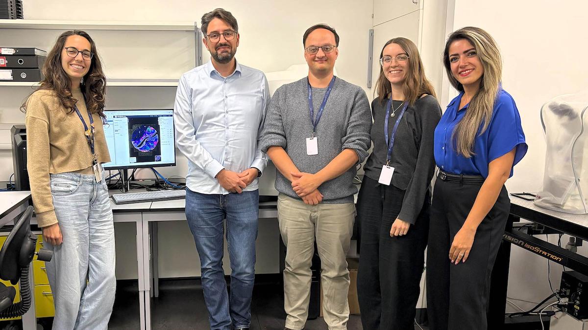 ZEISS Microscopy and Concept Life Sciences, a UK-based contract research organization (CRO), announced their collaboration aimed at pioneering new approaches to workflow automation and scalable image analysis in spatial biology. Key focus areas of the collaboration are: Automated high-throughput imaging workflow: Utilizing ZEISS Axioscan 7 spatial biology to streamline imaging processes...
 ZEISS partners with Kromnigon to launch multiplex IHC with 8 fluorescence channels using an accelerated heat-treatment-free TSA workflow. Kromnigon and ZEISS partner to develop and launch a tailored StreptaClick® HRP-based kit, specifically optimized for the ZEISS Axioscan 7 spatial biology automated slide scanner. This new kit targets the rapidly growing spatial biology market and delivers powerful multiplex immunofluorescence imaging of eight channels using Tyramide Signal Amplification (TSA).
 In early July, Carl Zeiss Microscopy GmbH has acquired all equity shares of Pi Imaging Technology SA, based in Lausanne, Switzerland. Pi Imaging Technology SA now operates as "Pi Imaging Technology SA – a ZEISS company". The Lausanne location with all employees will be retained. Pi Imaging Technology SA has been a trusted partner of ZEISS Research Microscopy Solutions for many years...
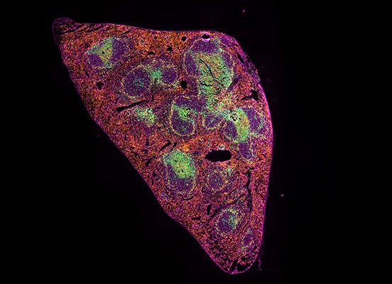 Zeiss simplify high-throughput multiplex IF imaging and analysis, making it robust, scalable and accessible. Our seamless tissue multiplexing workflow aims to empower routine histopathology labs, including those with no prior spatial biology expertise, to generate comprehensive, reproducible biomarker data across large sample cohorts. By combining automation, optimized imaging, and AI-powered analysis...
 ZEISS announces the introduction of its highly flexible and efficient software suite, ZEN core, for operating all ZEISS scanning electron microscopes (SEMs), including focused ion beam scanning electron microscopes (FIB-SEMs). ZEN stands for ZEISS Efficient Navigation and lets microscopists obtain more meaningful information. Users across various fields – including materials research, natural resources, electronics, and life sciences...
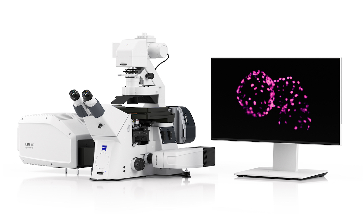 ZEISS announces the launch of Lightfield 4D, a new microscopy technology based on the light-field principle. As a new imaging mode integrated into the new ZEISS LSM 910 and LSM 990 confocal microscope systems, ZEISS Lightfield 4D redefines the way researchers observe living organisms, especially in the neurosciences, cancer research, developmental biology, and plant sciences...
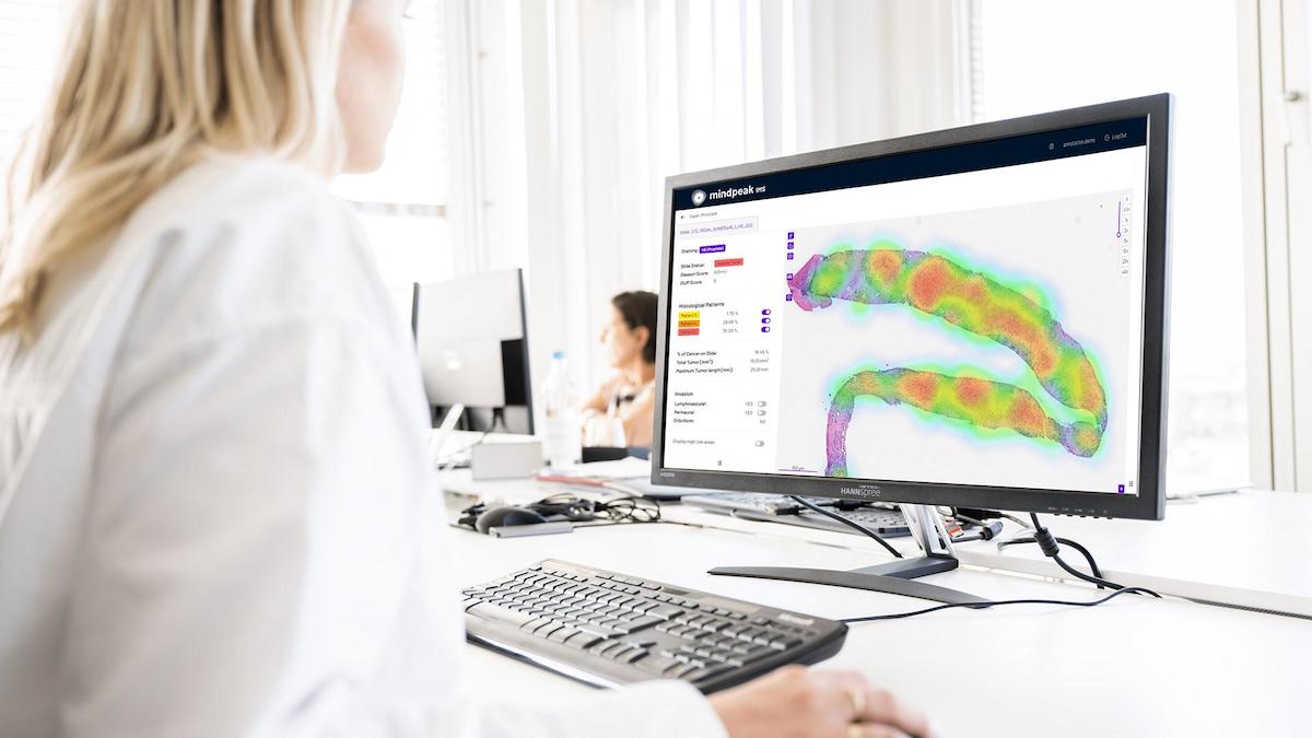 ZEISS partners with Mindpeak, a pioneer in AI-powered pathology solutions, to develop an integrated Multiplex Immunofluorescence (mIF) solution that serves pathologists across research, diagnostics, and clinical applications. Mindpeak’s expertise in AI-based image analysis of pathological tissues, particularly through its established algorithms for clinical routine applications, complements ZEISS's advanced instruments and software for research applications of mIF...
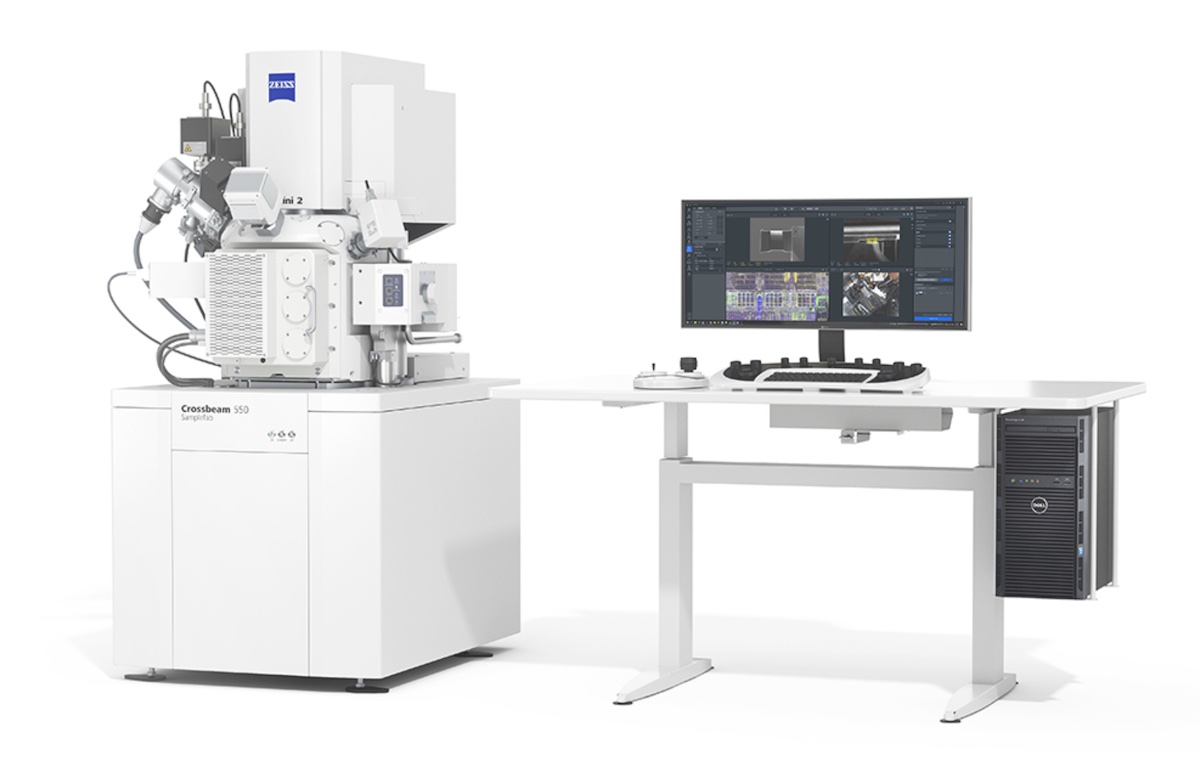 ZEISS announces the new ZEISS Crossbeam 550 Samplefab, a focused ion beam scanning electron microscope (FIB-SEM) optimized for fully automated preparation of transmission electron microscopy (TEM) samples (lamellae). Built for efficiency and throughput in the semiconductor lab, ZEISS Crossbeam 550 Samplefab provides recipe-based automation for the routine TEM sample preparation work of bulk milling, lift-out and thinning at any number of target points on the sample...
 ZEISS announces its full schedule of activities at the 50th annual ISTFA Conference, taking place at the Hilton San Diego Bayfront Hotel, Oct. 28-Nov. 1, 2024. ZEISS will deliver four tutorials and six paper presentations showcasing its expertise in artificial intelligence (AI)-powered fault isolation, failure analysis (FA) and yield enhancement. ZEISS will also participate in two panel discussions about the industry roadmap and AI and will be part of the Tools of the Trade tour...
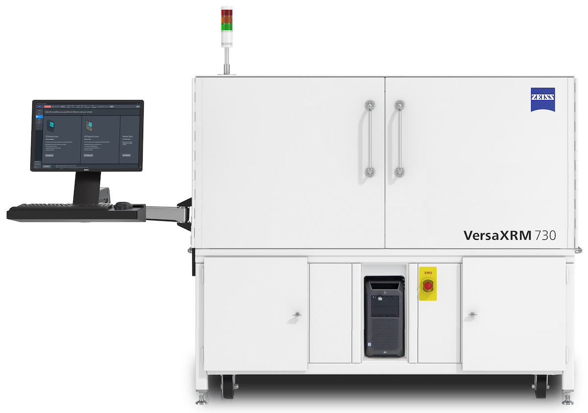 ZEISS launches its latest innovation in the field of X-ray microscopy: ZEISS VersaXRM 730®. The new system offers researchers an unprecedented level of performance, choice, and accessibility. As technology continues to evolve at a rapid pace, it is essential for researchers to have access to cutting-edge capabilities that can keep up with the changing landscape...
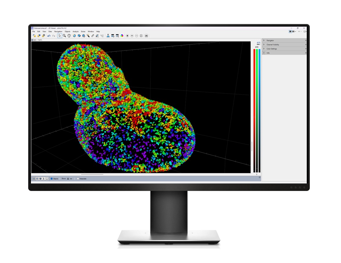 Flexibility in image analysis is key to addressing your diverse research questions. The ZEISS arivis software suite is designed to accelerate your research, offering advanced tools that ensure you reach reliable and reproducible results faster. No matter the size, format, or source of your image, ZEISS arivis can help you analyze it. And all without the need to code. Version 4.2 of arivis Pro is available now...
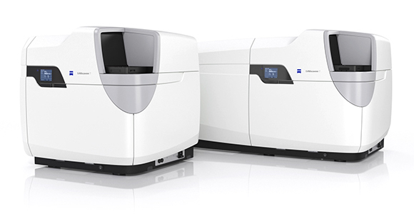 If your research requires explorative high-content imaging, you are often faced with a trade-off between the desired image quality and the need to capture large amounts of data efficiently. ZEISS Celldiscoverer 7 is your research companion for collecting statistically relevant data, giving you easy access to high-quality imaging, adaptability to demanding experiments, and stable long-term operation...
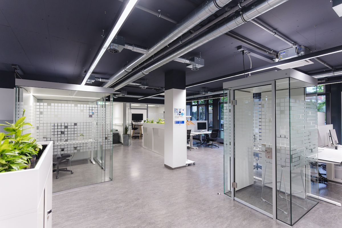 The new premises of the ZEISS Innovation Hub Dresden opened on 7 May. The new area offers the twelve employees more room for additional innovation projects and the opportunity for further growth. The team is moving from the premises in the building of the Else Kröner Fresenius Center (EKFZ) for Digital Health on the campus of the Carl Gustav Carus University Hospital in Dresden to a building in Blasewitzer Straße, directly opposite the campus...
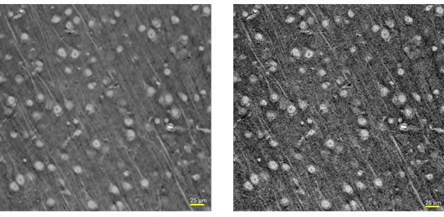 The ART is an innovative platform delivering state-of-the-art reconstruction technologies based on deep learning and cloud computing to enhance the performances of your ZEISS Xradia 3D XRM. It is indeed a set of image reconstruction technologies to significantly enhance XRM performances. Zeiss invite you to join their free online webinar on February 6th in order to learn more about this fascinating technology and its possibilities...
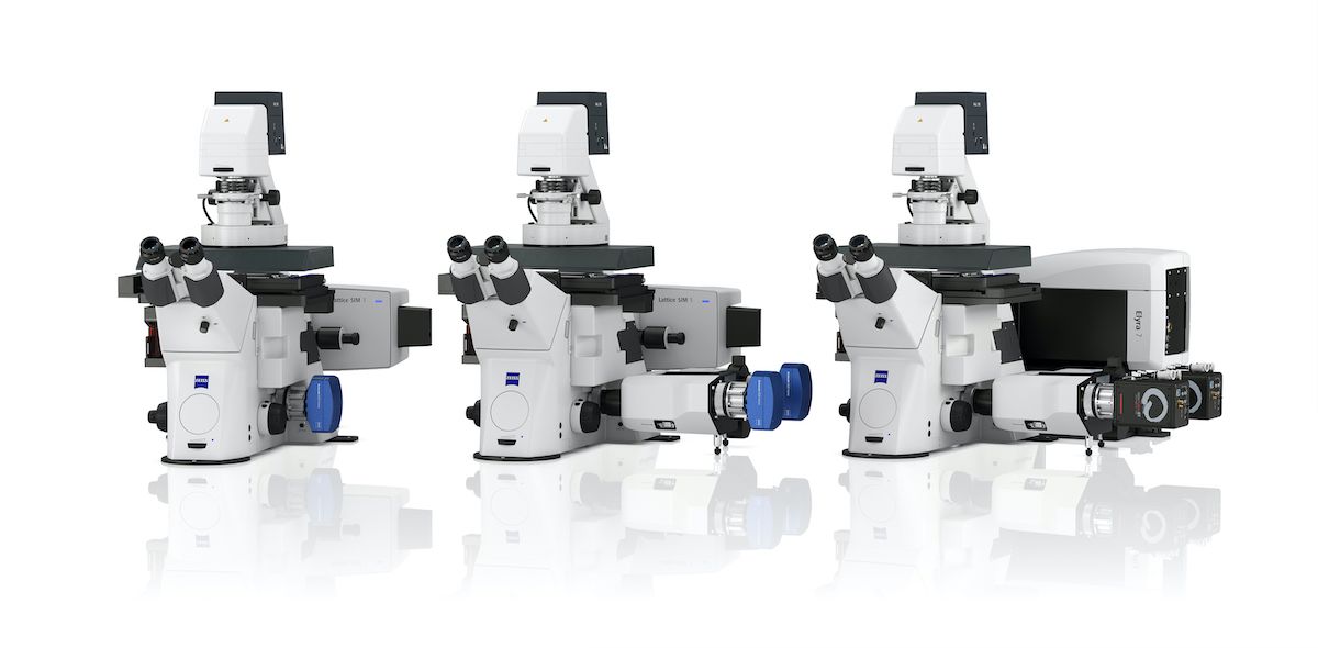 To help researchers unlock the full potential of structured illumination microscopy (SIM), two new systems will join the established super-resolution microscope ZEISS Elyra 7 to create a new Lattice SIM product family. Like ZEISS Elyra 7, the new members of the product family – ZEISS Lattice SIM 3 and ZEISS Lattice SIM 5 – use structured illumination to overcome the physical resolution limits of light microscopy. Each system has implemented this technology to address specific fields of application...
 The European Molecular Biology Laboratory (EMBL) and ZEISS have entered a long-term strategic partnership. The collaboration aims to close the gap between early-stage imaging technology development and its application in life science research. Through this collaborative approach, users of the EMBL Imaging Centre and related EMBL imaging services will have access to the latest microscopy technologies and expertise from ZEISS...
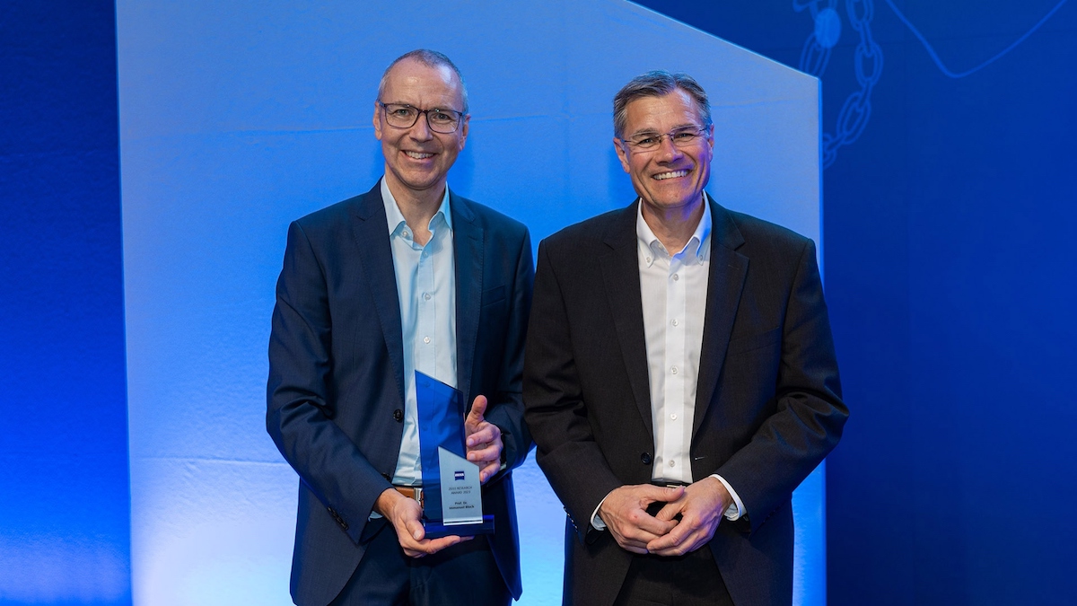 On Monday 27 June, ZEISS presented two research awards to a total of four scientists in ceremonial style. Prof. Dr. Immanuel Bloch was the recipient of the ZEISS Research Award, while Dr. Simon Baier, Dr. Arindam Ghosh and Dr. Dasha Nelidova all received the Carl Zeiss Award for Young Researchers. Through presenting such awards, the company can commend outstanding research in optics and photonics...
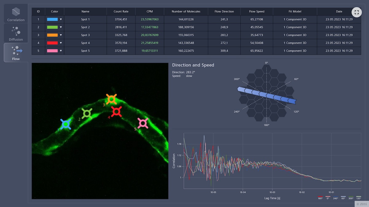 ZEISS Dynamics Profiler is adding a new functionality to ZEISS laser scanning microscopes (LSM) with Airyscan. It provides users with easy access to the molecular concentration and dynamics of fluorescently labelled molecules in living samples. Scientists can now explore their most delicate samples with little time needed and at low light intensity as well as boost data collection to enhance their research...
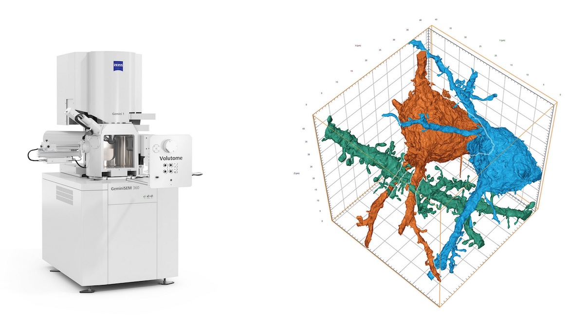 ZEISS Volutome is an end-to-end solution for serial block-face imaging from hardware to software including image processing, segmentation, and visualization. The ultramicrotome can be easily replaced by a conventional SEM stage, converting the 3D FE-SEM into a standard FE-SEM, making the system adaptable to a multi-purpose environment...
 Prof. Dr. Immanuel Bloch, who is considered a leading quantum physicist, is to be commended with the ZEISS Research Award for his outstanding research in the field of quantum simulation using ultracold atoms. The company has been recognizing outstanding research in optics and photonics since 1990. The ceremony will take place at the Deutsches Museum in Munich on 26 June 2023. Three young scientists from Germany, Austria and Switzerland will also receive awards. They will receive the Carl Zeiss Award for Young Researchers...
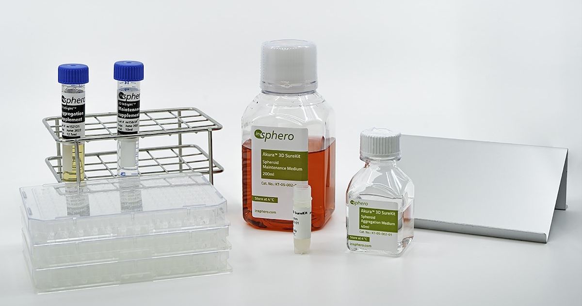 ZEISS is investing in life science start-up InSphero to further advance the adoption of 3D microtissues in research and drug development. InSphero AG, a pioneer in 3D spheroid and cell-based assays in pharmaceutical drug discovery and safety testing, has raised an eight-figure amount in its latest funding round. InSphero will utilize the proceeds to commercialize its cryo-preservation technology to enable further growth and scalability...
 ZEISS introduces the ZEISS Xradia 630 Versa X-ray microscope (XRM) for academic and industrial research. The newest addition to the ZEISS Xradia Versa family breaks resolution performance barriers, delivers an intuitive user experience, and accelerates productivity for all with easy, guided workflows and higher throughput. With the ZEISS Advanced Reconstruction Toolbox (ART) 3.0, the platform also leverages game-changing artificial intelligence (AI) for reconstruction...
 For high-resolution imaging, immersion media between the sample and objective is required. This can be a challenge for some experiments using water as immersion media. One application of immersion media might not be sufficient as the sample moves to different locations and water evaporates over long periods of time. Manual addition risks loss of data points, microscope damage, and is tedious and inefficient...
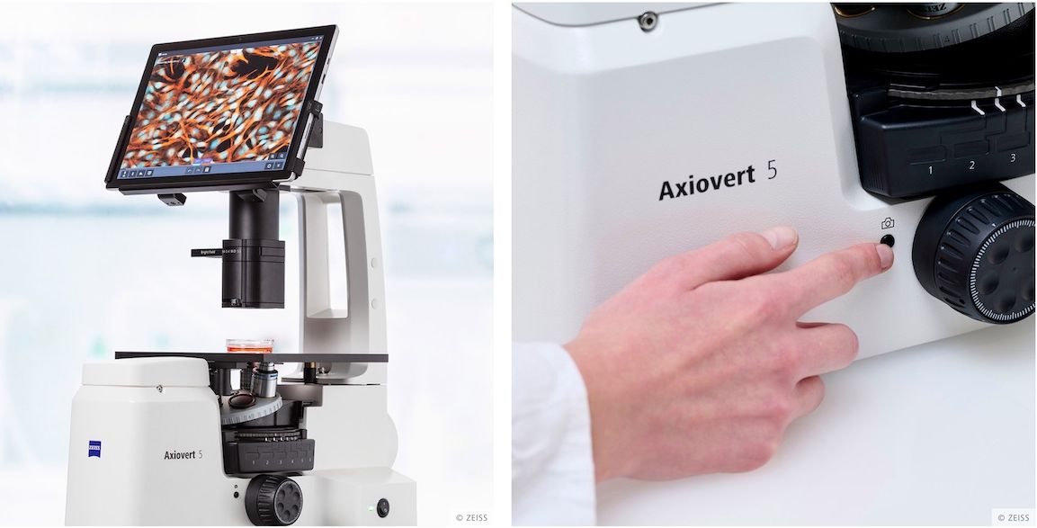 ZEISS introduces its new all-in-one cell imaging system. ZEISS Axiovert 5 digital uses artificial intelligence (AI) and automatic functions to ease daily work in the cell lab. From scientific routine to basic research, from phase contrast to multichannel fluorescence imaging, processes become more efficient and results more reproducible. The system comes with an intuitive operating concept...
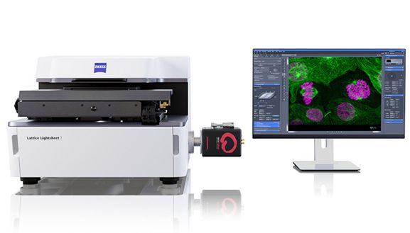 Live Webinar | December 13th, 2022 | 2 - 3.30 PM. For the development of the ZEISS Lattice Lightsheet 7, the ZEISS Microscopy team has received the " Deutscher Zukunftspreis" - the German President's Award for Technology and Innovation. This is a wonderful achievement and to mark the award ZEISS would like to give you the opportunity to learn more about the technology and how it is used to accelerate 3D Live Cell Fluorescence Microscopy...
 As we look ahead to next year, we would like to offer you the chance to request a free copy of the ZEISS Microscopy 2023 calendar. For the second year in a row, ZEISS celebrates the work of researchers using microscopy in various application fields. ZEISS users from all around the globe submitted almost 1,000 fascinating entries. We were honored by their participation and fascinated by the quality of images we received across different research interests and application types...
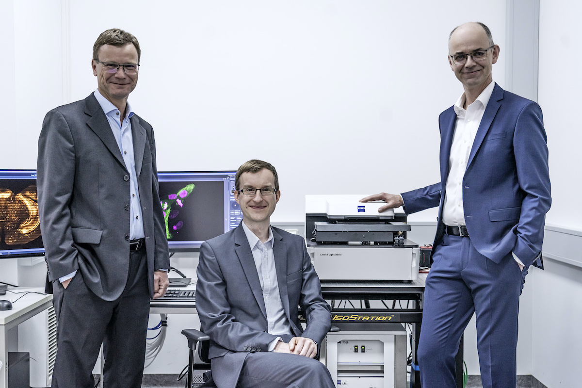 The development of the ZEISS Lattice Lightsheet 7 for the gentle 3D imaging of living cells was honored with the Federal President's Award for Technology and Innovation. The President of Germany, Frank-Walter Steinmeier, awarded the Deutscher Zukunftspreis 2022, The Federal President's Award for Technology and Innovation, to the team from ZEISS during a festive ceremony. The jury recognized the ZEISS experts...
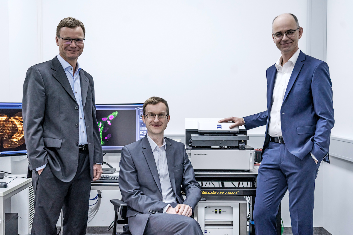 ZEISS experts Dr. Thomas Kalkbrenner, Dr. Jörg Siebenmorgen and Ralf Wolleschensky have been nominated for the 2022 German Future Prize. Their project: the innovative microscope ZEISS Lattice Lightsheet 7 for the gentle 3D imaging of living cells. By combining imaging that protects samples with high resolution, the system allows researchers to observe subcellular dynamics in 3D over hours and days, which was not previously possible with other microscopy technologies....
 What would a microscope be without the ability to capture the amazing details it reveals? ZEISS offers a wealth of microscope cameras as diverse as your imaging and documentation tasks. And because development never stops, we have once again updated and expanded the ZEISS Axiocam portfolio to better support your most demanding applications. Explore the new ZEISS Axiocam models...
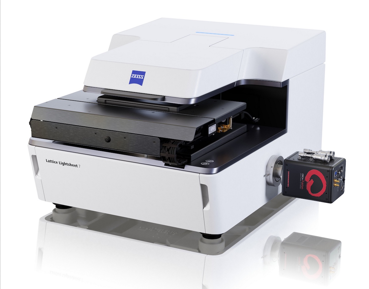 ZEISS releases the next generation of ZEISS Lattice Lightsheet 7. The microscope system was introduced to the market in December 2020 and opened up a new way for researchers to explore the dynamics of life at subcellular resolution. It was the first commercial, easy-to-use implementation of the lattice light-sheet technology known for enabling sample-preserving, long-term imaging of living cells...
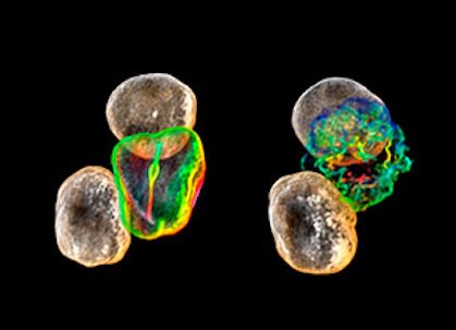 An overview of recent technologies for core facilities. With inverted lattice light sheet and lattice structured illumination microscopy, two new technologies provide access to novel and exciting live imaging experiments. They enable exploration of subcellular dynamics for longer periods of time without the need for special sample preparation – using standard dyes and standard sample carriers....
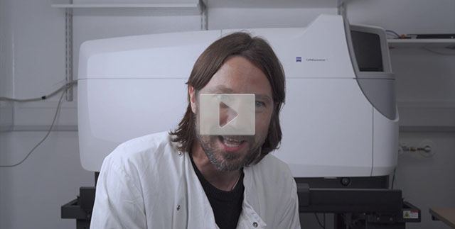 ZEISS recently caught up with the team from the Mostowy Lab at the London School of Hygiene & Tropical Medicine, UK. Watch the video below to hear how their ZEISS Celldiscoverer 7 has transformed the way they carry out research across the entire lab - from streamlining workflows to standardising results. ZEISS Celldiscoverer 7 is your automated microscope for live cell imaging, combining the ease-of-use of an automated high-end imaging system with the image quality and flexibility of a classic inverted research microscope...
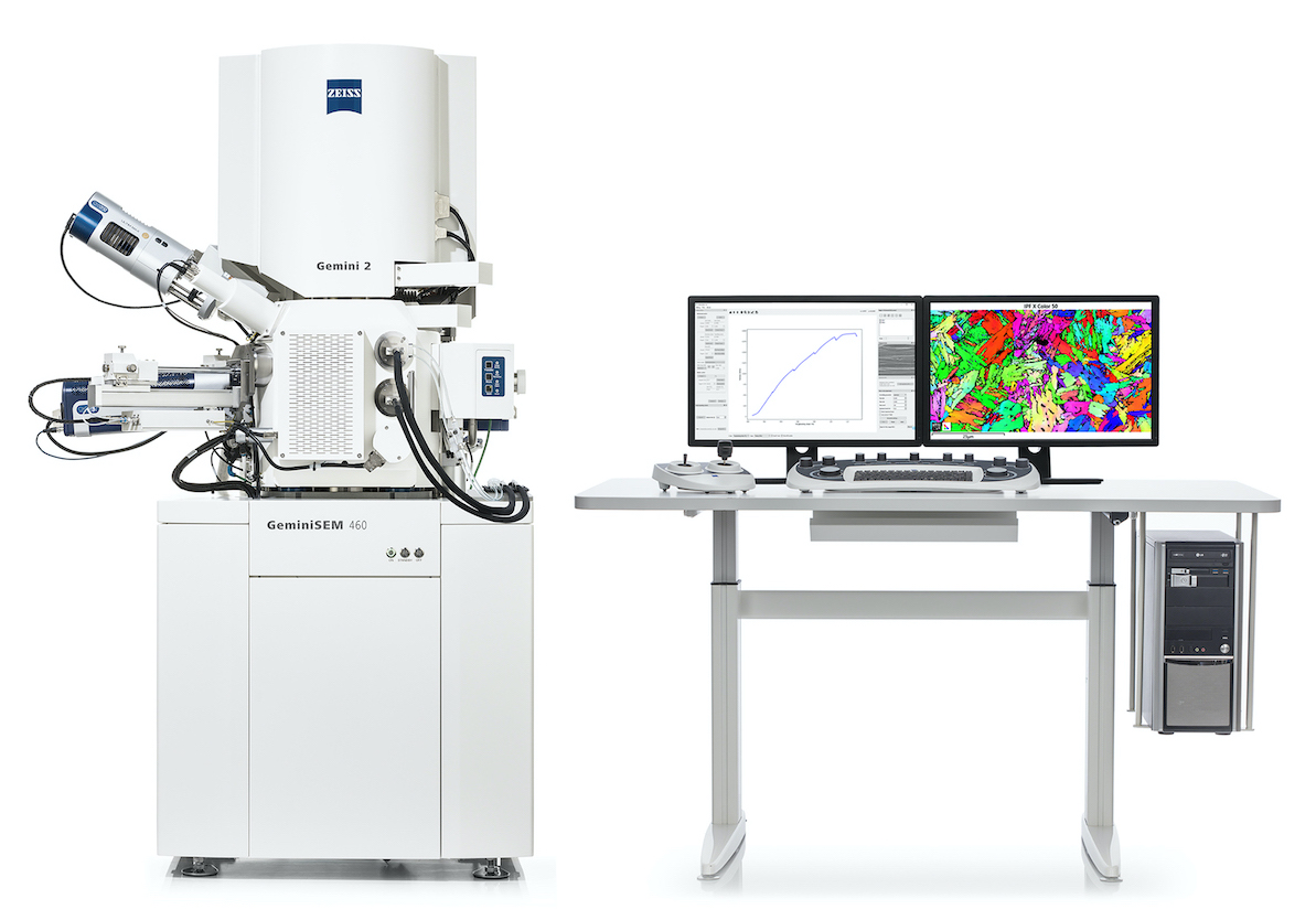 Deeper Insights into Material Properties with the In situ Lab for ZEISS Field Emission Scanning Electron Microscopes. ZEISS is introducing its new integrated in situ workflow for ZEISS field emission scanning electron microscopes (FE-SEM). When researchers need to link material performance to microstructure, which is essential for developing novel materials in a highly efficient way, they can now extend their ZEISS FE-SEM with an in situ solution for heating and tensile experiments...
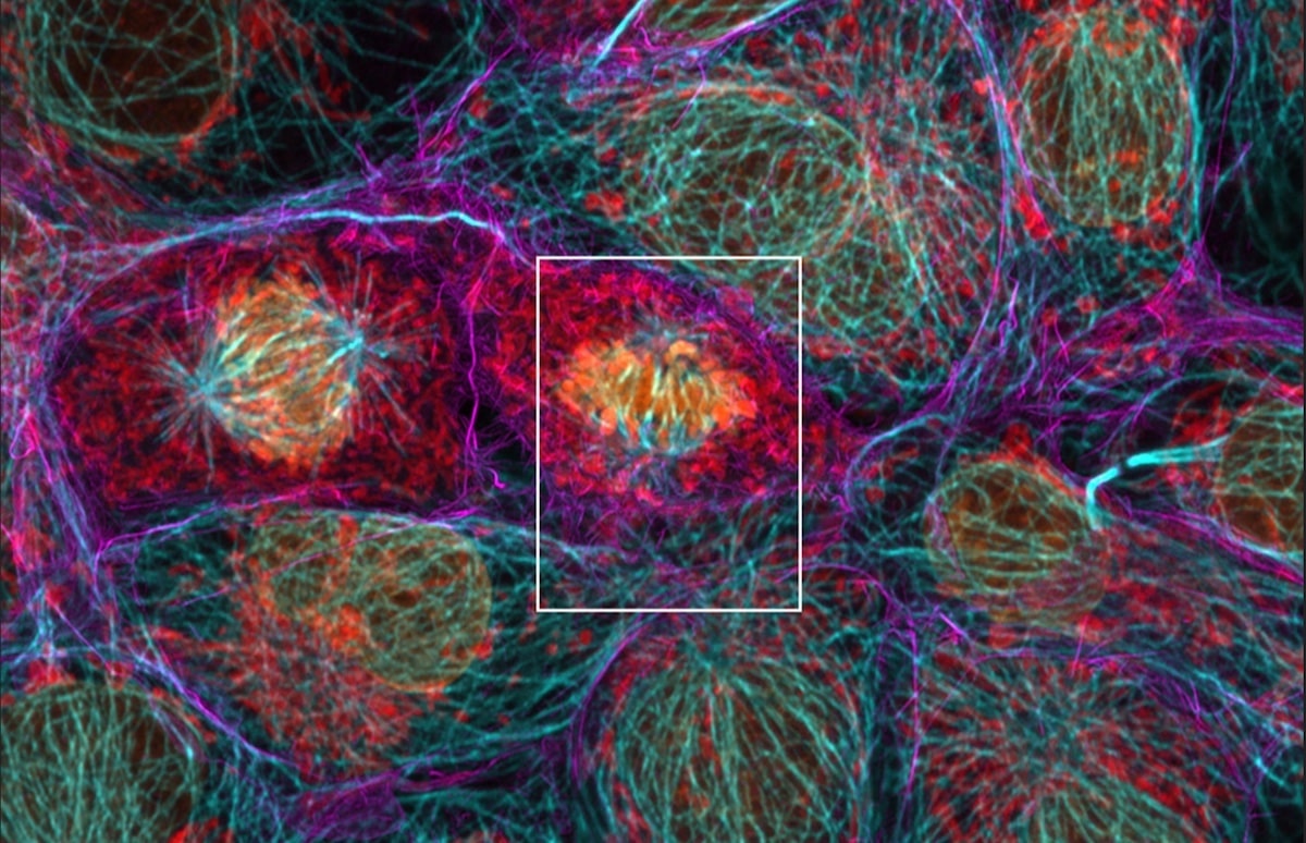 ZEISS introduces two new software functionalities: With ZEISS LSM Plus and ZEISS Airyscan Joint Deconvolution, users achieve better results in confocal microscopy. Laser scanning microscopes (LSMs) are the multifunctional high-end microscopes in every imaging facility, valued as much for their instant, high-quality imaging of optical sections as for their ease of use and application flexibility...
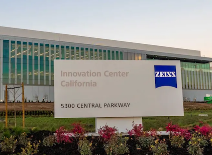 ZEISS announces the opening of its new ZEISS Microscopy Customer Center Bay Area (ZMCC BA), housed within the ZEISS Innovation Center California, a high-tech building designed to promote customer and employee collaboration. Unique to the industry, the ZMCC BA houses electron, light, and X-ray microscopes all in one location supported by resident application experts in life science, materials research, and electronics segments...
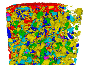 ZEISS introduces ZEISS Mineralogic 3D software for automated quantitative mineralogy that achieves greater productivity and increased efficiency for the mining industry. By understanding composition, mineral relationships, and fabric of the geological materials under scrutiny, including locked grains, miners are able to respond faster to critical production questions...
 Vision Engineering, British leading designer and manufacturer of high quality non-contact measurement, digital 3D visualisation, and ergonomic inspection technologies, is partnering with ZEISS Industrial Microscopy to add an extended depth of focus inspection system to its range of microscopy systems for the first time...
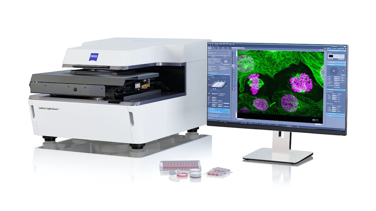 Imagine if the speed and gentleness of light sheet microscopy was combined with the near-isotropic resolution at confocal range for your live cell imaging. ZEISS Lattice Lightsheet 7 makes light sheet fluorescence microscopy available for live cell imaging at subcellular resolution – whilst also allowing you to use standard sample carriers. With this automated, easy-to-use system, gentle volumetric imaging of subcellular structures and dynamics over hours and days is now available to everyone....
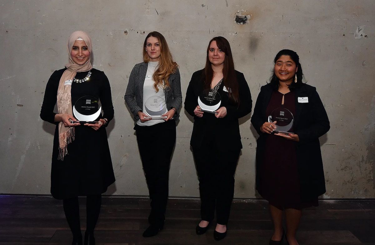 The eleventh ZEISS Women Award was awarded in Dresden. This prize honors outstanding female informatics students who are about to graduate. The award was offered throughout Germany and serves as a platform to publicly recognize successful young women in the digital sector and to encourage more women to consider a career in the field....
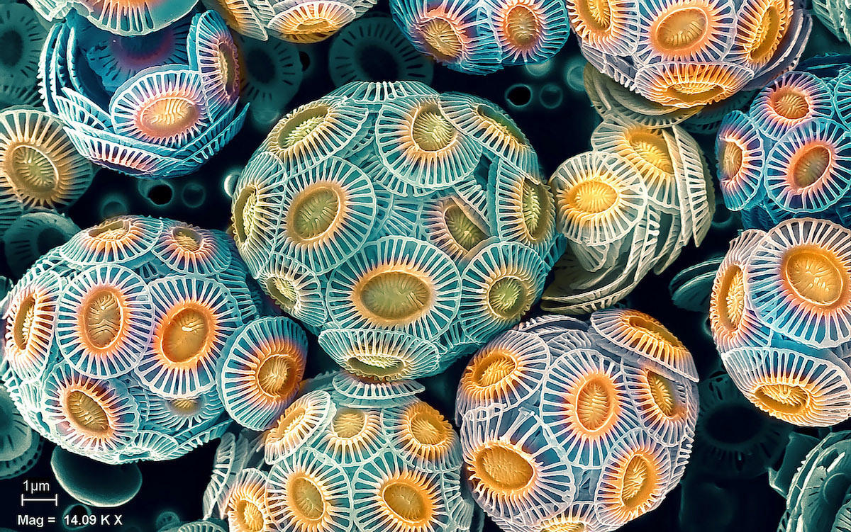 ZEISS has unveiled the winners of their ZEISS Microscopy Image Contest 2021. In its 175th anniversary year, ZEISS is celebrating the work of researchers using microscopy in various application fields with an image contest. Microscope systems, analytics and imaging capabilities all play a role in meeting many of our society’s most pressing challenges related to climate change, energy, health and food....
 For decades, fluorescence microscopy has been an invaluable tool in life sciences research, with new variations and implementations emerging almost every year. Laser scanning microscopy, airyscanning, structured illumination, light field and light sheet microscopy are among the many methods that have been developed through the years to overcome the limitations of the original widefield microscopy setups...
 Resolve Biosciences, the pioneer in Molecular Cartography™, and ZEISS have announced a co-development collaboration to advance spatial biology applications. The companies are working together to optimize advanced microscopy and 3D imaging solutions for subcellular spatial analysis. ZEISS microscopy technology is part of Resolve’s current Molecular Cartography service offering and will be incorporated into the commercial spatial transcriptomics system scheduled to launch later this year.
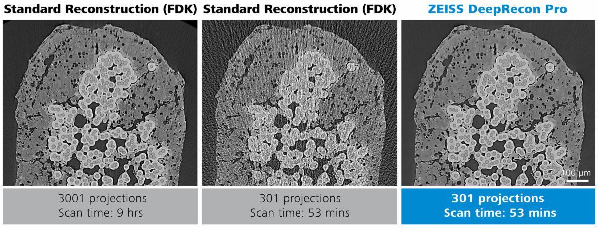 Two new reconstruction technologies introduced today by ZEISS use Artificial Intelligence (AI) to improve data collection and analysis, and speed up decision-making significantly. Now available for the Advanced Reconstruction Toolkit (ART) on ZEISS Xradia 3D X-ray platforms, ZEISS DeepRecon Pro and ZEISS PhaseEvolve modules increase throughput by up to 10x while producing better than ever image quality....
 ZEISS are thrilled to announce the ZEISS Microscopy Image Contest 2021. Do you take stunning microscopy images? Submit your best image now! The contest is open to all microscopy enthusiasts and professionals in any application field. Images can be taken with any ZEISS microscope (light, X-ray or electron microscope). Deadline for entry is Saturday 31st July, 2021...
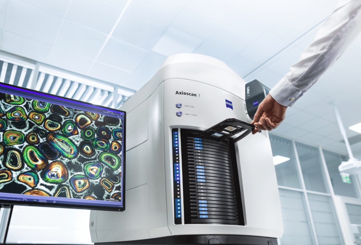 ZEISS is introducing a new ground breaking solution for petrographic analysis. ZEISS Axioscan 7 expands the possibilities of automated petrography by combining unique motorized polarization acquisition modes with unprecedented speed and a rich software ecosystem for visualization, analysis, and collaboration. Fully automated acquisition now comes with unprecedented speed across even the largest sample collections....
 The company will be celebrating its 175th anniversary through a variety of activities and events. Its close links to science are evident in projects such as the "ZEISS Beyond Talks" interview series. In these interviews, pioneers and eminent figures from across the globe, including climate researcher Professor Antje Boetius, speak about their work, their visions, their passion and the topics that are having a major impact on our world....
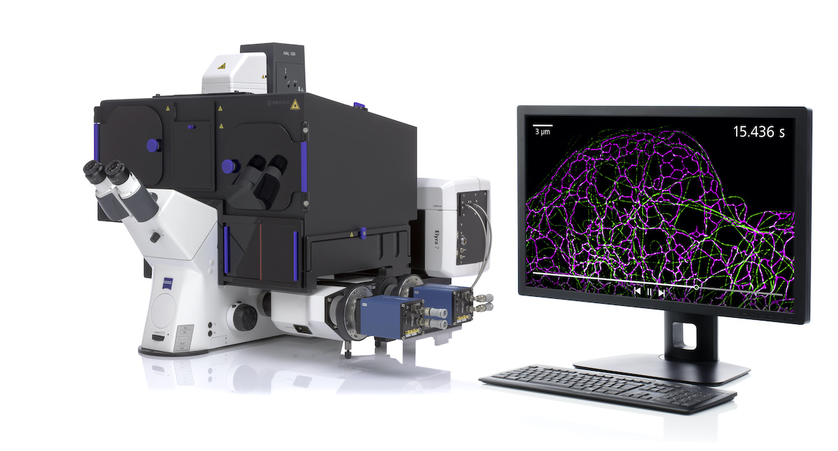 ZEISS introduces SIM², a groundbreaking image reconstruction algorithm that increases the resolution and sectioning quality of structured illumination microscopy (SIM) data. With SIM² on the microscope system ZEISS Elyra 7, life science researchers can now double the conventional SIM resolution and discriminate the finest subcellular structures of living and fixed samples, even those no more than 60 nm apart....
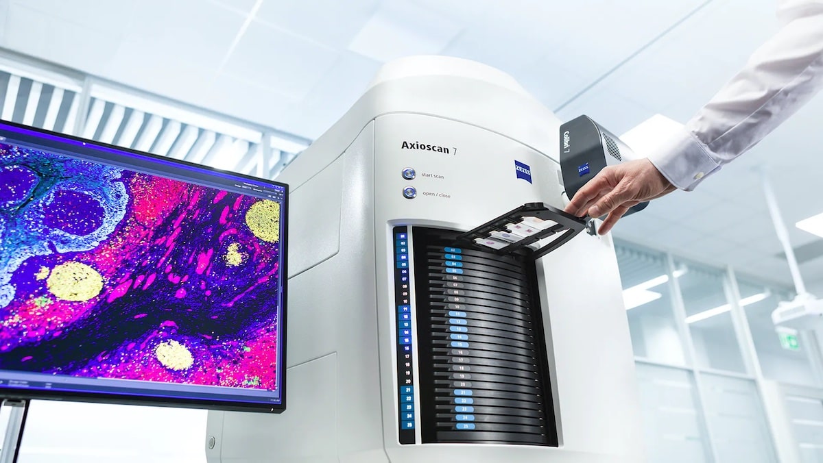 With the release of ZEISS Axioscan 7, ZEISS presents its next-generation slide scanner for the automated digitization of microscopy samples. Following its successful predecessor ZEISS Axio Scan.Z1, ZEISS Axioscan 7 provides significant improvements in almost every aspect: a new acquisition engine for higher scan speeds, a broader range of imaging modes for more application flexibility...
 With the release of ZEISS Correlative Cryo Workflow, ZEISS provides the life science research community with a new combined hardware and software solution for cryogenic microscopy. The workflow connects widefield, laser scanning, and FIB-SEM microscopes in a seamless and easy-to-use procedure....
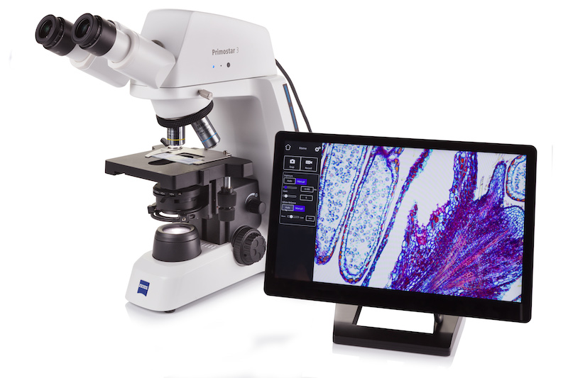 ZEISS has introduced a new compact microscope for digital teaching and routine lab work just in time for the International Day of Education, which took place yesterday. ZEISS Primostar 3 is a robust upright light microscope, which is made for daily work in a classroom or in a lab for tissue and sample examination in histology, cell biology, food or microbiology, etc. It is designed for long-term use and extreme durability....
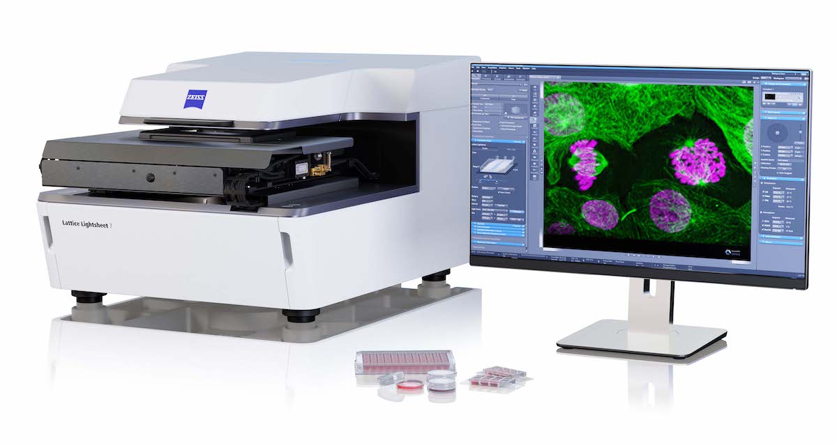 ZEISS announces that it has formed a research collaboration partnership with the Max Planck Florida Institute for Neuroscience (MPFI). Using an LSM 980 NLO next generation confocal microscope supplied by ZEISS, MPFI will investigate using implanted GRadient INdex (GRIN) lenses in combination with the Airyscan 2 area detector for deep brain functional neuroscience research....
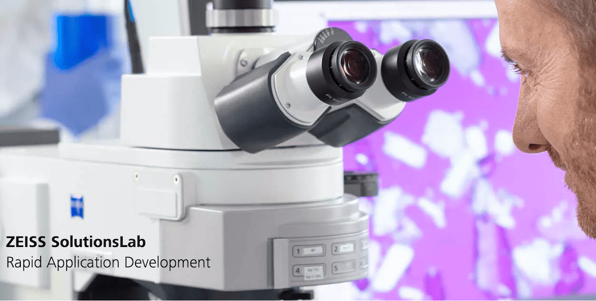 ZEISS introduces ZEISS Solutions Lab, an innovative service, that provides its customers with rapid access to new applications for their microscopy systems or correlative microscopy suites. ZEISS Solutions Lab enables customers to select, or to drive demand for module-based workflows built on ZEISS software packages for a wide variety of requirements specific to their research needs...
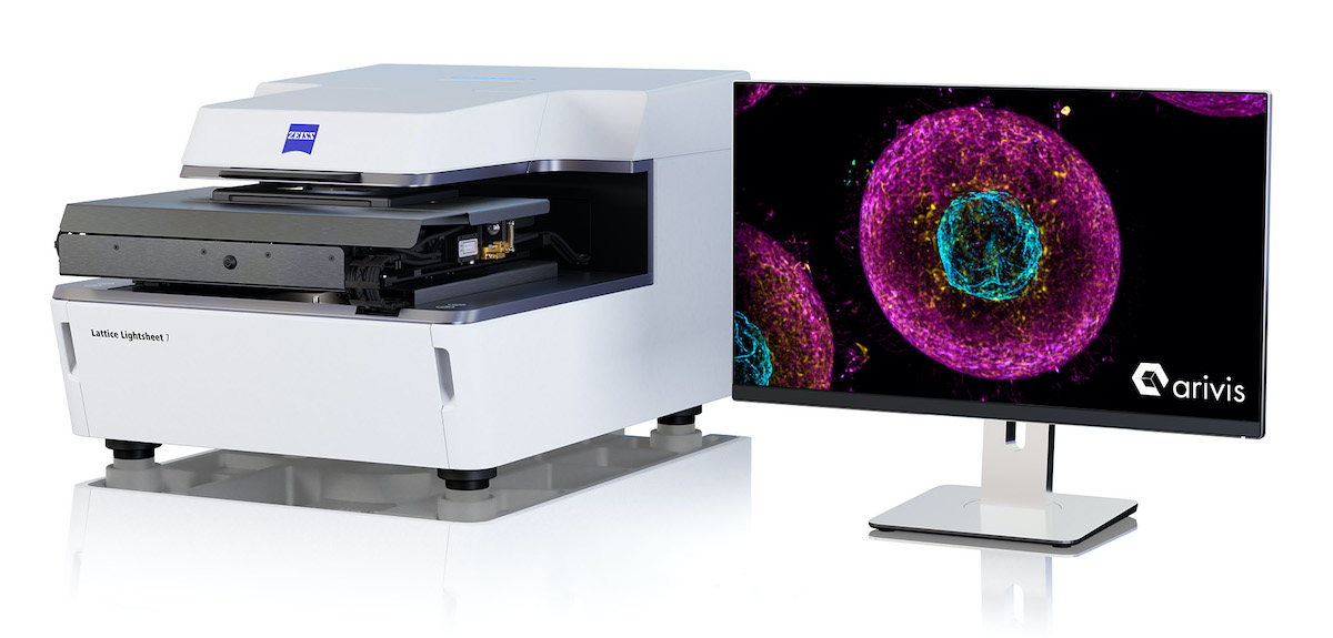 ZEISS is expanding its imaging technology capabilities with 3D and big image data software by acquiring a majority stake in the imaging business of arivis. With this investment, ZEISS is further strengthening its software competencies and market position in 3D image visualization, image processing, and analysis software for research microscopy. Both companies have a long-standing collaboration and strategic partnership, as arivis has been an important development partner for ZEISS for more than seven years..
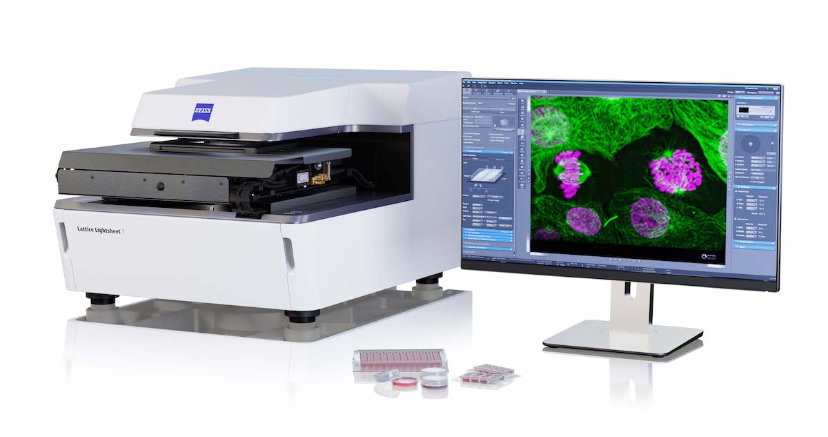 With the release of ZEISS Lattice Lightsheet 7, ZEISS places a turn-key lattice light sheet instrument at the disposal of the life science research community. Based on the pioneering research and developments of Ernst H. K. Stelzer while at EMBL, Heidelberg, on light sheet technology and of Nobel laureate Eric Betzig while at the Janelia Research Campus of HHMI on structuring light sheets as optical lattices to render them thinner and longer...
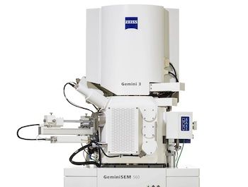 ZEISS presents a new generation of its field emission scanning electron microscope (FESEM) family ZEISS GeminiSEM. The new models ZEISS GeminiSEM 360, 460 and 560 are tailored for sub-nanometer imaging and effortless analytics. Users benefit from innovations in the electron optics and a new chamber design offering better image quality, usability and flexibility. ZEISS GeminiSEM 560 now being introduced brings the ZEISS Gemini 3 column to the market for the first time....
 Reduce your time to experiment from minutes to seconds. Microscopes are becoming increasingly automated. However, sample placement, focus adjustment, and identification of the relevant sample areas still require manual adjustments. The new ZEISS AI Sample Finder automates this sequence entirely, Placement of sample, Acquisition of an overview image, Identification of the sample carrier, Detection of relevant sample areas, Focus adjustment for each region of interest...
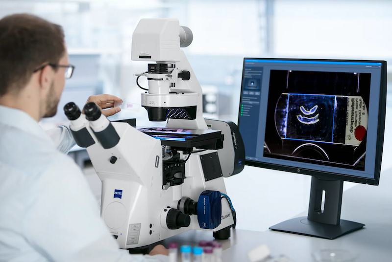 ZEISS presents the new AI Sample Finder for optimal user guidance and operation. With this feature, the open and flexible inverted microscope platform ZEISS Axio Observer makes sample placement easier than ever and significantly reduces the time to experiment. For researchers, it offers a completely different way to operate a microscope, greatly boosting both productivity and ease of use. Users are guided more efficiently and have to perform fewer manual steps....
 Learn about microscopy, its applications, technologies, tips and tricks, imaging methods, and new developments - from the basics to more advanced topics. On the new content platform Microscopy Insights Hub from ZEISS Microscopy, access on-demand webinar recordings, how-to videos and further information pieces about your field of interest....
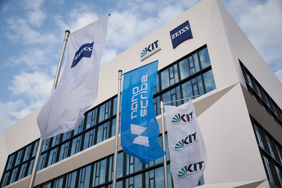 The ZEISS Innovation Hub on the campus of the Karlsruhe Institute of Technology (KIT) has seen a number of successful collaborations and projects since it opened in early 2020. ZEISS wants the hub to house high-tech and digital start-ups, as well as its own innovation and new business activities. KIT will thus join forces with ZEISS experts to pave the way for the technologies of the future....
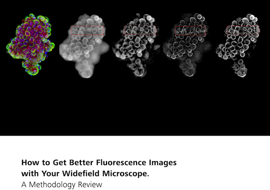 In this new 10-page Technology Note, learn how different image processing methods have the potential to make widefield microscope systems more powerful and versatile. Understand each methods limitations and pitfalls, as well as suitability for your specific applications. For decades, fluorescence microscopy has been an invaluable tool in life sciences research, with new variations and implementations emerging almost every year....
 When UCLA’s Dr. Araceli Espinosa-Jeffrey sent human brain cells into space, her goal was to gain a better understanding of how neural stem cells grow and develop in microgravity. That understanding is key to discovering more about the serious issues of intracranial hypertension affecting astronauts returning from space. The information may also one day be used to further cell replacement therapies...
 Jian-Wei Pan, Professor at the University of Science and Technology of China, in Hefei, is the winner of the prestigious ZEISS Research Award 2020. He is one of the world's leading researchers in the field of quantum technology. One of the most remarkable results of Jian-Wei Pan's research is the distribution of entangled photons over a distance of 1,200 km, by far the longest distance ever reached....
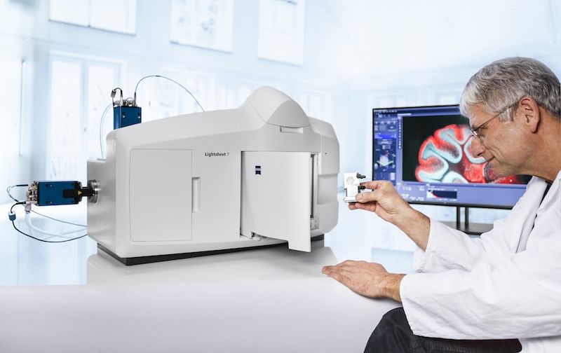 New Light Sheet Fluorescence Microscope Introduced. Light sheet fluorescence microscopy (LSFM) with its unique illumination principle allows fast and gentle imaging of whole living model organisms, tissues, and developing cells. The high stability of ZEISS Lightsheet 7 enables researchers to observe living samples over extended periods of time – even days – with less phototoxicity than ever before....
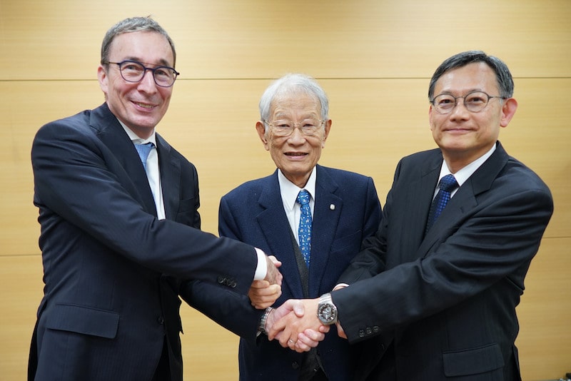 ZEISS and RIKEN Innovation Co., Ltd. have entered into a strategic collaboration agreement to jointly explore opportunities in aiming to identify projects where the combination of RIKEN’s expertise in bioengineering and image data manipulation and ZEISS’ leadership in optical solutions can accelerate bioengineering innovations and bring about a paradigm change in health care....
 Materials and Life Science researchers can now experience faster accessibility of deeper regions of interest when investigating samples in 3D. With the new extensions on ZEISS Crossbeam 350/550 and ZEISS Atlas 5, users benefit from improvements in speed and data quality when performing multi-scale, multi-modal studies in additive manufacturing, electronics, battery research, biomaterials and biological tissue investigations on resin-embedded biological specimens...
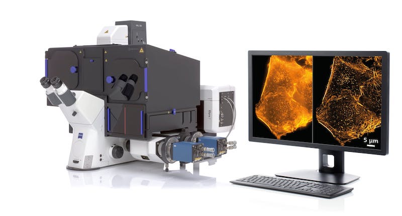 ZEISS is among the winners of the prestigious R&D 100 Awards 2019, with judges from R&D Magazine selecting ZEISS Elyra 7 with Lattice SIM for superresolution microscopy as an award recipient in the “Analytical/Test” category. ZEISS Elyra 7 with Lattice SIM is a microscope system for superresolution imaging in biomedical research. Its novel Lattice SIM technology is extremely light-efficient and fast. Researchers profit from less photodamage and high imaging speed....
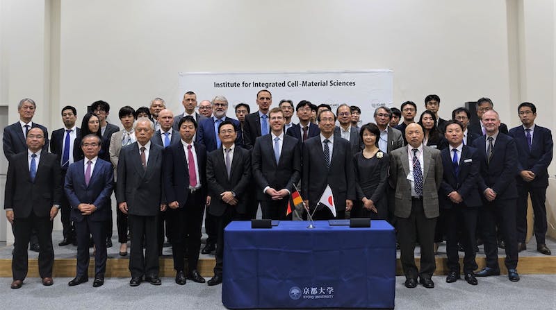 ZEISS and Kyoto University are intensifying their cooperation and have signed a new strategic research agreement. As part of the agreement, the partners are opening the ZEISS iCeMS Innovation Core – a collaborative laboratory at Kyoto University’s Institute for Integrated Cell-Material Sciences (iCeMS). As such, the contract aims to further solidify the long-standing partnership between the two institutions...
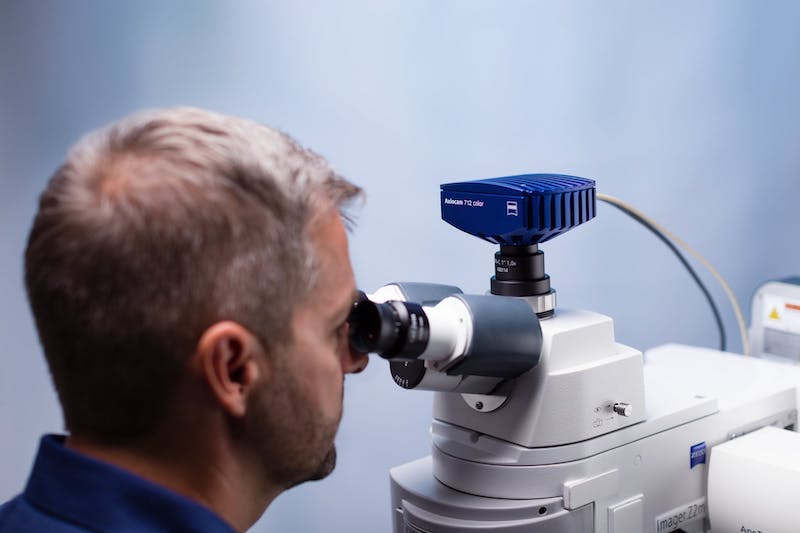 ZEISS introduces four new high-quality CMOS cameras for digital imaging in light microscopy. These cameras complete the portfolio of proven ZEISS Axiocam models which stand for excellent performance in demanding microscopy applications. The microscope cameras ZEISS Axiocam 705 color and 712 color deliver the best possible image quality for histology, pathology or material research and analysis, thanks to excellent color rendition and greatly improved dynamic range....
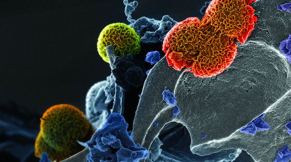 ZEISS introduces new capabilities for ZEISS ion beam microscopes, which cover advancements in analytics, tomography, sample preparation and data integrity. This brings new possibilities in engineering materials, energy materials, soft materials and geosciences covering megatrends in additive manufacturing, battery and photovoltaic research, building materials and nanomaterials....
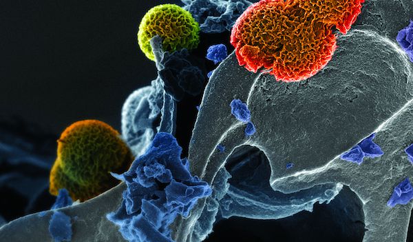 The proven ZEISS Celldiscoverer 7 is a fully integrated high-end imaging system with various incubation and detection options. It combines the easy-to-use automation of a boxed microscope with the image quality and flexibility of a classic inverted research microscope. To get better data from three-dimensional samples, it is now possible to add ZEISS LSM 900 with Airyscan 2 for confocal imaging....
 Smart Microscopy from ZEISS is a new concept for the routine digital documentation of microscopic samples. The microscopes ZEISS Axiolab 5 and ZEISS Axioscope 5, as well as the microscope cameras ZEISS Axiocam 202 mono and ZEISS Axiocam 208 color, can be combined to form a Smart Microscopy system that takes away a large share of the workload from the users. It automatically adjusts many of the required settings....
 The proven ZEISS Celldiscoverer 7 is a fully integrated high-end imaging system with various incubation and detection options. It combines the easy-to-use automation of a boxed microscope with the image quality and flexibility of a classic inverted research microscope. To get better data from three-dimensional samples, it is now possible to add ZEISS LSM 900 with Airyscan 2 for confocal imaging...
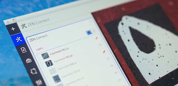 ZEISS ZEN Connect for the materials research lab. ZEISS ZEN Connect software module is now available for materials researchers at universities, multi-user facilities or industrial labs. It enables users to bring all imaging technologies together – ZEISS or otherwise – to answer their questions. Gaining unique insights and saving time. ZEISS ZEN Connect allows users to align and overlay images from any source. Researchers can use the workflow of...
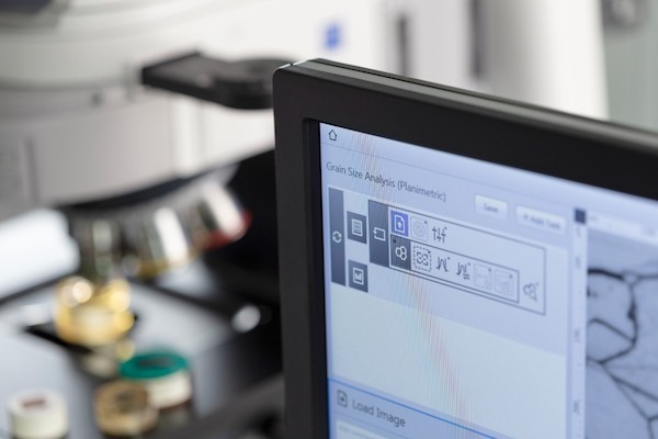 Making materials laboratories more productive. ZEISS ZEN core is a powerful software suite for microscopy imaging, automated analyses, and multi-modal workflows in connected materials laboratory environments. With the new release, materials researchers are now even more efficient. ZEISS ZEN core is not only used as a powerful tool for image analysis and interactive control of microscopes....
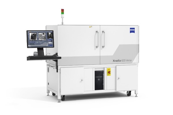 ZEISS Xradia 600-series Versa enables faster submicron-resolution imaging of intact samples. ZEISS introduces two new advanced models of the ZEISS Xradia Versa family: The ZEISS Xradia 610 & 620 Versa X-ray microscopes excel through faster non-destructive imaging of intact samples without sacrificing resolution and contrast over the full range of power and kV. Leading researchers and scientists across the world rely on the...
 For the first time ever, a technology developed by ZEISS has made it possible to utilize transparent glass or plastic in a whole range of different ways. The see-through surfaces with integrated, invisible microstructured optics permit a range of different applications, thus enabling innovations like gesture recognition or eye tracking without any visible optical systems. There are also plans to use the glass in smart homes....
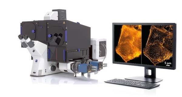 ZEISS is introducing ZEISS Elyra 7 with Lattice SIM, a new flexible platform for fast and gentle 3D superresolution. Lattice SIM expands the possibilities of structured illumination microscopy (SIM): illuminating the sample with a lattice pattern rather than grid lines gives higher contrast and allows a more robust image reconstruction. Scientists can use 2x higher sampling efficiency to lower the illumination dosage to observe fast cellular processes in superresolution....
 ZEISS announces its biggest new release of the ZEISS Mineralogic software at the Process Mineralogy '18 conference in Cape Town, South Africa. This is the 7th instalment of ZEISS Mineralogic since the software was brought to the market in July 2014 and represents a significant advancement in both features and productivity. The software is already well known for providing quantitative mineralogy with the...
 New microscopy technologies, such as light sheet fluorescence microscopy (LSFM) or clearing methods, allow the imaging of large samples at high resolution or high frame rates. Handling, processing, and analyzing these multi-terabyte data sets has become increasingly difficult for scientists, e.g. in developmental biology and neuroscience. New tools are needed to overcome these challenges. Therefore, EISS has teamed up with arivis AG to offer complete solutions from initial image acquisition to final results...
 At the Annual Meeting of the Society for Neuroscience 2018 (Nov 03 – 07), ZEISS presented new workflows in collaboration with Inscopix (Palo Alto, CA) where ZEISS’s Airyscan confocal imaging technology is leveraged with the Inscopix freely behaving microscopy systems. The joint workflow development utilizes the increased spatial resolution, signal-to-noise and sectioning of ZEISS Airyscan to further inform the freely behaving signaling data collected with the Inscopix system....
 Joint further development of 3D visualizations of multidimensional image data. ZEISS and arivis have now expanded and deepened their cooperation, which began in 2014. The companies are pursuing the common goal of enabling new findings in life sciences and in materials research by improving the presentation and analysis of digital data. Modern microscope systems with high-resolution optics record ever increasing amounts of data, which can only be displayed and analyzed with the help of advanced software...
 High resolution 3D block face imaging for biological samples with fast acquisition rates and minimal sample damage. In collaboration with the National Center for Microscopy and Imaging Research (NCMIR) at the University of California San Diego, ZEISS releases a new Focal Charge Compensation module for block face imaging with ZEISS GeminiSEM and 3View® from Gatan, Inc. Focal Charge Compensation...
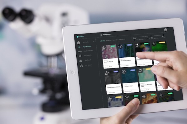 ZEISS presents the initial release of its cloud-based digital microscopy platform, known under the name APEER at the Microscopy & Microanalysis conference (M&M) in Baltimore, USA. Microscopy users will be able to automatically process images in the cloud by leveraging application workflows for 3D reconstructions, staining or segmenting...
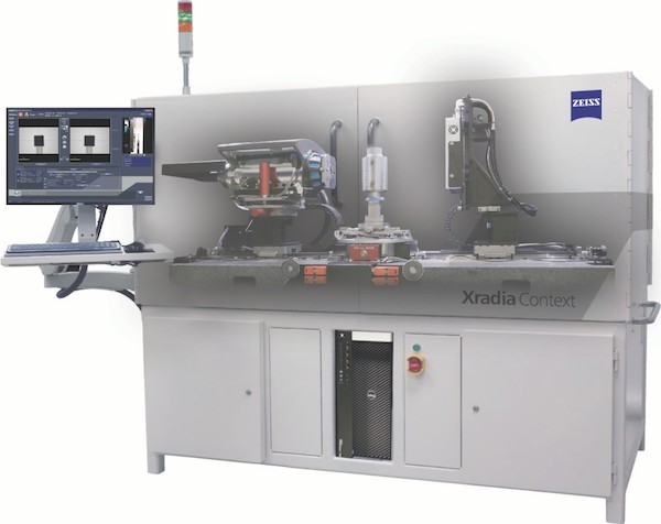 ZEISS expands X-ray imaging portfolio with ZEISS Xradia Context microCT. Built on renowned ZEISS Xradia platform and field-convertible to ZEISS Xradia Versa X-ray microscope. ZEISS introduces a new X-ray imaging instrument, ZEISS Xradia Context microCT, a large field-of-view, non-destructive 3D X-ray microcomputed tomography system. ZEISS Xradia Context is built on time-tested ZEISS Xradia technology, reaping crossover benefits of years of...
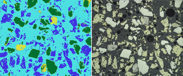 First ZEISS ZEN Intellesis solution enables segmentation of correlative microscopy datasets.
 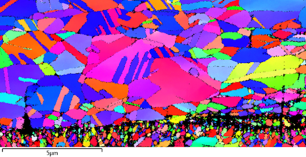 Addressing the highest demands in imaging and analytics from any sample. ZEISS introduces its new field emission scanning electron microscope (FE-SEM) ZEISS GeminiSEM 450. The instrument combines ultrahigh resolution imaging with the capability to perform advanced analytics while maintaining flexibility and ease-of-use...
 ZEISS introduces new software modules with enhanced imaging technology: ZEISS ZEN Connect is especially useful for, but not limited to, structural analysis, examination of cellular processes, localization of cells, and much more. The proven ZEISS ZEN software combines numerous functions for day-to-day work with microscopes and stands for scientific success.....
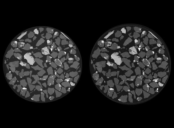 ZEISS introduces a new module for the ZEISS Xradia Versa 500-series of 3D X-ray microscopes (XRM) that will allow users to acquire high quality images in one-quarter the time. ZEISS OptiRecon enables users to make the optimal choice for their requirements: same quality images four times faster, or superior quality in the same amount of time as standard image acquisition using an advanced new...
 Prof. Tobias J. Kippenberg and Prof. Jean-Pierre Wolf are the 2018 winners of the prestigious ZEISS Research Award. The jury was impressed by their exceptional work. Tobias Kippenberg, Professor at the Laboratory of Photonics and Quantum Measurements at the École Polytechnique Fédérale de Lausanne (EPFL), is a pioneer in the field of cavity optomechanics and microresonator-based optical frequency combs....
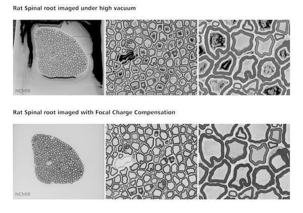 In collaboration with the National Center for Microscopy and Imaging Research (NCMIR) at the University of California San Diego, ZEISS releases a new Focal Charge Compensation module for block face imaging with ZEISS GeminiSEM and 3View® from Gatan, Inc. Focal Charge Compensation expands versatility and considerably increases data quality without prolonging acquisition times and enables easy imaging of even the most charge-prone samples....
 Modular platform for intuitive operation, routine investigations and research applications. ZEISS presents the new generation of its proven high-performance scanning electron microscope (SEM): The new instruments of the ZEISS EVO family come with a variety of improvements regarding usability, image quality and seamless integration into multimodal workflows. With its comprehensive range of available options, the ZEISS EVO family can be tailored precisely to requirements in life sciences, material sciences, or routine industrial quality assurance.....
 ZEISS and BOSELLO HIGH TECHNOLOGY have announced that the ZEISS Group will acquire a majority stake in the provider of industrial X-ray solutions.
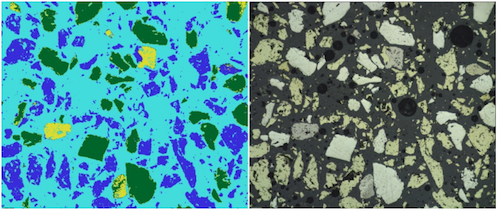 First ZEISS ZEN Intellesis solution enables segmentation of correlative microscopy datasets
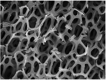
 Improved optical sectioning delivers higher resolution without the need to acquire a z-stack
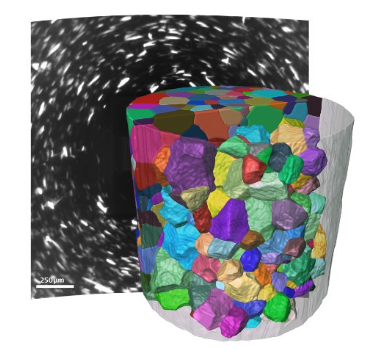 Advanced analytical module for tomography in the laboratory provides 3D grain imaging
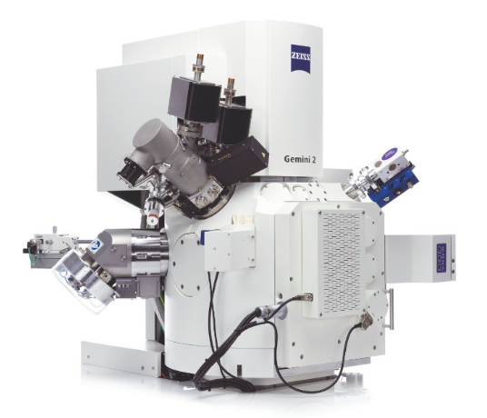 A jury of experts from the German Design Council has voted the ZEISS Crossbeam 550 focused ion beam scanning electron microscope (FIB-SEM) as a winner of the German Design Award 2018 in the "Material and Surfaces" category.
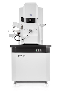 The new instruments of the ZEISS EVO family come with a variety of improvements regarding usability, image quality and seamless integration into multimodal workflows. With its comprehensive range of available options, the ZEISS EVO family can be tailored precisely to requirements in life sciences, material sciences....
ZEISS will invest over €300 million in a new integrated high-tech site. The world’s technology leader in the optics and optoelectronics industries unveiled its plan today in Jena.
 In line with this plan, ZEISS will bring together its existing sites in Jena by 2023. To do this, ZEISS will acquire partially unused premises from SCHOTT, which it will renovate and use to construct a new facility for its second-largest site worldwide. Jena is thus an integral part of ZEISS’s global investment strategy....
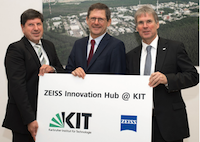 Construction is scheduled to begin in early 2018; the €30 million hub will cover 12,000 square meters. With the ZEISS Innovation Hub, the global technology leader in optics and optoelectronics is making a sustainable investment in Germany as a place for innovation.....
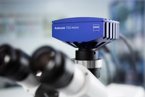 ZEISS introduces two new digital microscope cameras; ZEISS Axiocam 702 mono and ZEISS Axiocam 512 color complement the current portfolio of high-speed USB 3.0 microscope cameras. With ZEISS Axiocam 702 mono ZEISS for the first time introduces a microscope camera with a scientific CMOS sensor...
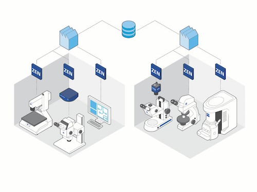 ZEISS presents a networked lab for quality control and quality assurance. Connected systems, data and workflows enable more efficient collaboration - even across lab boundaries....
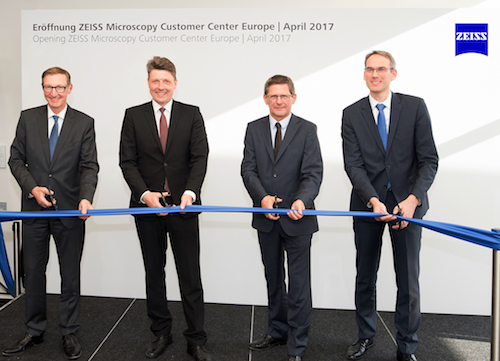 On 24 April 2017 ZEISS opened the new ZEISS Microscopy Customer Center Europe at the Oberkochen site. This is the most comprehensive ZEISS Customer Center including light, electron and X-ray microscopy in a single location...
 ZEISS presents a new generation of focused ion beam scanning electron microscopes (FIB-SEMs) for high-end applications in research and industry. ZEISS Crossbeam 550 features a significant increase in resolution for imaging and material characterization and a speed gain in sample preparation....
 ZEISS introduces a new system for automated microscopy in life sciences research. ZEISS Celldiscoverer 7 combines the user-friendly automation features of a boxed microscope with the image quality and flexibility of a classic inverted research microscope. Scientists acquire better data in shorter times with 2D or 3D cell cultures, tissue sections or small model organisms.
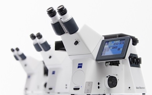 ZEISS introduces a new inverted microscope platform for life science research. The ZEISS Axio Observer family consists of three stable and modular microscope stands for flexible and efficient imaging. Scientists benefit from reproducible results from their experiments and high quality image data from a whole range of samples in a variety of conditions....
 ZEISS has announced a new variant of the world’s fastest scanning electron microscope: ZEISS MultiSEM 506 features 91 beams working in parallel and increases the throughput of the ZEISS MultiSEM 505 by a factor of three. The unrivaled net acquisition speed of more than 2 Tera pixel per hour enables large-scaled experiments such as imaging of cubic millimeters of brain tissue at nanometer resolution for the analysis of neural circuits.
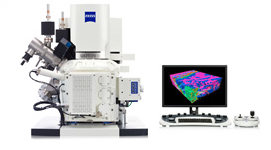 ZEISS Microscopy announces that it will be demonstrating its new ZEISS Crossbeam Focused Ion Beam Scanning Electron Microscope (FIB-SEM) at M&M 2014, August 3-7 at the Connecticut Convention Center in Hartford, CT. ZEISS Crossbeam FIB-SEM increases throughput using a high beam current of up to 100 nA. While milling samples at unprecedented precision at all currents, users acquire images and analytical data in up to...
 At the trade show for quality assurance Control from May 6–9, 2014, the Microscopy business group of ZEISS presented its first automated digital microscope. The device particularly stands out thanks to its one-of-a-kind operating concept, which makes operation as easy as possible for the user. In an industrial environment, optical inspection is an important part of production-related quality testing. Parts such as screws, printed circuit boards, and metal elements need to be examined for defects...