Channels
Special Offers & Promotions
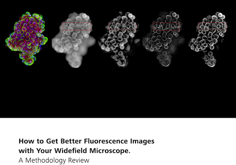
How to Get Better Images with Your Widefield Microscope
In this new 10-page Technology Note, learn how different image processing methods have the potential to make widefield microscope systems more powerful and versatile. Understand each methods limitations and pitfalls, as well as suitability for your specific applications.
For decades, fluorescence microscopy has been an invaluable tool in life sciences research, with new variations and implementations emerging almost every year. Laser scanning microscopy, airyscanning, structured illumination, light field and light sheet microscopy are among the many methods that have been developed through the years to overcome the limitations of the original widefield microscopy setups.
These more recent techniques have brought new insights into biological samples and enabled new discoveries. However, we should not forget that even comparably simple and inexpensive widefield microscopes can help to extract meaningful data from many samples. In this article we focus on some popular image processing methods. These techniques have the potential to make widefield microscope systems more powerful and versatile so long as their limitations and pitfalls are known. Simple deblurring methods—for example, background subtraction, computational clearing, unsharp masking and the like—deliver a quick and clearer preview of the sample while more accurate deconvolution models yield higher resolution, fewer artefacts and more quantitative results
Find out more about
- Sharp images in 2D
- Solutions for 3D volumes
- Unsharp masking
- No-neighbor / Nearest-Neighbours
- Background subtraction
- Limitations of widefield microscopy
- Conclusion
View | Download Technology Note
Media Partners


