
Channels
Special Offers & Promotions
Carl Zeiss SMT AG
Products
Contact Carl Zeiss SMT AG
All articles from Carl Zeiss SMT AG
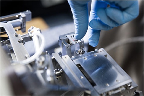
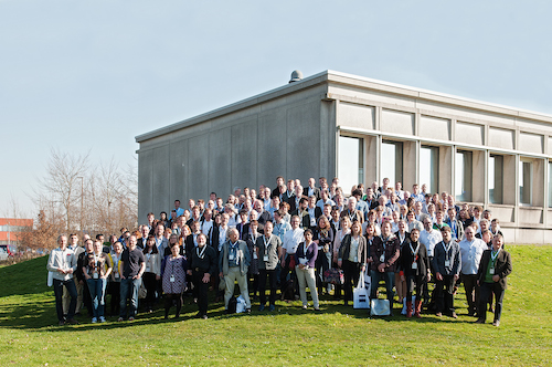 In cooperation with the Flanders Institute of Biotechnology (VIB) and Core for Life, ZEISS invited the international community of correlative microscopists for 3 days to a joint workshop at the Bioimaging Core of the VIB Inflammatory Research Centre (IRC) in Ghent, Belgium. The workshop followed the first meeting of the community at the ZEISS Microscopy Labs Munich in 2012. From 9-11 March 2014, 150 scientists from 23 countries took the opportunity to discuss the current state of the art in correlative microscopy and 3D imaging techniques....
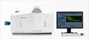
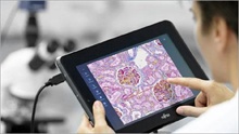
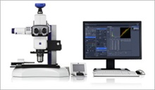
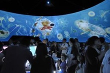
 ZEISS introduces a new imaging system for material analysis at the Laser World of Photonics China 2014 trade show in Shanghai. ZEISS Primotech is used both in industrial quality control as well as in the education of geologists and mineralogists. In routine applications, customers can quickly and reproducibly analyze their products within the production process, in an educational setting, Primotech and the iPad imaging app Matscope transform classical learning environments into digital classrooms.


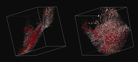
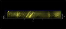
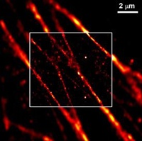
 Expanding its 3D microcopy portfolio, ZEISS is introducing superresolution photoactivated localization microscopy (PALM) in 3D at the Society for Neuroscience Annual Meeting in San Diego, California. With a new module, ELYRA P.1 now enables superresolution photoactivated localization microscopy (PALM) in 3D for endogenously-expressed photo-switchable fluorescent proteins. Users can capture highly resolved structures in 3D, while treating the sample so gently it stays fit for long-term observation...
 Over forty international scientists gathered for the 5th Annual Light Sheet Fluorescence Microscopy Workshop in September in Thornwood, New York. The meeting was held at the U.S. headquarters of Carl Zeiss Microscopy, LLC. Researchers presented data and shared ideas regarding instrument development, standards, applications and specific applications of light sheet fluorescence microscopy (LSFM) and related technologies to various biological model organisms such as zebrafish, drosophila, mouse and others...


 At the awards ceremony on 1 July 2013, the ZEISS Microscopy business group received the renowned red dot award: product design for the Axio Zoom.V16 zoom microscope. In 19 categories, the jury selected the product with the most successful product concept from the total 4,662 entries. Following the ceremony, the product will be on display in a special four-week exhibition at the red dot design museum at the Aalto Theater in Essen...

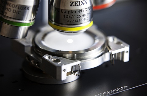
 At the 2013 Control international trade fair for quality assurance, ZEISS presented its new Hardware Auto Focus and cleanroom kit for the proven Axio Imager Vario microscope system. An integratable autofocus system ensures fast and precise focusing. The Hardware Auto Focus is ideal for surface inspections on reflective, low-contrast samples. In transmitted and reflected light (brightfield, darkfield, polarization contrast and oblique illumination), the focus system ensures high precision down to 0.3 of the depth of field of the objective lens....

 At the American Association for Cancer Research (AACR) Annual Meeting 2013 in Washington, D.C., USA, SelectScience announced that ZEISS Lightsheet Z.1 has been awarded with the Best New Life Science Product of 2012 by members of SelectScience. The winning product is recognized as having significantly contributed towards laboratory efforts in 2012...
European Patent Office Grants Patent for PAL-M Superresolution Technology Exclusively Licensed by Carl Zeiss
Mar 21, 2013
 Based on the same invention, several patents were issued in the years from 2009 to 2011 by the US Patent Office.The patent EP 1894010 B1, whose issue was announced on 27 February 2013, protects methods as well as devices for PAL-M. The basic idea of the invention is that only a low number of fluorescent dye molecules in a sample may be activated and excited to fluoresce at a time. This enables the physical position of these individual fluorescent molecules to be exactly determined to within a few nanometers....
 The UK manufacturing and sales divisions of Carl Zeiss have cemented their roots in Cambridge with an expansion of their existing production site and the construction of new application laboratories, sales and service offices and customer facilities. Coldhams Lane in Cambridge has been home to Carl Zeiss Microscopy Ltd for some years. Due to the sustained growth in international orders, the business has acquired neighbouring premises in order to increase production of their scanning electron microscopes, including EVO, ∑IGMA and process specific tools...
Carl Zeiss Receives Second Prize at 2013 CLEAN! Fraunhofer Cleanliness Technology Award
Feb 14, 2013
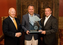
 On 6 February 2013, the Microscopy business group of Carl Zeiss received second prize in the 2013 CLEAN! Fraunhofer Cleanliness Technology Award for its Correlative Particle Analyzer (CAPA). The judges of the Fraunhofer Institute for Production Technology and Automation (IPA) honored the three award winners on day two of the Cleanroom Lounge trade fair in Karlsruhe. "By bridging scanning electron and light microscopy, Correlative Particle Analyzer is revolutionizing cleanliness technology," said the Fraunhofer Institute commenting on its success....
 The winner of this year's Carl Zeiss Research Award, one of the most renowned honors in optics, goes to Professor Anne L’Huillier from Lund University in Sweden. L’Huillier is being honored for her pioneering work in the field of high harmonic generation which has laid the foundation for the generation of attosecond impulses and enabled key advances in attosecond physics."Professor L’Huillier not only described the theory of attosecond technology, but also verified it experimentally”, stated the jury in announcing its decision. Her works enables further development and application of this technology...


 The Carl Zeiss Microscopy business group is presenting two new options for particle analysis at this year's parts2clean trade fair in Stuttgart, Germany. Using Axio Zoom.V16, testers can investigate a component's cleanliness quickly and efficiently, while correlative microscopy provides chemical information and material classifications on individual particles...
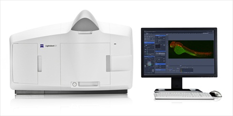 The Microscopy business group at Carl Zeiss is presenting a new microscopy technology at the Society for Neuroscience Annual Meeting in New Orleans, Louisiana. Lightsheet Z.1 provides biologists with a new method of imaging dynamic processes in living organisms...
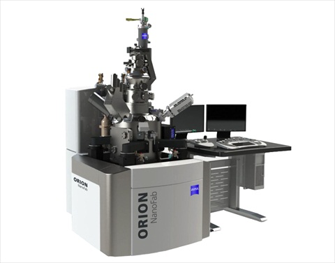 Carl Zeiss Microscopy has recently introduced ORION NanoFab at the European Microscopy Congress (EMC) in Manchester, UK. It is the first multi-ion-beam tool based on Gas Field Ion Source (GFIS) technology. As a major enhancement to the existing helium ion microscope, ORION NanoFab also utilizes neon ions. The system is therefore capable of providing a complete sub-10 nanometer nanofabrication and sub-nanometer imaging solution for industry, government, and academic research laboratories. An optional gallium focused ion beam (FIB) column can also be integrated...
 Prior to the final voting, a jury of scientists had examined all submissions and made a selection. Readers of G.I.T. Laboratory Journal, customers and visitors of Analytica 2012 in Munich and of Achema 2012 in Frankfurt then selected their favorites. Carl Zeiss Microscopy launched the FIB SEM in March this year. The system combines the advantages of the CrossBeam workstation with the capabilities of a pulsed micro-focus laser for fast ablation of material...
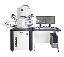 SIGMA HD offers customers high resolution, fast imaging and easy sample navigation for nanoscale analytics in addition to the performance of the established SIGMA series...
 Carl Zeiss Microscopy, a company of the Carl Zeiss Group and leading provider of light, laser-scanning and electron and ion beam microscopes, announces a new program which allows users to trade in their current Zeiss systems for credit towards a new 7-series laser scanning, spectral confocal system. The old products that are being accepted for trade in are the LSM 510, LSM 5 LIVE, LSM 5 Pascal or Bio-Rad confocal. The credit from trading in your old systems can be applied to Zeiss' LSM 780, LSM 710 or LSM 700 systems.
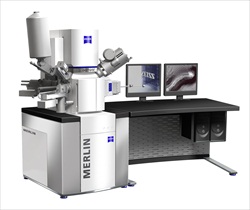 Customers can now choose between the systems MERLIN Compact, MERLIN VP Compact and MERLIN and configure them individually. The Plug-and-Play feature allows the customer to add and change detectors with minimum effort to handle tasks ranging from simple image capture to extensive material analysis. A large frame store of 32k x 24k pixels allows imaging of very large areas. New features include in-situ 3D surface reconstruction and calculation of 3D data from 2D data...
 Today, the collaboration between Carl Zeiss Microscopy and Gatan, Inc. was officially announced. Carl Zeiss Microscopy is a leading supplier of light and electron microscopes. Gatan, Inc. is the world's leading manufacturer of instrumentation and software for the enhancement of electron microscopes' operation and performance. In this joint project, the companies will promote the development and sales of a system which provides high-resolution 3D data of resin embedded cell and tissue samples...
 Multiphoton confocal microscopes from Carl Zeiss now permit the simultaneous use of two NLO lasers or one laser with an optical parametric oscillator (OPO). Both components are fully integrated and expand the functionality of the multiphoton systems. In dual laser systems different laser wavelengths simultaneously excite several fluorescent dyes or proteins. Without time loss, users can image specimens with one wavelength and manipulate them in the multiphoton mode with another...
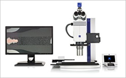 With Axio Zoom.V16, Carl Zeiss is defining a new instrument class in microscopy. For the first time, zoom microscopes are now combining typical benefits of stereomicroscopes such as zoom optics and long working distances with the higher resolutions of light microscopes. In comparable image fields, Axio Zoom.V16 offers a 2.5 times higher resolution than stereomicroscopes. With a 16x zoom range, Axio Zoom.V16 surpasses all comparable microscopes currently available...
Carl Zeiss has received two orders for the extreme ultraviolet lithography system AIMS EUV
Apr 26, 2012
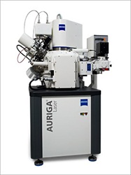
AURIGA Laser: Innovative Combination of FIB/SEM Technology with Laser Ablation for Fast Sample Preparation
Apr 5, 2012
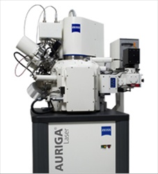 Carl Zeiss has launched the AURIGA® Laser, a new advanced system combining the specific advantages of the AURIGA® CrossBeam (FIB-SEM) workstation with the capabilities of a pulsed micro-focus laser for fast ablation of material. AURIGA® Laser is particularly useful for the examination of samples where the target structure is deeply buried under material layers. To gain access to the target structure this material needs to be removed - a procedure which is difficult to conduct with conventional techniques...
 A microsite dedicated to the natural resource industries has been launched today by Carl Zeiss. The sustainable use of natural resources is dependent upon improving the efficiency of the mining, refining and end production industries. The new microsite from Carl Zeiss addresses the challenges for natural resource industries and demonstrates how pioneering microscopy solutions can overcome these...
 The Axio Vert.A1 is an inverted microscope from Carl Zeiss with extensive performance capabilities. For the first time, a compact cell culture microscope combines all standard contrasting techniques in a single stand. In transmitted light, the Axio Vert.A1 offers brightfield, phase contrast, polarization contrast, PlasDIC, VAREL and improved Hoffmann modulation contrast (iHMC). The microscope is the only system in its class to also offer differential interference contrast (DIC). This makes it possible to record very fine structures in cells, enabling users to better assess cell cultures...
 Tab4Lab comprises a touch pad and the required software. In combination with the AxioCam ERc 5s camera, the two components form an image recording system. The software is based on the established ZEN imaging software. It has been specially adapted to tablet PCs and can be operated via touchscreen. This minimizes both the learning curve and the time required for routine applications. The workflows are modeled on those used in clinical labs and in quality assurance in industry. The user interface is available in various languages...
 ZEN software was launched by Carl Zeiss for confocal laser scanning microscopes in 2007. The extremely logical structure of the ZEN user interface has received much praise and recognition from customers all over the world. Highly complex experiments can be designed and executed by users with minimal training and support...
 The completely redesigned plan-apochromatic 20x/1.0 VIS-IR objective lens from Carl Zeiss now makes it possible to acquire 3D images of nerve cells down to depths of 5.6 mm in intact tissue using a confocal laser scanning or multiphoton microscope, without having to section the brain. This permits the three-dimensional visualization of the branches of individual nerve cells and the imaging of their connections. In untreated tissue, the penetration depth now achievable is five to ten times more than that of a multiphoton microscope featuring traditional optics...
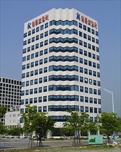 Carl Zeiss Semiconductor Metrology Systems (SMS) Division has opened an office in the Bundang province of South Korea. The new location south of Seoul enables Carl Zeiss SMS to provide optimized customer service with faster delivery of spare parts and on-site activities...
 University of Delaware receives tenth anniversary shipment Carl Zeiss Microscopy, a company of the Carl Zeiss Group and leading provider of light, laser-scanning and electron and ion beam microscopes, today announced the celebration of its tenth anniversary of the CrossBeam© FIB-SEM technology. In honor of the tenth anniversary, a ceremony was held at the Center for Composite Materials at the University of Delaware, who have acquired an AURIGA 60 CrossBeam workstation. The system is the first major acquisition...
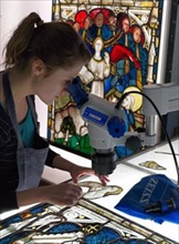 Britain's oldest and largest specialist stained glass conservation studio has recently embarked upon the conservation of York Minster's Great East Window with help from Carl Zeiss microscopes. The York Glaziers Trust chose a pair of Stemi DV4 Spot microscopes to investigate surface corrosion on Britain's largest expanse of medieval stained glass and the Zeiss AxioCam to record the phenomena for future reference...
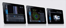 Light Lab, the first accompanying app for ZEN 2011 imaging software from Carl Zeiss, is now available for iPad, iPhone and iPod touch. Light Lab supplements the ZEN 2011 controlling and image processing software functions within the experimental lab workflow. The app supports the configuration of fluorescence experiments in both confocal and light microscopy. The user can also access all product brochures, social media channels and microscopy activities of Carl Zeiss. Light Lab utilizes information on the current location and notifies the user of the closest contact person for Support and Service...
 With the Axio Zoom.V16, Carl Zeiss is defining a new instrument class in microscopy. For the first time, zoom microscopes are now combining typical benefits of stereomicroscopes such as zoom optics and long working distances with the higher resolutions of traditional light microscopes. In comparable image fields, the Axio Zoom.V16 offers a 2.5 times higher resolution and ten times brighter fluorescence than stereomicroscopes...
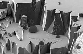
 Scientists from Lewis and Clark College, working collaboratively with the Joint School of Nanoscience and Nanoengineering (JSNN) in the USA have examined the fascinating climbing skills of geckos and spiders using the ORION helium ion microscope from Carl Zeiss. High resolution images displaying unique definition and detail rendition reveal even the finest structures on the feet of these animals. This imaging advances the understanding of how the masterly climbing skills of these creatures are attributable to adhesive forces or, in other words, intermolecular attractions...
 Carl Zeiss Nano Technology Systems, a company of the Carl Zeiss Group and leading global provider of electron and ion beam microscopes, today announced that it has been awarded the NorthFace ScoreBoard Award from Omega Management Group Corp. in recognition of achieving excellence in customer service and support in 2010...
 Images from Royal Society Research Fellow, Dr Richard Kirby, selected by Royal Photographic Society for their International Images for Science Exhibition 2011 and Royal Society Summer Exhibition...
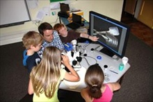 Children at Longworth Primary School in Oxfordshire were treated recently to the sight of everyday objects from the world around them in exquisite detail, thanks to a live microscopy workshop organised by Carl Zeiss. The event was the brainchild of Dr Oliver Clarke, a Business Manager at Carl Zeiss, and was held as part of the school's Summer Fete. A Stemi 2000 C stereo microscope complete with the Educational version of the AxioCam ERc5s digital camera was set up in one of...
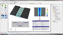 Carl Zeiss and Digital Surf announce the signing of an agreement enabling Carl Zeiss to provide ConfoMap® software. ConfoMap® is a surface imaging and analysis software for confocal microscopes and compound microscopes for topographical research...
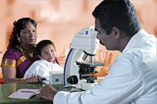 For the efficient and rapid detection of tuberculosis bacteria the World Health Organization WHO now recommends LED-fluorescence microscopy. With Primo Star iLED Carl Zeiss developed, in cooperation with FIND (Foundation for Innovative New Diagnostics), a microscope aligned for this application...
 The United States Patent and Trademark Office has granted Dr. Eric Betzig and Dr. Harald Hess another patent for their invention of super-resolution localization microscopy. In 2007, the microscopy from Carl Zeiss received the exclusive rights to market the technology.
 After a two-week final stretch the winners of the first Carl Zeiss Nano Image Contest have now been chosen. The winners are Heinrich Badenhorst from the University of Pretoria, South Africa (category Scanning Electron Microscopy (SEM), Norman Hauke and Arne Laucht of the Walter Schottky Institute of Munich Technical University, Germany (category CrossBeam (FIB-SEM), ...
Carl Zeiss Introduces Correlative Light and Electron Microscopy Solution for Life Sciences
Aug 27, 2010
Exciting Nanofabrication Capabilities Are Now Available on Carl Zeiss Helium Ion Microscope
Aug 4, 2010
Media Partners