
Channels
Special Offers & Promotions
Marking the 300th anniversary of the death of Dutch microscopist Antoni van Leeuwenhoek
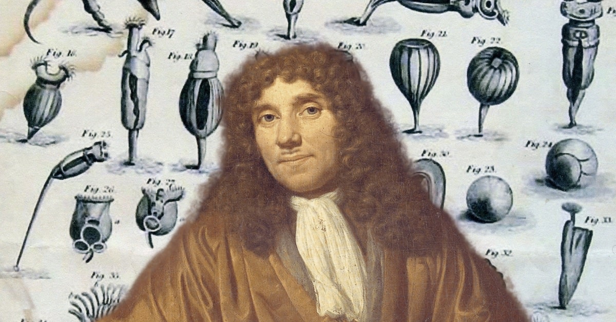
LEEUWENHOEK’S THIRD CENTURY - Revealing the Sights of the Seventeenth Century by Brian J Ford Hon FRMS, Hon FLS
There was much excitement around the world when I revealed one of the most extraordinary revelations of my entire academic life – the original microscope specimens, prepared by Antony van Leeuwenhoek in the 1600s, had survived intact. They lay secure in folded paper envelopes hidden among his letters to London, and carefully preserved at the Royal Society.
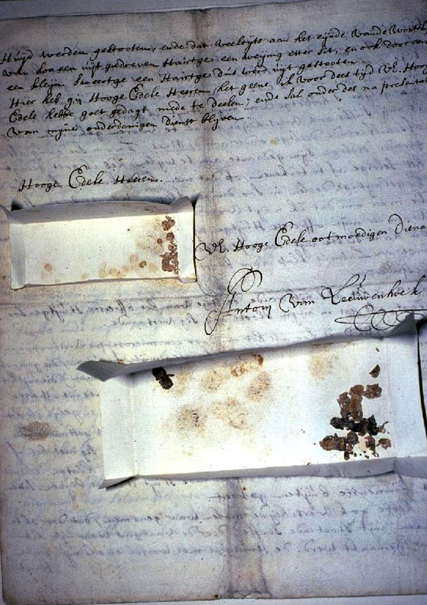
Fig 01: Leeuwenhoek’s letter to the Royal Society of 2 April 1686 contained cotton seed specimens, including the first serial sections. Dutch scholars had misinterpreted copies of the letters as having “drawn rectangles” on the page. In fact, they were packets containing priceless specimens.
August 26 marks the 300th anniversary of that great pioneer’s death. He was an extraordinary man; born in Delft on October 24, 1632, and never trained in science or philosophy, this cloth merchant and town official devoted himself to probing the microscopic realm. He had visited London around 1667, where he was shown a copy of Robert Hooke’s great book Micrographia that had been published in 1665, and (as Samuel Pepys reported) was the talking-point of the time. No scholar had realised, until I drew attention to the fact, but the fine detail revealed in Hooke’s exemplary engravings cannot be seen through his compound microscope.
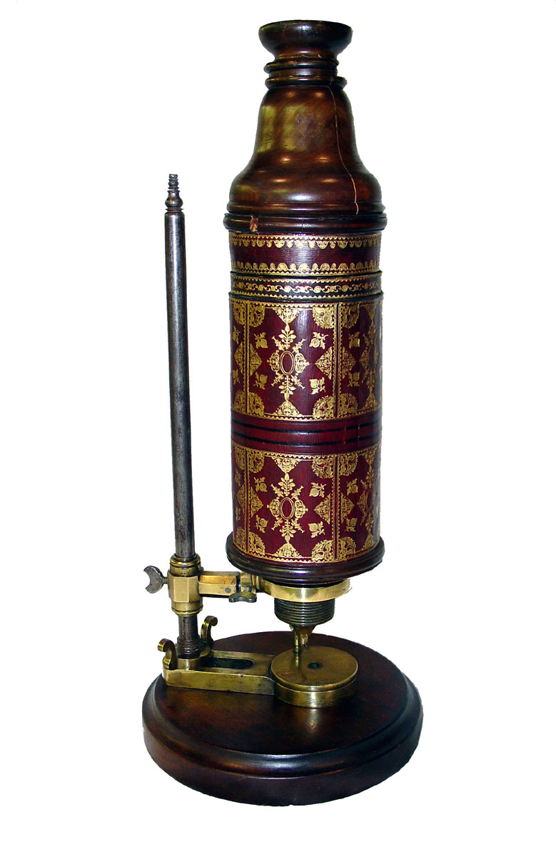
Fig 02: Robert Hooke used this decorated and beautifully crafted compound microscope to examine his specimens. However, I showed it could not resolve the fine details shown in his engravings. For these, he used a single-lensed microscope, and this design was adopted by Leeuwenhoek.
I found the reason hidden in the unnumbered pages of Hooke’s preface. For observing fine detail, he used a single-lensed microscope. Indeed, he described exactly how to make one. Hooke would draw out a fine fibre of glass in the flame, then allow it to form a semi-molten droplet at the end. This was not to be the lens, however; he describes how (with point uppermost) you’d fix it onto a plate with sealing-wax, and then grind away the glass, finishing with a fine abrasive like jeweller’s rouge, so you end up with a plano-convex lens a millimetre or two in diameter. The finished lens was then mounted in a hole cut in a small plate of metal (brass, lead, or pewter was recommended) and an object viewed through this diminutive instrument could be seen in astonishing detail. It would make “Objects more distinct than any of the great Microscopes,” wrote Hooke.
He was right. This simple method of construction produced a magnifier that could resolve bacteria. Compound instruments (“great” microscopes, as Hooke called them) magnified the aberrations more than the image. And this was the design that Leeuwenhoek was to adopt. He had not looked through a microscope by the time he was forty, yet he went on to devote a half-century of endeavour to unveiling the microscopic world.
Fig 03: My purpose-built adaptor for an Olympus OM2n 35 mm camera (top left) lies on a bench in Utrecht with the body of their Leeuwenhoek microscope (right) with its mechanism carefully laid to one side. With test objects for focussing (centre), we also see his original cork sections (bottom).
My finding nine specimen packets was a neat counterpoint to the nine microscopes that were linked to Leeuwenhoek’s name. Two of his chosen specimens were of cork, and of elder pith, the same samples Hooke had described in his book. In each case he had cut remarkably fine sections (you could be proud to emulate his results today). There is nothing new about observing these materials. Cork has been used as a test object since Micrographia first appeared. Robert Hooke, when examining the tiny square features he observed in a section of cork, thought of them as like little cells in a monastery or jail. This is why he gave them that name, and it has stuck with us ever since. Elder pith is familiar, too; this medullary tissue comes from the core of a young Sambucus stem and has long been a favourite supporting medium for hand-cutting sections of leaves.
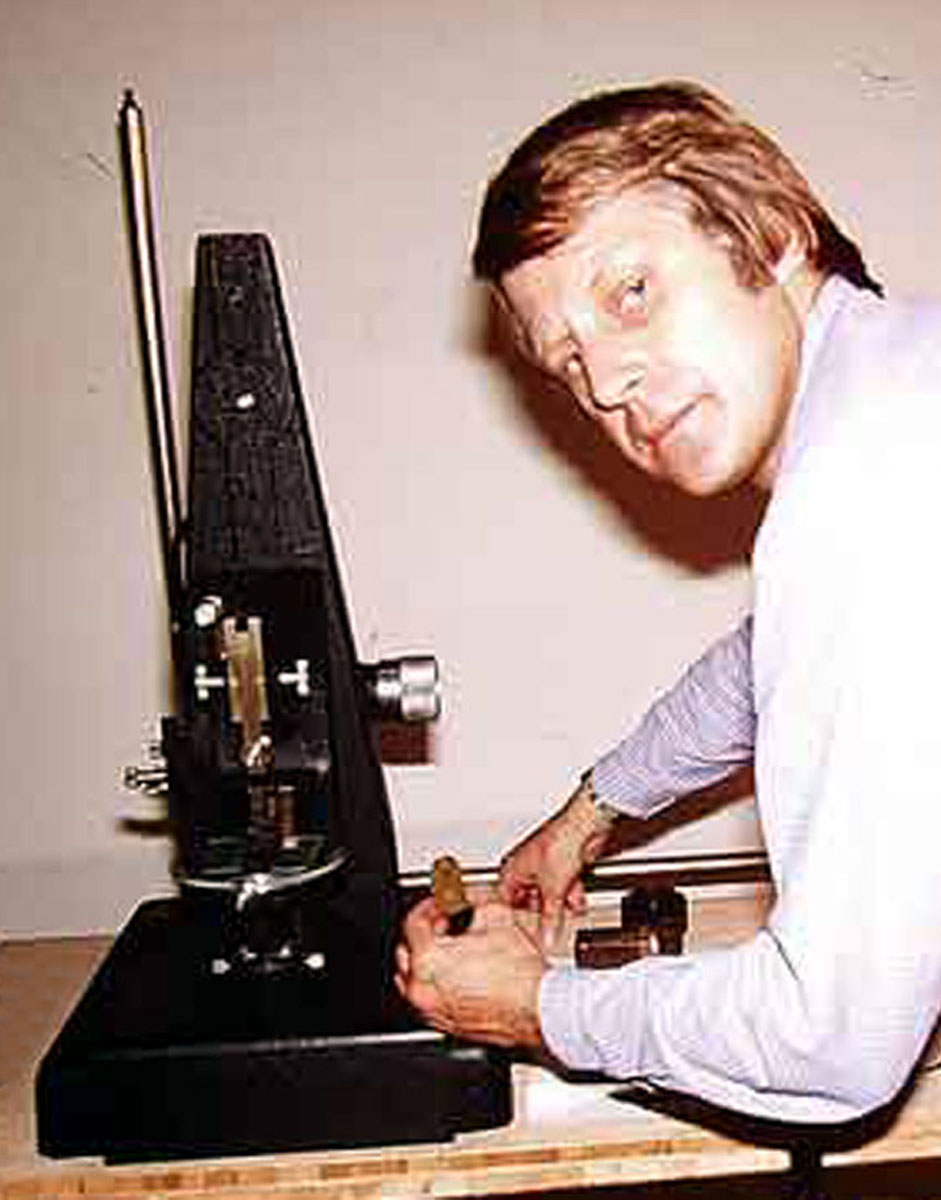
Fig 04: Test run – a replica microscope is being used to test the system. The Utrecht museum technician had created a brass bracket to support my camera assembly, using a design I’d drawn on an envelope. Once all was tested, the original Leeuwenhoek microscope was carefully installed.
So both are familiar microscopical subjects – but how would they have appeared to Leeuwenhoek? I hastened to the Netherlands to image his own sections through the original Leeuwenhoek microscope held at the university Museum in Utrecht. I inspected each under his microscope from the late 1600s, using a purpose-built adaptor that held the tiny microscope body in position, in place of the camera lens. The results were breathtaking – I have seen worse images presented by present-day researchers using modern microscopes.
The best regions of the cork sections were no more than 10 µm thick, and the structure could be beautifully resolved under the original Leeuwenhoek microscope. The cells of cork measure about 40 µm across while Elder cells are larger (often over 100 µm). As well as the cell structure one would expect, I could make out a fine tracery of fungal hyphae across the surface of the cell walls, testimony to three centuries languishing in a store-room. In each case I took several 35 mm images. The earliest microscopes are too delicate to handle more than necessary. In each case I was resolved to acquire a few images; just enough to prove the point.
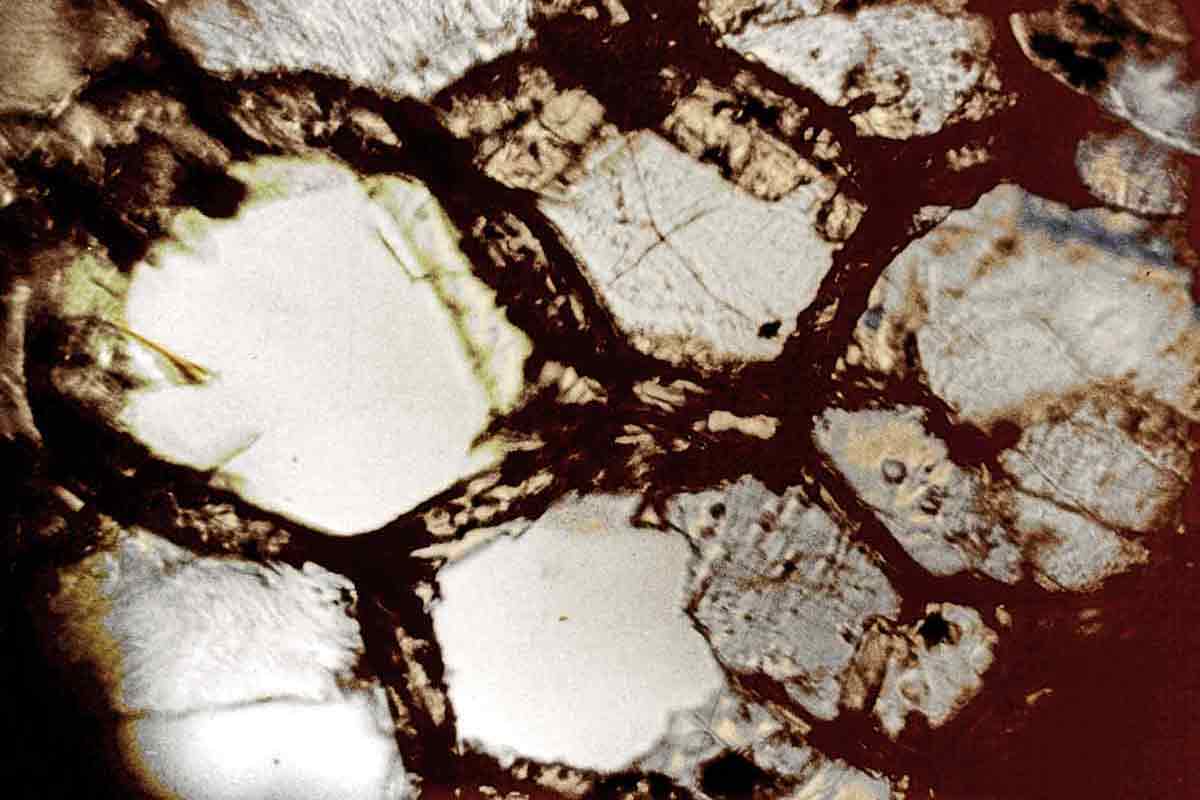
Fig 05a: The first view of an elder-pith section. Leeuwenhoek’s exquisitely prepared section is revealed in remarkable detail through the original lens from c1690. We can see cell-wall pitting, typical of Sambucus medulla, and traces of fungal hyphae from over three centuries of storage.
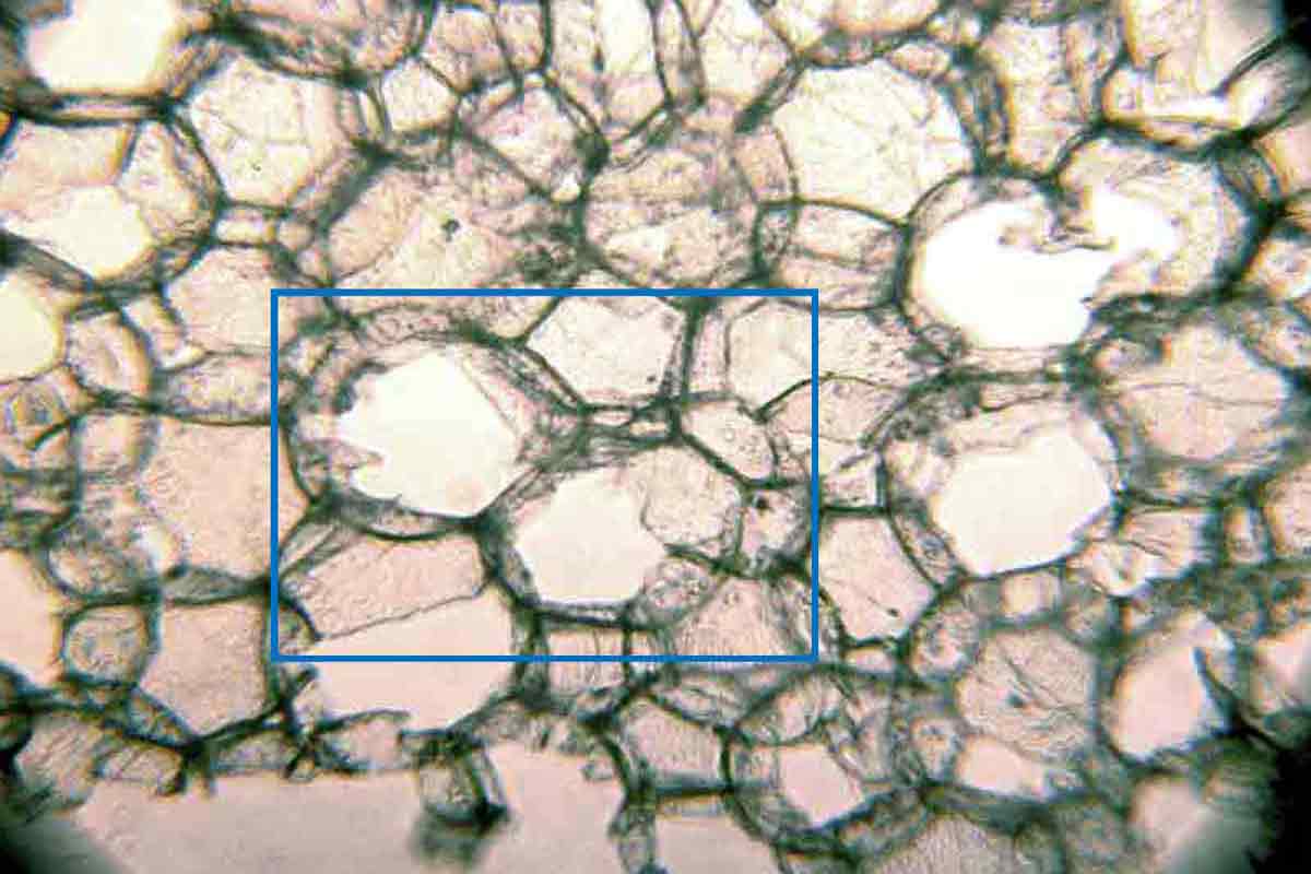
Fig 05b: Locating the same field of view among the thousands of cells in the elder-pith section was a demanding undertaking. Tracking methodically across the specimen under a Leitz Dialux microscope eventually led me to identify the same cells (within the blue rectangle).
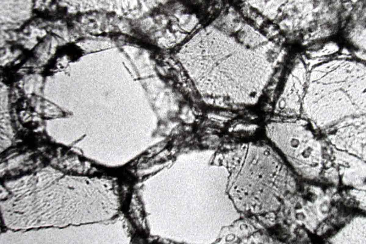
Fig 05c: The modern view of the section belies conventional scholarship which insists early microscopes produced indistinct images that were compromised through aberrations. The sectioned cell walls, pitting, and phycomycete hyphae were all resolved by the Leeuwenhoek lens.
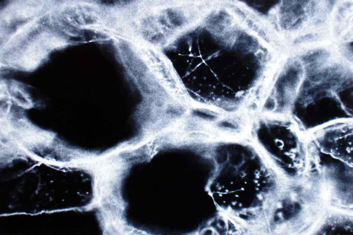
Fig 05d: Dark-ground illumination of Leeuwenhoek’s hand-cut section temporarily immersed in xylene (RI 1.5) displayed the hyphae and pitting with greater contrast. Correlated microscopy, embracing a range of disparate techniques, can alone reveal the details of such specimens.
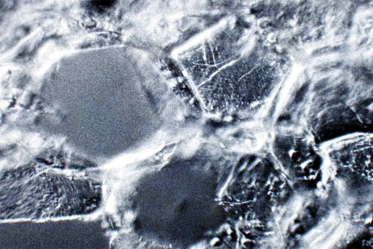
Fig 05e: Fine detail of the cell-wall structures and hyphae can be further visualised through phase-contrast microscopy. The liquid mountant rapidly evaporates after this procedure, allowing further perusal to take place without the original specimen being significantly altered.
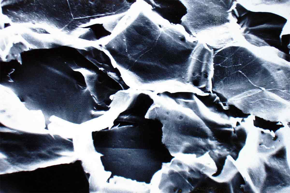
Fig 05f: After these investigations, the section was mounted on an aluminium stub and gold sputtered for scanning electron microscopy (SEM). This technique allowed the Leeuwenhoek image to be accurately quantified. The finest fibril the original lens resolved measured 7 µm.
This was exacting, but much more was to follow. I decided to take correlated micrographs under disparate conditions. The first task was to use a modern microscope to locate the identical cells I’d imaged with the Leeuwenhoek lens. There are thousands of cells, even in a small section, and to this day I cannot grasp the enormity of that decision. Eventually I was able to find the same cells. I recorded them with the microscope camera, then took a further picture at higher magnification. The cell walls were clearly visible, and the pits that characterise elder pith could also be seen. So could the fungal hyphae. These would prove to be important, as they gave an intimation of how well an ancient lens could resolve such fine details.
I was eager to compare this light-ground view with dark-ground illumination, a technique that would display the hyphae (and many other surface features) with greater vividness. Ordinarily, this would involve placing the specimen in a mounting medium of suitable refractive index (RI), around 1.5. By chance, this is the RI of xylene, a widely-used laboratory solvent, so a small droplet was irrigated by capillarity between slide and coverslip, enabling dark-ground micrographs to be obtained. The hyphae stood out brilliantly against the velvet-black field of view. I also took the opportunity to image the same cells with phase-contrast. Finally, the tiny specimen was allowed to dry, and was subsequently gold-sputtered on an aluminium stub for the final perusal, study by scanning electron microscopy (SEM) at Cardiff University. The result is a series of spectacular images of exactly the same cells, visualised under a range of microscopes from Leeuwenhoek’s lens of the late 1600s through to the scanning electron microscope of the present day. Nothing like it has ever been attempted before or since. I’d not recommend you to try; it was an exasperating exercise in diligence.
In separate programmes of research, details were discovered in the specimens that threw further light on Leeuwenhoek’s life and methods. The SEM allowed me to find the microscopic mites about which he complained, and even traces of blood cells left by his cutting sections with a razor. Reconstituting some of the dried algal films he had prepared gave me sight of diatoms and algae, and even preserved water-fleas. When future scientists apply as yet undiscovered new methods of analysis, imagine what else might be found. This is why I was meticulous to ensure that the bulk of each specimen remained intact, uncontaminated, and untouched. These specimens take of back to the dawn of the modern scientific age. Investigating them offers a priceless resource for the future.
Fig 06: The results were formally published in the journals – from Nature and the British Medical Journal to New Scientist and Scientific American – and Leeuwenhoek’s view of nature was also unveiled to international scholarship at the Royal Society’s annual Conversazione and their Soirée.
The initial results were revealing. The single lens of the 1600s could resolve smaller features than has been thought. The finest fibrillar structure that could be visualised under the Leeuwenhoek lens proved to measure 0.7 µm in width. The standard research microscope cannot resolve details smaller than 0.2 µm, so we can conclude that a single lens comes within a factor of four of a modern microscope. This is truly remarkable, and belies the conventional view that early microscopists must have been bedevilled by chromatic and distorted images, until the achromatic lens was perfected. It also destroys the patronising attitude that the pioneering investigators cut their material clumsily, or crudely tore specimens apart to expose what lay within.
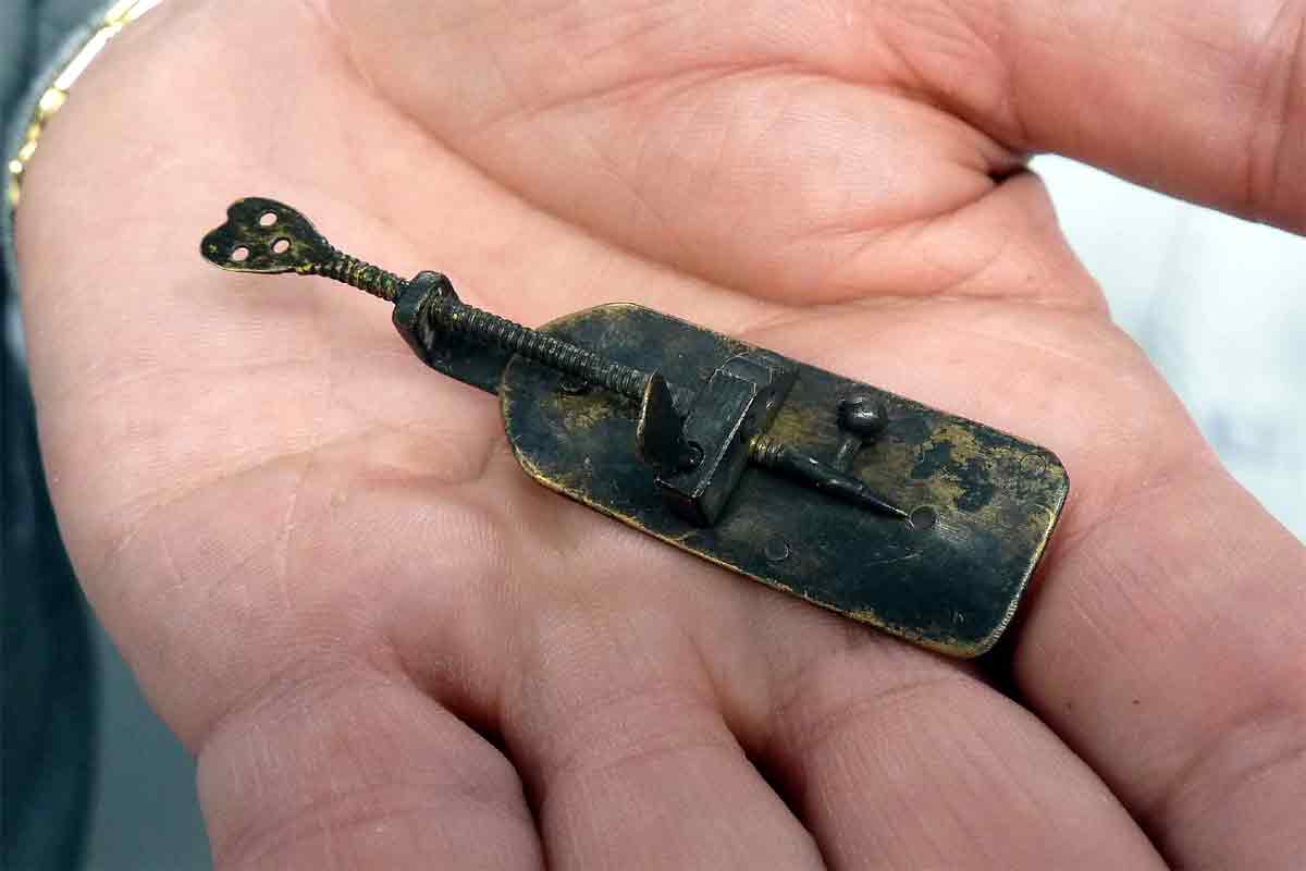
Fig 07: The brass Leeuwenhoek microscope found in mud from a Delft canal, and identified by the author from a set of “weird drawing instruments” advertised on eBay. It was purchased privately by Dr Tomás Camacho, and is the first scientific instrument to be examined by SEM.
More was to follow. Early in 2014 I was invited to call in to see a curio that had been brought into Christie’s, the London auction house. In a small, private room I was presented with a diminutive silver object on a baize topped table … an unknown Leeuwenhoek microscope. With my pocket lens (I never go anywhere without a loupe) I could check the manufacture of the screw thread, and the details of construction, all typical of Leeuwenhoek’s handiwork. It was later purchased privately by a vintage car collector and dealer in the Netherlands. It never reached auction. Within a year another appeared; a little microscope found in mud excavated from a Delft canal was advertised on ebay. This too never went to auction; a Spanish collector purchased it privately before the sale was concluded.
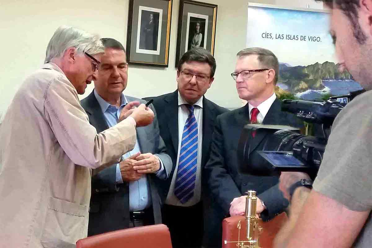
Fig 08: (L-R) The author, with Dr Abel Álvarez, the mayor of Vigo, Spain; with Councillor Carlos López and Dr Tomás Camacho at a reception to announce the new Leeuwenhoek microscope. It has since been declared an Asset of Cultural Importance in the Spanish national collections.
Since its earliest days, microscopy has been an exacting art and the legacy those investigators have bequeathed to us can teach us lessons still. Leeuwenhoek was no dabbling amateur, no dilletante; he was a meticulous man of consummate skill and rare diligence. Investigating the new microscopes, and exposing the secrets hidden within his original specimens, has taught us much. Everyone should know about his achievements. Leeuwenhoek opened the door to the world we now know, and he will always deserve our respect.
About Brian J Ford Hon FRMS, Hon FLS
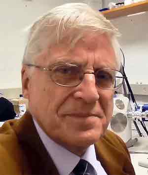 Brian J Ford is a research biologist and lecturer who has published numerous papers, articles, and books on microscopy. He is an authority on the history of the microscope, and on the pioneering microscopist, Antony van Leeuwenhoek. Forty years ago he discovered Leeuwenhoek’s original specimens, hidden in the archives of the Royal Society, and his extensive investigations have laid the groundwork for our understanding of the origins of microscopy. He recently identified two previously unknown Leeuwenhoek microscopes and has used scanning electron microscopy to reveal many details of manufacture. Professor Ford is associated with several universities, including Leicester, Cardiff, the Open University, and Cambridge.
News Channels
- Latest News
- New Laboratory Products
- Industry News
- Laboratory Automation | IT Solutions
- Microscopy | Image Analysis
- Separation Science
- Research | Case Studies
- Video Presentations
- Events | Conferences
Subscribe to any of our newsletters for the latest on new laboratory products, industry news, case studies and much more!

Popular this Month
Top 10 most popular articles this month
Today's Picks
Looking for a Supplier?
Search by company or by product
Please note Lab Bulletin does not sell, supply any of the products featured on this website. If you have an enquiry, please use the contact form below the article or company profile and we will send your request to the supplier so that they can contact you directly.
Lab Bulletin is published by newleaf marketing communications ltd.
Media Partners