
Channels
Special Offers & Promotions
Powering a greener future through material innovation
How electron microscopy accelerates the development of low-carbon technologies
With critical deadlines approaching for the 2026–2031 Nationally Determined Contributions (NDCs), countries are under increasing pressure to deliver stronger climate commitments. As key pillars of the Paris Agreement, NDCs outline each nation's targets for reducing emissions and advancing sustainability efforts. Here, Kate Vanderburgh, Sr. Product Specialist, Thermo Fisher Scientific, explains how electron microscopy (EM) drives material innovation to support sustainable, low-carbon technologies to combat climate change.
The advancement of materials is vital for developing low-carbon technologies. In renewable energy, semiconductors boost solar efficiency, while advanced composites improve wind turbine durability. Furthermore, in energy storage, breakthroughs in electrodes and electrolytes enhance battery performance, lifespan and stability.
Electron microscopy (EM) plays a crucial role in these advancements, providing nanoscale resolution for detailed characterization of material structures, interfaces and defects. This optimizes properties such as conductivity, durability and catalytic efficiency, essential for energy storage and renewable energy systems.
Advancing sustainable technology
Several technologies that are core to advancing sustainable progress reap the benefits of EM throughout their lifecycle.
For instance, EM drives advances in solar panel materials by revealing microscopic defects and grain boundary structures with sub-nanometer precision. In perovskite solar cells, EM uncovers variations in crystallinity and composition that affect charge transport and recombination. This precise insight allows researchers to optimize material formulations and processing methods, resulting in higher efficiency and improved device stability.
EM is indispensable throughout the entire lifecycle of battery materials — from early R&D and manufacturing to quality control and recycling. It enhances the lifespan of lithium-ion and solid-state batteries by identifying structural weaknesses at the nanoscale, detecting defects such as dendrite formation, which can cause short circuits, and monitors chemical changes during charging. These insights help engineers develop safer, longer-lasting, more environmentally-friendly battery materials that support circular economy principles.
Hydrogen technology also benefits from EM’s capabilities. EM is used to analyze hydrogen storage alloys and fuel cell materials by revealing nanoscale structural features and elemental distributions critical for efficient hydrogen uptake and release. This information guides the optimization of material design, leading to faster hydrogen absorption, improved storage capacity and enhanced long-term stability — key factors for scaling hydrogen-powered transport and renewable energy systems.
ChemiSEM: A step forward in sustainable materials research
Developing advanced materials for low-carbon technologies require precise structure and compositional analysis. However, modern materials — such as solid-state battery electrolytes and hydrogen storage alloys — present challenges that traditional EM methods struggle to address.
Conventional scanning electron microscopy (SEM) provides high-resolution imaging of material surfaces, revealing grain boundaries and nanoscale defects. Meanwhile, energy dispersive X-ray spectroscopy (EDS) analyzes elemental composition.
In traditional workflows, these techniques require sequential analysis, with EDS performed after, or instead of, imaging, often under different conditions. This can lead to inconsistencies, making it difficult to link structure to composition. Furthermore, emerging sustainable materials usually have complex, heterogeneous compositions in which subtle elemental variations impact performance and longevity.
Thermo Fisher ChemiSEM presents a single, streamlined solution that integrates SEM imaging and EDS analysis into one user interface. This not only enables faster result times and live structural and compositional analysis but also provides accurate quantification, complete with live peak deconvolution and automated removal of sum and escape peak artifacts.
As an integrated data management platform combining advanced elemental analysis with real-time electron imaging, ChemiPhase within ChemiSEM enables the discovery of material phases through advanced statistical analysis. This allows users to perform SEM and EDS analysis simultaneously, reducing response times and minimizing redundancies and downtime typically associated with using separate SEM and EDS systems.
Multiple different types of analysis can be performed in ChemiSEM, including point and region analysis, line scans, gross count mapping, quantitative mapping and phase mapping. Furthermore, with ChemiSEM, chemical analysis is always on. This helps to provide the user with insightful information about a sample’s structural, chemical and material properties as it is analyzed.
The system is also highly user-friendly, making it accessible to a broader range of researchers, including those who are not highly trained in advanced material science. This accessibility helps democratise high-level microscopy techniques, accelerating innovation across industries and nations.
Case Study: Ag/Zn Si Photovoltaic
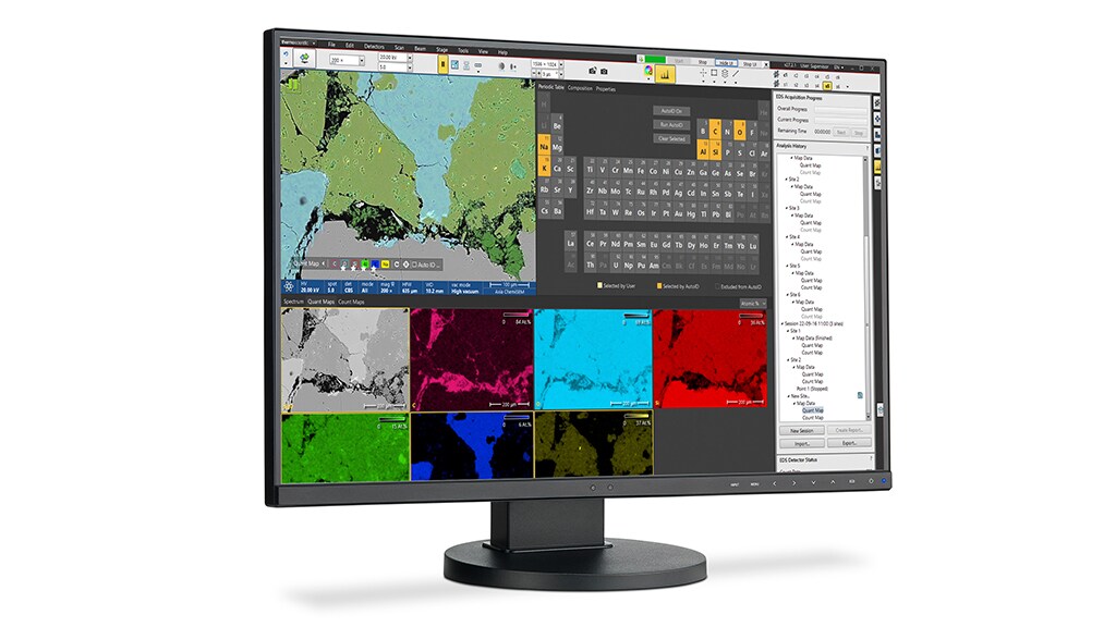
Solar energy is a critical form of renewable energy and is primarily harvested by photovoltaics which convert sunlight into electricity. Materials research and characterization is at the forefront of making current solar cell technology cheaper, more efficient and more reliable. EM is an important technique to view photovoltaic materials at micro and nanoscales to examine the material structure and composition. Figure 1 shows an Ag/Zn Si photovoltaic simultaneously imaged with four different detectors on a Thermo Fisher Apreo ChemiSEM.
At first inspection, there appeared to be differences in material structure and compositional contrast throughout the sample surface. ChemiSEM made it possible to deconvolute these differences due to topography versus chemical structure with a click of the ChemiSEM color wheel and spectra buttons and without having to change any imaging conditions. Figure 2 illustrates a ChemiSEM image of the A) Ag/Zn Si photovoltaic and individual quantitative elemental maps of B) silver (blue), C) zinc (pink), and d) silicon (green). Being able to identify the elemental distribution helps to better understand material properties to progress photovoltaic research for a cleaner energy future.
Electron microscopy is proving indispensable not only for early-stage research and development but throughout the entire lifecycle of sustainable materials. Such comprehensive capabilities are critical as nations work to meet the ambitious climate targets set forth in the upcoming 2026–2031 NDC deadlines, ultimately supporting the transition to a sustainable, low-carbon future.
About Thermo Fisher Scientific
Thermo Fisher Scientific Inc. is the world leader in serving science, with annual revenue over $40 billion. Our Mission is to enable our customers to make the world healthier, cleaner and safer. Whether our customers are accelerating life sciences research, solving complex analytical challenges, increasing productivity in their laboratories, improving patient health through diagnostics or the development and manufacture of life-changing therapies, we are here to support them. Our global team delivers an unrivaled combination of innovative technologies, purchasing convenience and pharmaceutical services through our industry-leading brands, including Thermo Scientific, Applied Biosystems, Invitrogen, Fisher Scientific, Unity Lab Services, Patheon and PPD.
Recent news from Thermo Fisher Scientific
News Channels
- Latest News
- New Laboratory Products
- Industry News
- Laboratory Automation | IT Solutions
- Microscopy | Image Analysis
- Separation Science
- Research | Case Studies
- Video Presentations
- Events | Conferences
Subscribe to any of our newsletters for the latest on new laboratory products, industry news, case studies and much more!
Popular this Month
Top 10 most popular articles this month
Today's Picks
Looking for a Supplier?
Search by company or by product
Please note Lab Bulletin does not sell, supply any of the products featured on this website. If you have an enquiry, please use the contact form below the article or company profile and we will send your request to the supplier so that they can contact you directly.
Lab Bulletin is published by newleaf marketing communications ltd.
Media Partners