Members Login

Channels
Special Offers & Promotions
Agar Scientific rewards imaging ability at emc2012
 |
Agar Scientific, a leading supplier of microscopy accessories and consumables, recently took part at the UK's largest ever microscopy event – emc2012, the 15th European Microscopy Congress. Here, the company reports on results of two microscopy imaging competitions
Agar Scientific have been serving microscopists with the supply of accessories and consumables for forty years. In celebration of this anniversary, the company ran a photographic competition at the European Microscopy Congress in Manchester. Entrants were given the opportunity to test their knowledge and imagination. The goal was to identify twenty colour enhanced micrographs from the world of microscopy. Clues to each image were provided and two of the entrants got the maximum score.
After a drawing conducted by the RMS organiser, Allison Winton, the microscopist to win the latest Canon PowerShot S100 digital camera was Dr Lucy Collinson of the London Research Institute of Cancer Research UK. Lucy, runs the Electron Microscopy suite where her team have access to three transmission electron microscopes and one scanning electron microscope along with a comprehensive range of sample preparation equipment and image analysis workstations. The EMU team has five post-doctoral researchers, whose backgrounds cover cell biology, neurobiology, plant biology, microbiology and bioinformatics, with over 100 years' experience in electron microscopy techniques and technologies between them.
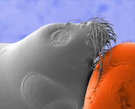
Agar also support the biennial International Micrograph Competition organised by the Royal Microscopical Society. Their sponsorship comes in the form of a cash prize and this year it was awarded to Dr Rok Kostanjsek from the University of Ljubljana. His image is a micrograph depicting the anterior part of the cephalothorax of money spider Walckenaeria cucullata (family Linyphiidae) under FE-SEM.
Rok describes his image: Unique cuticular structures in anterior and dorsal face of the cephalothorax are one of distinctive features in male spiders in genus Walckenearia. In the case of W. cucullata cuticular protuberances and tufts of cuticular hairs closely resemble the head of a seal on large ball, when observed under appropriate angle. The specimen was fixed in glutaradahyde, postfixed in osmium, dehydrated in series of ethanol, critical point dried and coated by platinum prior observation under JEOL JSM-7500F cold cathode FESEM. Picture was taken at 22,2 mm working distance, acceleration voltage of 2 kV and depicts area of approximately 270 x 225 µm. Obtained monochromatic picture was colourized by Corel Paint Shop Pro X2.
Agar provides a comprehensive catalogue and price list on accessories and consumables for microscopy. To receive your free copy, please visit www.agarscientific.com and register today.
Media Partners


