Channels
Special Offers & Promotions
Quantifying Receptor-Ligand Binding to GPCRs in Live Cells - A new HTS format Replaces Radioactive Ligand Studies

Ligand binding affinities at G-protein coupled receptors (GPCRs) have historically been determined using a radioligand that competes for receptor binding sites against an unlabeled drug-like compound. But the potential hazards of open-source radioisotope handling, and the environmental impact of radioisotope disposal, make this a less desirable, costly technology
Fluorescent Ligand Binding Assays in Live Cells Use High Content Analysis (HCA)
New methods, such as the development of fluorescent ligands for GPCRs (CellAura Tehnologies, Ltd.) provide a safer method for determining ligand binding affinities. However, measurement of fluorescent ligands binding to cells relies on capturing and processing complex high resolution images. This approach can be lengthy and can result in 96-well plate read times of approximately 30 minutes or more. Cell-based assays that take too long can undergo changes that could affect the assay results. Therefore, these High Content Analysis (HCA) assays are too slow to be useful for High Throughput Screening (HTS).
HTS Microplate Reader is Sensitive Enough for Live Cell Assays
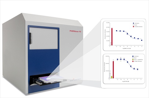
Here enters the PHERAstar FS microplate reader from BMG LABTECH for HTS and Core Lab facilities. Using the adjustable focal height and advanced bottom reading features on the PHERAstar FS, live cells are quickly and easily quantified for fluorescent ligand binding to the adherent cell layer in a 96-well plate format. Depending on several variables, a 'read to IC50 pKi curve plot' times were between 2.5 and 10 minutes for a 96-well plate. This is in stark contrast to a similar HCA assay, which can take between 30-60 minutes depending on the measurement variables.
Here is example data outlined in the application note Quantifying Fluorescent Ligand Binding to GPCRs in Live Cells using the PHERAstar FS - a new format for HTS. The PHERAstar FS performs GPCR ligand binding assays using two fluorescent ligands from CellAura for adenosine (A1 and A3) and dopamine (D1) receptors. Derived pKi curve plots were comparable to previous radioligand binding assays and good Z’ values show that these fluorescent binding assays with this microplate reader are amendable for high throughput screening.
Safer, Faster Receptor-Ligand Binding Assays
The PHERAstar FS and CellAura fluorescent ligands provide a rapid and robust way to quantify receptor-ligand binding in live cells. Using a 96-well format and a simple 'add > mix > wash > read' assay protocol, whole-cell radioligand binding assays are replaced with this safer and more cost effective alternative. In addition, the time saving advantages over HCA assays make this format suitable for high throughput screening (HTS). In addition, multiplexing with other fluorophores can be used to normalize for cell number such (i.e. Hoechst or DAPI) or to measure downstream second messenger effects of like calcium flux or cAMP production.
Find out more about fluorescent ligands at CellAura Technologies Ltd.
To find out more about the PHERAstar FS HTS microplate reader please visit www.bmglabtech.com.
Media Partners


