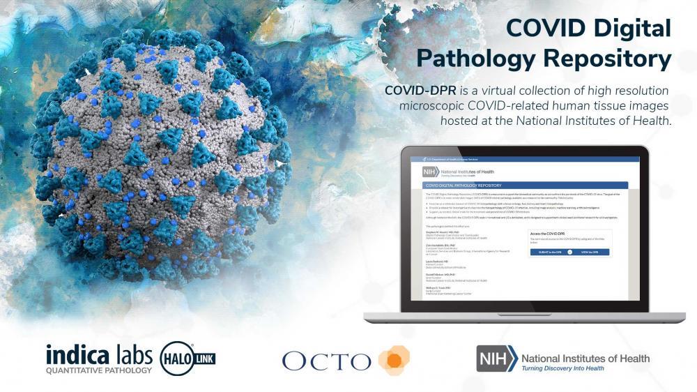
Indica Labs, a leading provider of computational pathology software, and Octo, a premiere information technology systems provider to the U.S. Federal Government, are pleased to announce the online COVID Digital Pathology Repository (COVID-DPR), a virtual collection of high resolution microscopic COVID-related human tissue images hosted at the National Institutes of Health....
When UCLA’s Dr. Araceli Espinosa-Jeffrey sent human brain cells into space, her goal was to gain a better understanding of how neural stem cells grow and develop in microgravity. That understanding is key to discovering more about the serious issues of intracranial hypertension affecting astronauts returning from space. The information may also one day be used to further cell replacement therapies...
Jian-Wei Pan, Professor at the University of Science and Technology of China, in Hefei, is the winner of the prestigious ZEISS Research Award 2020. He is one of the world's leading researchers in the field of quantum technology. One of the most remarkable results of Jian-Wei Pan's research is the distribution of entangled photons over a distance of 1,200 km, by far the longest distance ever reached....
To capture the importance of this initiative, and in anticipation of the grand opening of the Ellison Institute’s new building, Olympus has created a video, highlighting the relationship between imaging analysis tools and precision cancer medicine. One focus of the partnership is encouraging projects that engage translational oncology and precision anti-cancer drug screening...
Linkam has recently seen an increased interest in its stage that was specifically designed for the analysis of catalytic reactions: the CCR1000. Catalysis is a technique for improving the yield or rate of a chemical reaction using a catalyst material. A wide range of catalytic materials are available; including aluminosilicates, which are used in the petrochemical industry to reduce natural materials to smaller hydrocarbons...
The sensitivity of Beckman Coulter Life Sciences’ CytoFLEX Flow Cytometer is underpinning a pioneering multinational research initiative. This is designed to transform the lives of those suffering from a disfiguring and potentially life threatening parasitic disease. The research team has just secured a grant of eight million Euros from the European & Developing Countries Clinical Trials Partnership...
Vision Engineering, a 61 year old British leading designer and manufacturer of high-quality visual measurement and inspection technologies, has attained ISO 17025:2017 accreditation from UKAS and is now a UKAS accredited calibration laboratory No. 7706. The award of ISO 17025:2017 by UKAS Accreditation, which is globally recognised, is the only mechanism that determines the technical competence and integrity of the organisations offering testing and calibration services....
For use in advanced medical research into inflammation, oncology and nephritis the 7T PET-MR imaging system from MR Solutions provides a combination of PET and MRI images either simultaneously or separately. This ground breaking combination technology allows researchers to have superior soft tissue contrast and molecular imaging together.
Olympus cellSens imaging software improves the efficiency of your research with accurate object detection and segmentation. Leveraging the power of deep learning, Olympus cellSens imaging software for microscopy offers significantly improved segmentation analysis, such as label-free nucleus detection and cell counting, for more accurate data and efficient experiments....
Labs working to combat COVID-19 will benefit from this initiative, as CytoSMART aims to reduce the huge workload currently facing researchers on projects vital to controlling the disease. “We aim to do our part to assist researchers in minimizing the time they have to spend in high-contamination labs, by providing them with remote video access to evaluate the status of their cell cultures. The video data is used to remotely monitor the cytopathic effect, this way researchers know when it’s the right time to harvest the virus....
The movements of cell muscles in the form of tiny filaments of proteins have been visualised at unprecedented detail by University of Warwick scientists. In a study published in the Biophysical Journal, scientists from the University’s Department of Physics and Warwick Medical School have used a new microscopy technique to analyse the molecular motors inside cells that allow them to move and reshape themselves, potentially providing new insights that could inform the development of new smart materials....
FLIR Systems has announced the FLIR A400/A700 Thermal Smart Sensor and Thermal Image Streaming fixed camera solutions for monitoring equipment, production lines, critical infrastructure, and screening for elevated skin temperatures. The FLIR A400/A700 Thermal Smart Sensor solution initially will be prioritized for those responding to COVID-19....
The new Cytation™ 7 Cell Imaging Multi-Mode Reader from BioTek Instruments combines an automated upright microscope, an inverted microscope, and multi-mode microplate detection in a single instrument. This combination of microscopes and multi-mode reading enables applications that would typically require multiple instruments. The inverted microscope supports...
The new Cell Count & Viability Starter Kit automates cell counting and viability measurements in a mammalian cell suspension. When used with BioTek’s Cytation™ Cell Imaging Multi-Mode Readers or Lionheart™ Automated Imagers in a workflow, researchers may quickly and automatically image and analyze cell counts in multiple samples. The Starter Kit includes the Cell Count & Viability App...
Olympus is proud to announce the winners of its first Global Image of the Year Life Science Light Microscopy Award, a competition that recognizes the best in life science imaging worldwide. Ainara Pintor from Spain was selected as the global winner for her vibrant image of an immunostained mouse brain slice with two fluorophores. She named her winning image “Neurogarden” because it reflects the brain’s complexity....
New Light Sheet Fluorescence Microscope Introduced. Light sheet fluorescence microscopy (LSFM) with its unique illumination principle allows fast and gentle imaging of whole living model organisms, tissues, and developing cells. The high stability of ZEISS Lightsheet 7 enables researchers to observe living samples over extended periods of time – even days – with less phototoxicity than ever before....
Collaboration to initially focus on the development and global commercialisation of a suite of CelLuminate dyes, specifically created to revolutionise live cell imaging by enabling cells to remain functional and viable for up to 14 days. Abcam plc, a global innovator in life science reagents and tools, and SomaServe Ltd, a Pharma service and specialist reagent business exploiting PolyNaut® Technology, have announced a partnership to commercialise the cell delivery potential of...
Syngene, a world-leading manufacturer of image analysis solutions, has introduced a far-red HI-LED lighting option for its new design of G:BOX Chemi and G:BOX mini multi application gel and blot imaging systems. This quick-fit, environmentally friendly lighting enables fast workflow and precise detection of IR fluorescently labelled proteins on gels and blots....
JEOL USA, a leader in developing instruments used to advance scientific research and technology, is pleased to showcase the latest updates to its core product range at Pittcon 2020. JEOL has 70 years of expertise in the field of electron microscopy, more than 60 years in mass spectrometry and NMR spectrometry, and more than 50 years of e-beam lithography leadership. JEOL expanded its mass spectrometer product line with the development of a GC-triple quadrupole mass spectrometer system....
MR Solutions will be displaying its latest dry magnet 3T to 9.4T preclinical, multi-modality MRI (with PET or SPECT) and new benchtop PET/CT systems to the imaging science community at the 15th European Molecular Imaging Meeting (EMIM 2020) 25-28 August 2020 in Thessaloniki, Greece. This is an annual event organised by the European Society for Molecular Imaging, of which MR Solutions is a member...
Optical Surfaces Ltd. reports how a motorised UV-Vis-NIR collimator and interferometric alignment system it supplied to the Deutsches Zentrum für Luft- und Raumfahrt (DLR) in Berlin, Germany has been used to set-up the ground breaking DLR Earth Sensing Imaging Spectrometer (DESIS). On 23 October 2019, DLR the renowned German Aerospace Center, and the U.S. company Teledyne Brown Engineering (TBE) announced the start of routine operations for DESIS on the International Space Station...
Governing molecular mechanism for chromosome condensation identified 140 years after mitosis first described. The molecular mechanism underlying chromosome condensation, one of the key steps in cell division, has been discovered from three-dimensional (3-D) refractive index (RI) reconstructions of individual mitotic cells. Writing in Nature Communications1, the authors describe using the Tomocube HT-1S holotomographic microscope to quantify the structural and biochemical parameters of the cytoplasm and chromosomes within the individual mitotic cells...

 Indica Labs, a leading provider of computational pathology software, and Octo, a premiere information technology systems provider to the U.S. Federal Government, are pleased to announce the online COVID Digital Pathology Repository (COVID-DPR), a virtual collection of high resolution microscopic COVID-related human tissue images hosted at the National Institutes of Health....
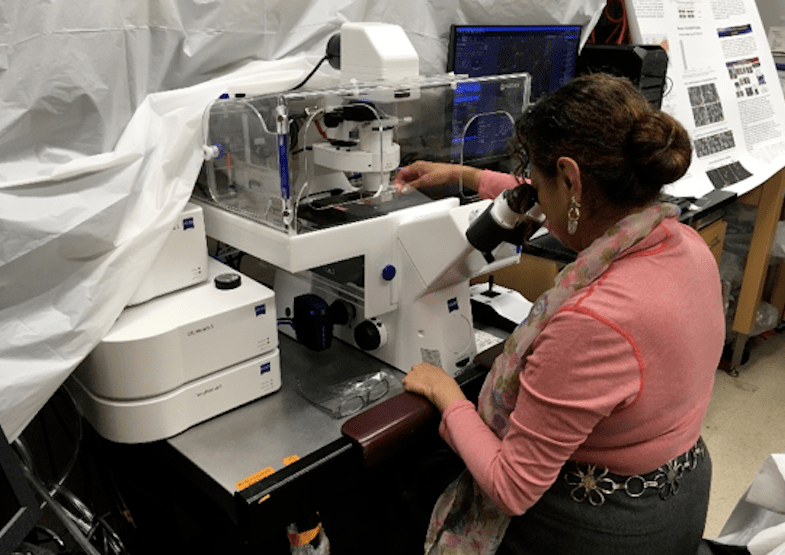 When UCLA’s Dr. Araceli Espinosa-Jeffrey sent human brain cells into space, her goal was to gain a better understanding of how neural stem cells grow and develop in microgravity. That understanding is key to discovering more about the serious issues of intracranial hypertension affecting astronauts returning from space. The information may also one day be used to further cell replacement therapies...
 Jian-Wei Pan, Professor at the University of Science and Technology of China, in Hefei, is the winner of the prestigious ZEISS Research Award 2020. He is one of the world's leading researchers in the field of quantum technology. One of the most remarkable results of Jian-Wei Pan's research is the distribution of entangled photons over a distance of 1,200 km, by far the longest distance ever reached....
 To capture the importance of this initiative, and in anticipation of the grand opening of the Ellison Institute’s new building, Olympus has created a video, highlighting the relationship between imaging analysis tools and precision cancer medicine. One focus of the partnership is encouraging projects that engage translational oncology and precision anti-cancer drug screening...
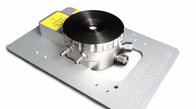 Linkam has recently seen an increased interest in its stage that was specifically designed for the analysis of catalytic reactions: the CCR1000. Catalysis is a technique for improving the yield or rate of a chemical reaction using a catalyst material. A wide range of catalytic materials are available; including aluminosilicates, which are used in the petrochemical industry to reduce natural materials to smaller hydrocarbons...
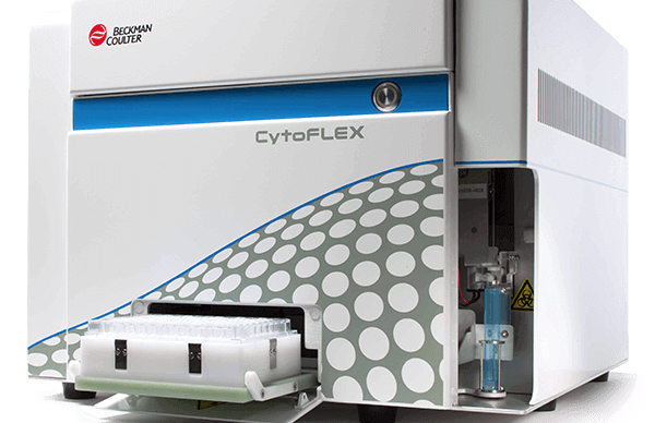 The sensitivity of Beckman Coulter Life Sciences’ CytoFLEX Flow Cytometer is underpinning a pioneering multinational research initiative. This is designed to transform the lives of those suffering from a disfiguring and potentially life threatening parasitic disease. The research team has just secured a grant of eight million Euros from the European & Developing Countries Clinical Trials Partnership...
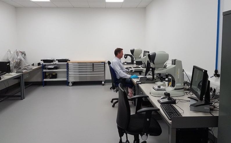 Vision Engineering, a 61 year old British leading designer and manufacturer of high-quality visual measurement and inspection technologies, has attained ISO 17025:2017 accreditation from UKAS and is now a UKAS accredited calibration laboratory No. 7706. The award of ISO 17025:2017 by UKAS Accreditation, which is globally recognised, is the only mechanism that determines the technical competence and integrity of the organisations offering testing and calibration services....
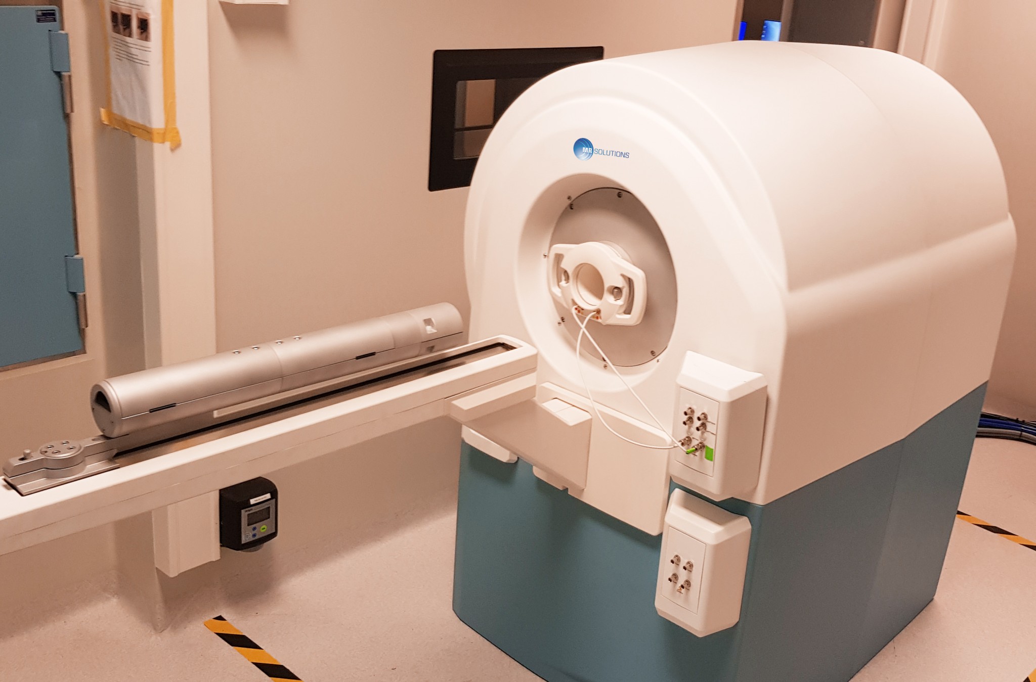 For use in advanced medical research into inflammation, oncology and nephritis the 7T PET-MR imaging system from MR Solutions provides a combination of PET and MRI images either simultaneously or separately. This ground breaking combination technology allows researchers to have superior soft tissue contrast and molecular imaging together.
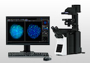 Olympus cellSens imaging software improves the efficiency of your research with accurate object detection and segmentation. Leveraging the power of deep learning, Olympus cellSens imaging software for microscopy offers significantly improved segmentation analysis, such as label-free nucleus detection and cell counting, for more accurate data and efficient experiments....
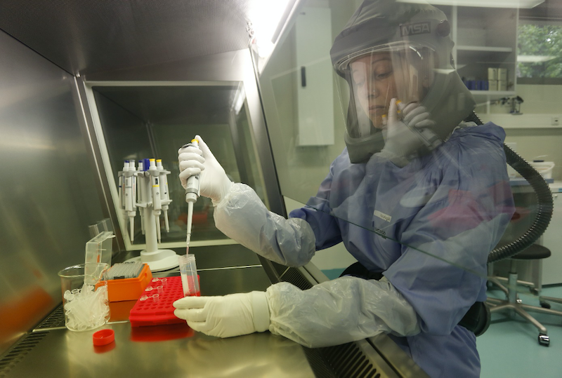 Labs working to combat COVID-19 will benefit from this initiative, as CytoSMART aims to reduce the huge workload currently facing researchers on projects vital to controlling the disease. “We aim to do our part to assist researchers in minimizing the time they have to spend in high-contamination labs, by providing them with remote video access to evaluate the status of their cell cultures. The video data is used to remotely monitor the cytopathic effect, this way researchers know when it’s the right time to harvest the virus....
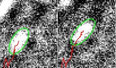 The movements of cell muscles in the form of tiny filaments of proteins have been visualised at unprecedented detail by University of Warwick scientists. In a study published in the Biophysical Journal, scientists from the University’s Department of Physics and Warwick Medical School have used a new microscopy technique to analyse the molecular motors inside cells that allow them to move and reshape themselves, potentially providing new insights that could inform the development of new smart materials....
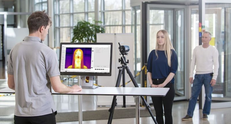 FLIR Systems has announced the FLIR A400/A700 Thermal Smart Sensor and Thermal Image Streaming fixed camera solutions for monitoring equipment, production lines, critical infrastructure, and screening for elevated skin temperatures. The FLIR A400/A700 Thermal Smart Sensor solution initially will be prioritized for those responding to COVID-19....
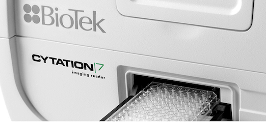 The new Cytation™ 7 Cell Imaging Multi-Mode Reader from BioTek Instruments combines an automated upright microscope, an inverted microscope, and multi-mode microplate detection in a single instrument. This combination of microscopes and multi-mode reading enables applications that would typically require multiple instruments. The inverted microscope supports...
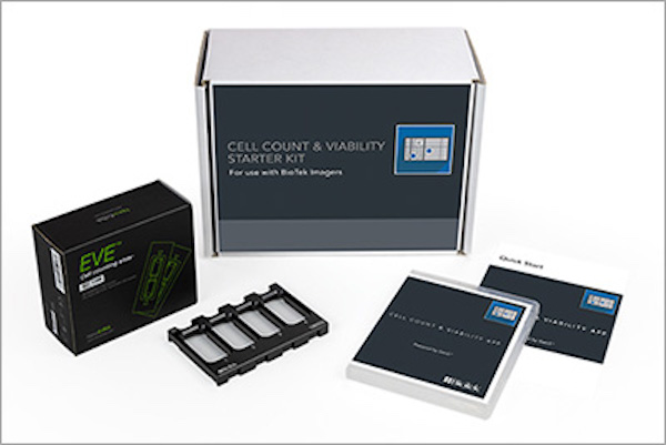 The new Cell Count & Viability Starter Kit automates cell counting and viability measurements in a mammalian cell suspension. When used with BioTek’s Cytation™ Cell Imaging Multi-Mode Readers or Lionheart™ Automated Imagers in a workflow, researchers may quickly and automatically image and analyze cell counts in multiple samples. The Starter Kit includes the Cell Count & Viability App...
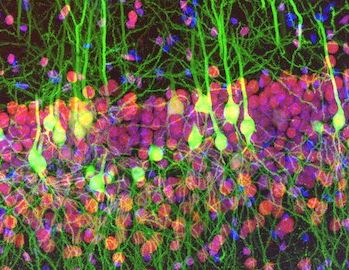 Olympus is proud to announce the winners of its first Global Image of the Year Life Science Light Microscopy Award, a competition that recognizes the best in life science imaging worldwide. Ainara Pintor from Spain was selected as the global winner for her vibrant image of an immunostained mouse brain slice with two fluorophores. She named her winning image “Neurogarden” because it reflects the brain’s complexity....
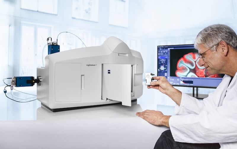 New Light Sheet Fluorescence Microscope Introduced. Light sheet fluorescence microscopy (LSFM) with its unique illumination principle allows fast and gentle imaging of whole living model organisms, tissues, and developing cells. The high stability of ZEISS Lightsheet 7 enables researchers to observe living samples over extended periods of time – even days – with less phototoxicity than ever before....
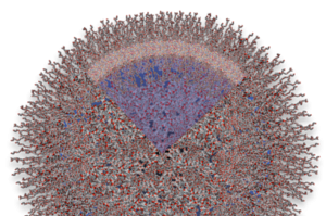 Collaboration to initially focus on the development and global commercialisation of a suite of CelLuminate dyes, specifically created to revolutionise live cell imaging by enabling cells to remain functional and viable for up to 14 days. Abcam plc, a global innovator in life science reagents and tools, and SomaServe Ltd, a Pharma service and specialist reagent business exploiting PolyNaut® Technology, have announced a partnership to commercialise the cell delivery potential of...
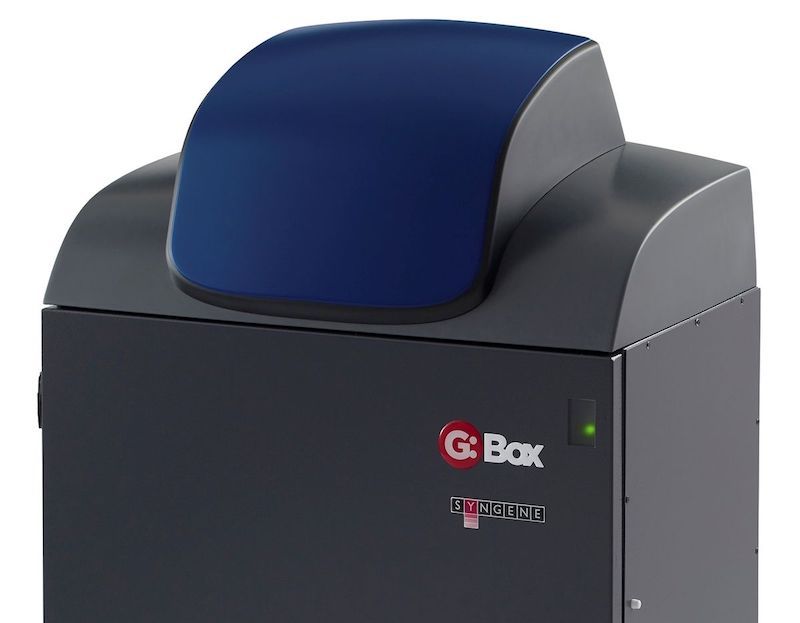 Syngene, a world-leading manufacturer of image analysis solutions, has introduced a far-red HI-LED lighting option for its new design of G:BOX Chemi and G:BOX mini multi application gel and blot imaging systems. This quick-fit, environmentally friendly lighting enables fast workflow and precise detection of IR fluorescently labelled proteins on gels and blots....
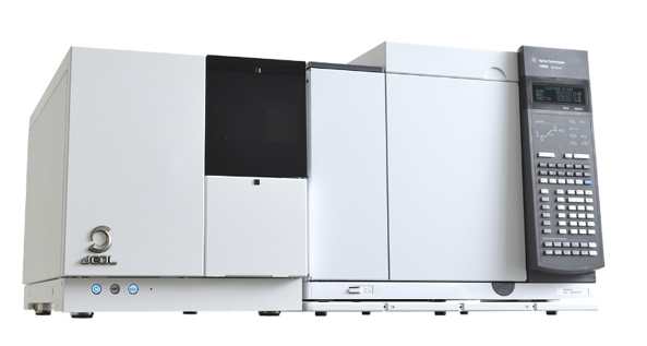 JEOL USA, a leader in developing instruments used to advance scientific research and technology, is pleased to showcase the latest updates to its core product range at Pittcon 2020. JEOL has 70 years of expertise in the field of electron microscopy, more than 60 years in mass spectrometry and NMR spectrometry, and more than 50 years of e-beam lithography leadership. JEOL expanded its mass spectrometer product line with the development of a GC-triple quadrupole mass spectrometer system....
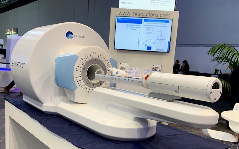 MR Solutions will be displaying its latest dry magnet 3T to 9.4T preclinical, multi-modality MRI (with PET or SPECT) and new benchtop PET/CT systems to the imaging science community at the 15th
  Optical Surfaces Ltd. reports how a motorised UV-Vis-NIR collimator and interferometric alignment system it supplied to the Deutsches Zentrum für Luft- und Raumfahrt (DLR) in Berlin, Germany has been used to set-up the ground breaking DLR Earth Sensing Imaging Spectrometer (DESIS). On 23 October 2019, DLR the renowned German Aerospace Center, and the U.S. company Teledyne Brown Engineering (TBE) announced the start of routine operations for DESIS on the International Space Station...
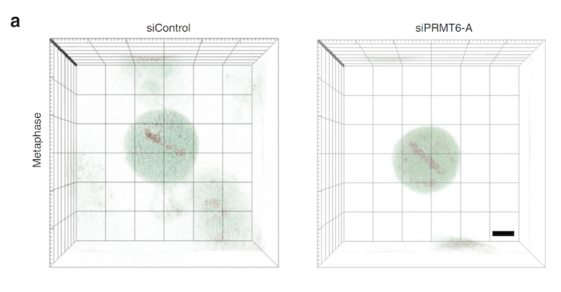 Governing molecular mechanism for chromosome condensation identified 140 years after mitosis first described. The molecular mechanism underlying chromosome condensation, one of the key steps in cell division, has been discovered from three-dimensional (3-D) refractive index (RI) reconstructions of individual mitotic cells. Writing in Nature Communications1, the authors describe using the Tomocube HT-1S holotomographic microscope to quantify the structural and biochemical parameters of the cytoplasm and chromosomes within the individual mitotic cells...