
Channels
Special Offers & Promotions
New imaging technique unlocks secrets of the zebrafish heart
A new type of microscopy is helping to unlock the secrets of the zebrafish’s heart – which could also teach us more about how human hearts form, grow and heal.
In a new paper published recently in the journal Nature Communications, researchers from the Universities of Glasgow and Edinburgh describe how they have developed a new method to capture 3D video images of the growing hearts of zebrafish embryos for the first time.
Zebrafish hearts are surprisingly similar to those of humans, making them useful in cardiac research. The British Heart Foundation – one of the funders of the study – hope that the new method of visualising zebrafish hearts will provide scientists with valuable new insights into the cellular and subcellular processes which occur during the earliest stages of heart development.
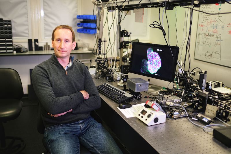
The development of the imaging breakthrough was led by Dr Jonathan Taylor of the University of Glasgow’s School of Physics and Astronomy, working closely with colleagues at the University of Edinburgh’s BHF Centre for Cardiovascular Science.
Their novel fluorescence time-lapse imaging technique uses a very thin sheet of laser light to build up images into a full 3D image of the heart layer by layer.
While 3D images of the beating heart had been recorded in the past, there was still more work to be done to turn a 3D freeze-frame of a single point in time into what cardiac researchers really wanted to study: a time-lapse video of the heart growing over a full day. The new technique also allows researchers to see for the first time individual heart cells growing and dividing in the beating heart in astonishing detail over 24 hours
Dr Taylor said: “Zebrafish embryos’ hearts beat three times a second, and this constant motion makes them incredibly challenging targets to image accurately in three dimensions. By the time you’ve taken an image of one layer of the heart, it has moved on to another part of its cardiac cycle. To add to the challenge, heart cells can be damaged by over-exposure to laser light that is needed for fluorescence imaging”.
To overcome those challenges, the researchers developed a computer program which uses real time image analysis to control exactly when to fire the lasers for imaging the heart.
Dr Taylor explained: “Our microscope uses visible light to ‘watch’ the beating heart of the zebrafish through its transparent skin, allowing our computer programs to know exactly when to selectively fire laser light at the heart to capture specific moments in its cycle.
“This lets us watch continuously as the heart forms over the course of a whole day, without causing any harm to the fish. It’s pulled back the veil on processes such as cell division within the heart which we just didn’t have any way to visualise before.”
Dr Martin Denvir of the University of Edinburgh worked with Dr Taylor to develop the new technique and is one of the paper’s co-authors. Dr Denvir said: “This exciting breakthrough, which brings together state-of-the-art research in physics and engineering at Glasgow and Durham with our heart research here in Edinburgh, has allowed us to see live processes in heart formation which we’ve simply had no way to observe before.
“There are two very promising potential applications of this new technique. Firstly, now that we can see detailed images of the growth of the heart on a cellular level, we hope to be able to apply that new knowledge to develop treatments for abnormal heart formation in the future.
“Secondly, we’ve been able to observe immune cells travelling to injured areas of the heart, which could help us guide the modification of immune response to more effectively treat cardiac inflammation and heart disease.”
Professor John Mullins, who directed the Edinburgh BHF Centre of Research Excellence from 2008-18 and is another of the paper’s co-authors, said: “The synergy between optical physics and biomedical research has delivered an entirely novel approach to imaging complex biological processes.
“The broader applications will enable new insights into a wide range of biological mechanisms, especially where movement and changes of shape have previously made long term imaging extremely challenging. The vision of the British Heart Foundation to enable such innovative collaborations will continue to transform our understanding of the cardiovascular system. “
The paper, titled ‘Adaptive prospective optical gating enables day-long 3D time-lapse imaging of the beating embryonic zebrafish heart’, is published in Nature Communications. The research was supported with funding from the British Heart Foundation (New Horizons award), the Engineering and Physical Sciences Research Council, the Royal Society of Edinburgh .
Funding for the single plane illumination microscope (SPIM) and advanced transgenic zebrafish used in these studies was provided by The BHF Centre of Research Excellence Award, University of Edinburgh.
News Channels
- Latest News
- New Laboratory Products
- Industry News
- Laboratory Automation | IT Solutions
- Microscopy | Image Analysis
- Separation Science
- Research | Case Studies
- Video Presentations
- Events | Conferences
Subscribe to any of our newsletters for the latest on new laboratory products, industry news, case studies and much more!
Popular this Month
Top 10 most popular articles this month
Today's Picks
Looking for a Supplier?
Search by company or by product
Please note Lab Bulletin does not sell, supply any of the products featured on this website. If you have an enquiry, please use the contact form below the article or company profile and we will send your request to the supplier so that they can contact you directly.
Lab Bulletin is published by newleaf marketing communications ltd.
Media Partners