Channels
Special Offers & Promotions
Tomocube launches World

Ground-breaking technology unlocks label-free 3D and 4D live cell imaging on standard imaging plates for higher-throughput and automated screening applications
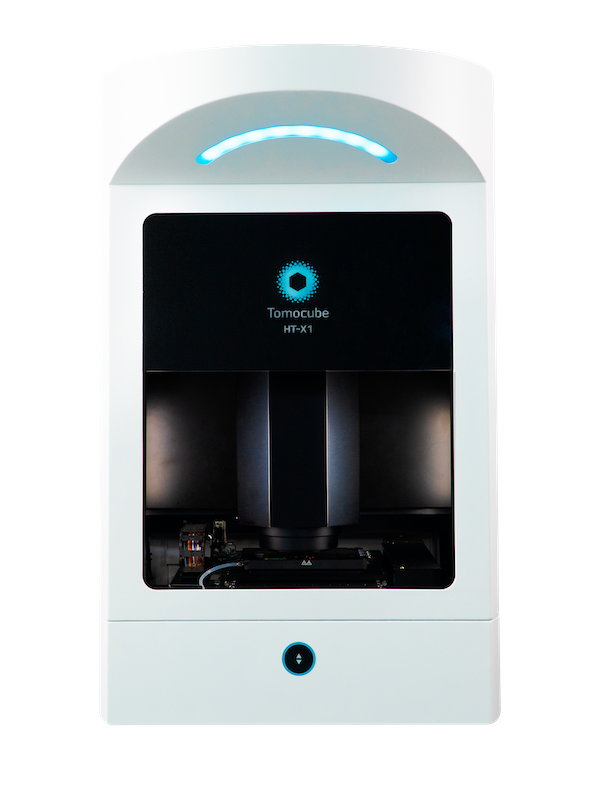
A novel optical microscope utilizing incoherent light to generate holographic images of unlabelled live cells is now available from Tomocube.
Called HT-X1, the new microscope is ideally suited to higher-throughput and automated screening applications with its ability to image multi-well plate formats, large field-of-view, laser autofocus system, and very high performance 0.95NA objective. Core imaging facilities will also value its integrated gassed incubator for long-term, time-lapse studies and its software-driven approach, allowing multiple users to access its formidable performance simply and quickly through its intuitive GUI, TomoStudio X.
“Much of life science research depends on the study of living cells, the interactions between them, and the ways that they respond to external stimuli,” says Aubrey Lambert, Tomocube’s Chief Marketing Officer. “The majority of microscopy techniques limit the quantity and quality of information available to researchers, constrain the study of thicker specimens, and even harm the cells during long-term studies.
“The new HT-X1 overcomes these limitations, allowing work with confluent cell sheets, significantly reducing laser-induced speckle noise for enhanced contrast, delivering enhanced levels of fluorescence illumination, and enabling the study of thicker specimens.”
The new microscope builds on the optical diffraction technology (ODT) of Tomocube’s award-winning HT-1 and HT-2 microscopes. Like them it presents morphological, mechanical, and chemical properties of individual living cells through the 3D refractive index (RI) tomograms quickly and simply without any sample preparation, and molecular specificity information through fluorescence imaging. However, the new HT-X1 uses a conventional single-beam white light LED instead of a laser light source. Instead of rotating the laser beam to illuminate the sample at various incidence angles it illuminates the sample with various beam patterns specifically designed to retrieve the refractive index and captures a sequence of holograms from different positions.
This unique single-beam technique simplifies the imaging process by eliminating the need for background calibration and allowing imaging in confluent samples without an increase in light intensity. As well as being easily combined with complementary imaging modalities, it is mechanically more stable and less sensitive to speckle noise for high contrast imaging.
The HT-X1 incorporates a 450 nm LED light source for transmitted light together with a customisable five-channel fluorescence filter engine, with any 3 of the channels being captured for overlaying. The large, motorized stage will easily accommodate 24-, 12- and 6-well plates and standard 35mm imaging dishes in the integrated incubator, which is equipped with a sealing lid to allow gassing and combines with a motorized laser-powered autofocus module for accurate and repeatable monitoring of locations during long-term studies. A 40x 0.95 NA air objective lens allows rapid imaging of multiple locations with a lateral resolution of 156 nm. The extremely high numerical aperture also allows more illumination for fluorescence imaging while the large 160 x 160 micron field-of-view allows rapid and easy imaging of multiple locations.
Benefits
- Unique single-beam technique for mechanically-stable, high-performance, enhanced-contrast imaging free of laser-induced speckle.
- Increased throughput with multi-well plate imaging
- Large 160x160 micron field of view reduces the number of tiles required for large area capture
- Laser auto-focus for faultless operation and well-to-well reproducibility
- Integrated 5-channel fluorescence engine
- Automated correlative microscopy - high-quality 3D quantitative images with 3D fluorescence for each sample
- Z-stack imaging enables imaging of thicker samples.
- No labelling or sample preparation – rapid and convenient
Specifications
- Objective lens: 40x 0.95 NA air
- HT light source: 450 nm LED
- Lateral resolution: 156 nm
- Field of View: 160 µm x 160 µm
- Fluorescence: Up to 5 channels – 4 channels built-in with blank position for 5th filter of choice
- Stage: motorized with Laser auto-focus module
- Size: 9000x500x600 mm (h, w, d)
- Weight: 90 kg
Tomocube is dedicated to delivering products that can enhance biological and medical research via novel optical solutions that can assist in understanding, diagnosing, and treating human diseases. Our microscope platform enables researchers to measure nanoscale, real-time, dynamic images of individual living cells without the need for sample preparation through the measurement of 3D refractive index tomograms. This enables researchers and clinicians to work with primary cells and non-invasively observe label-free 3D dynamics of live cells and tissues, make quantitative measurements, and retrieve unique cell properties such as cell volume, cytoplasmic density, and surface area. Founded in 2016, Tomocube provides a series of HT microscopy to the market and won the 2019 Microscopy Today 2019 Innovation Award for the HT-2, the world’s first correlative microscope to combine the quantitative phase imaging (QPI) approach of label-free, 3-D refractive index (RI) tomography with 3-D fluorescence imaging
Media Partners


