Channels
Special Offers & Promotions
Tomocube Label-free Holotomography Microscope opens up Advanced Platelet Research

Image
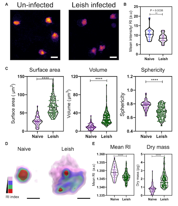
Morphological and biochemical differences in platelets from naïve and Leishmania infected mice. (A,B) Max RI projections of the 3D phase images of platelets from Naïve and L. donovani infected mice n = 23 platelets. (C) Morphological changes seen in platelets in surface area, volume, and sphericity. n = 62 platelets, 10 platelets from each WT mice in study (6 mice) and n = 172 platelets from 17 L. donovani infected mice, median represented by black line in the violin plot. Scale bar 2 mm. (D) 3D rendered iso-surface image of platelets from naïve and L. donovani infected mice based on RI. Scale bar 1 mm. (E) Changes in mean RI (unpaired two-tailed t-test, P = < 0.0001), and dry mass (unpaired two-tailed t-test, P = < 0.0001) of the platelets. RI index scales; Pink: 1.3406–1.3525, blue: 1.3525–1.3621, green: 1.3622–1.3735, red: 1.3736–1.3822.
Tomocube’s cutting-edge 3D quantitative phase imaging is set to play a crucial role in platelet research according to a new paper from scientists at the University of York.
Using holotomography images generated by the Tomocube HT-2H microscope, the team were able to identify and quantify in single unlabelled, live platelets clear disparities in activation status and potential functional ability in disease states without experimental interference, such as from fixation or labelling.
The ability to use minute volumes of blood is often a vital consideration in platelet research, where mouse platelets are commonly substituted for human platelets. Although they provide an excellent experimental model for functional comparisons, traditional methods of studying platelet phenotype, function, and activation status rely on using large numbers of whole isolated platelet populations. The large volumes required severely limits the number and type of assays that can be performed with mouse blood.
According to Aubrey Lambert, Tomocube’s Chief Marketing Officer, “Platelet biology has benefited from advances in super-resolution imaging and the quantitative information provided about internal structure and cell function. However, fluorescence-based imaging almost always requires samples to be genetically modified or immuno-labelled to provide the contrast required for visualization. In this study, Tomocube’s 3D holotomography imaging technique eliminates the need for labelling or post isolation processing and enables alterations in cell morphology and changes in biophysical parameters of unaltered cells to be studied and compared in healthy and diseased platelets.
“Also, standard in vitro functional assays, such as lumi-aggregometry or western blotting, demand many platelets and, as a result, researchers are often limited to a single assay for each mouse. The assays outlined at York require just 10ml to sample 100–200 platelets at single cell level to generate quantitative information relating to platelet morphology and RI/dry mass. Therefore, holotomography can serve as a valuable additional or complementary technique to other in vitro platelet function assays.”
Platelets play a key role in maintaining haemostasis and preventing excessive blood loss following injury, rapidly activating to form a platelet plug to reduce blood loss and initiating secondary haemostasis to promote the formation of a stable fibrin-rich thrombus. More recently, they have also been implicated in innate immunity and inflammation by directly interacting with immune cells and releasing proinflammatory signals. It is likely therefore that in certain pathologies, such as chronic parasitic infections and myeloid malignancies, platelets can act as mediators for haemostatic and proinflammatory responses.
Reference
- Quantitative Optical Diffraction Tomography Imaging of Mouse Platelets. Tess A. Stanly, Rakesh Suman, Gulab Fatima Rani, Peter O’Toole, Paul M. Kaye and Ian S. Hitchcock. doi:10.3389/fphys.2020.568087
About Tomocube, Inc.
Tomocube is dedicated to delivering products that can enhance biological and medical research via novel optical solutions that can assist in understanding, diagnosing, and treating human diseases. Our microscope platform enables researchers to measure nanoscale, real-time, dynamic images of individual living cells without the need for sample preparation through the measurement of 3D refractive index tomograms. This enables researchers and clinicians to work with primary cells and non-invasively observe label-free 3D dynamics of live cells and tissues, make quantitative measurements, and retrieve unique cell properties such as cell volume, cytoplasmic density, and surface area. Founded in 2016, Tomocube provides a series of HT microscopy to the market and won the 2019 Microscopy Today 2019 Innovation Award for the HT-2, the world’s first correlative microscope to combine the quantitative phase imaging (QPI) approach of label-free, 3-D refractive index (RI) tomography with 3-D fluorescence imaging.
Media Partners


