
Channels
Special Offers & Promotions
Tomocube Holotomography Microscopy drives forward rapid diagnosis of blood cancers
Uniquely shaped red blood cells observed for the first time in patient blood could be breakthrough in diagnosis of Myelodysplastic Syndrome (MDS)
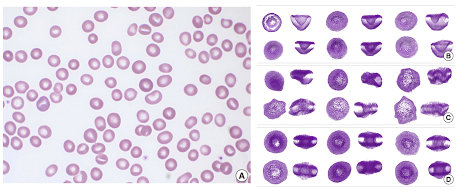
Although the three-dimensional (3-D) shape of erythrocytes is strongly associated with various malignant blood disorders, conventional optical imaging approaches only provide information on two-dimensional morphology. Now, a Korean research team has used the Tomocube holotomography microscope to observe uniquely shaped red blood cells (RBCs) for the first time in the peripheral blood of a patient diagnosed with myelodysplastic syndrome (MDS) (1).
According to Aubrey lambert, Tomocube’s chief marketing officer, “this could be a breakthrough for the rapid diagnosis of MDS, a group of cancers that disrupt the production and maturation of blood cells within the bone marrow. Not only may it eliminate the need to carry out a confirmatory bone marrow examination under local anaesthetic but the ability to quickly screen blood samples may help with early diagnosis of MDS, a disease that progresses to acute myeloid leukaemia (AML) in approximately one third of patients.”
The research team was led by Dr Seongsoo Jang of the University of Ulsan College of Medicine and Asan Medical Center in Seoul, Korea. Using blood samples left over from the complete blood cell count of a patient originally diagnosed with MDS who had subsequently undergone an allogeneic hematopoietic stem cell transplantation (HSCT), the 3-D RI tomograms of reconstructed RBCs revealed unique cup-shaped RBCs in approx. one-third of the RBCs. No cup-shaped RBCs were found in a control sample and the normal 2-D light microscopy examination was unable to differentiate between normal and cup-shaped RBCs.
The myelodysplastic syndromes (MDS) are a group of fast-progressing diseases affecting the elderly in which most of the immature blood cells in the bone marrow are of poor quality and are destroyed before they leave the bone marrow. Red cells, white cells and platelets are affected and, although the bone marrow in MDS sufferers is more active than normal, fewer blood cells are found in the circulation, which is an indication for MDS. Typically, there are no early symptoms but tiredness, shortness of breath, frequent infections and easy bleeding are found later, and a significant proportion of sufferers go on to develop acute myeloid leukaemia. Risk factors include previous chemotherapy or radiation therapy, exposure to heavy metals, and exposure to certain chemicals, such as tobacco smoke and pesticides.
The Tomocube holotomography microscope delivers quantitative, nanoscale, real-time, 3-D images of individual living cells quickly and simply without any sample preparation. The holotomography images also deliver vital information on unique cell properties, including cell volume, shapes of sub-cellular organelles, cytoplasmic density, surface area, and deformability. Although not used in the MDS study, the latest HT-2 model combines the quantitative phase imaging (QPI) approach of label-free, 3-D refractive index (RI) tomography with 3-D fluorescence imaging. Winner of the Microscopy Today 2019 Innovation Award, this microscope enables long-term tracking of specific targets in live cells while minimising stress. The capability to easily deliver holotomography and fluorescence correlative analysis in 2D, 3D and 4D will enable researchers and clinicians to open new frontiers in bioscience and better understand, diagnose, and treat disease.
Reference
(1) Se-eun Koo, Seongsoo Jang, Chan Jeoung Park, Young-Uk Cho, and YongKeun Park. Unique Red Blood Cell Morphology Detected in a Patient with Myelodysplastic Syndrome (MDS) by Three-dimensional Refractive Index Tomography. Lab Med Online. 2019 Jul;9(3):185-188 https://doi.org/10.3343/lmo.2019.9.3.185
About Tomocube, Inc.
Tomocube is dedicated to delivering products that can enhance biological and medical research via novel optical solutions that can assist in understanding, diagnosing, and treating human diseases. Our microscope platform enables researchers to measure nanoscale, real-time, dynamic images of individual living cells without the need for sample preparation through the measurement of 3D refractive index tomograms. This enables researchers and clinicians to work with primary cells and non-invasively observe label-free 3D dynamics of live cells and tissues, make quantitative measurements and retrieve unique cell properties such as cell volume, cytoplasmic density, and surface area. Founded in 2016, Tomocube provides a series of HT microscopy to the market and won the 2019 Microscopy Today 2019 Innovation Award for the HT-2, the world’s first correlative microscope to combine the quantitative phase imaging (QPI) approach of label-free, 3-D refractive index (RI) tomography with 3-D fluorescence imaging.
News Channels
- Latest News
- New Laboratory Products
- Industry News
- Laboratory Automation | IT Solutions
- Microscopy | Image Analysis
- Separation Science
- Research | Case Studies
- Video Presentations
- Events | Conferences
Subscribe to any of our newsletters for the latest on new laboratory products, industry news, case studies and much more!

Popular this Month
Top 10 most popular articles this month
Today's Picks
Looking for a Supplier?
Search by company or by product
Please note Lab Bulletin does not sell, supply any of the products featured on this website. If you have an enquiry, please use the contact form below the article or company profile and we will send your request to the supplier so that they can contact you directly.
Lab Bulletin is published by newleaf marketing communications ltd.
Media Partners