Channels
Special Offers & Promotions
Tomocube Holotomography Microscopy enables Label-free, Non-invasive Study of individual Spermatozoon
Rapid 3D imaging and in-depth structural analysis opens up quality monitoring and spermatozoa selection for artificial insemination in humans and animals
A key determinant for success in artificial insemination for humans and animals is the quality of the spermatozoa. However, current 2D label-free imaging methods can only detect abnormal spermatozoa with significant morphological changes or incomplete structures and are unable to provide information on their internal structure or composition.
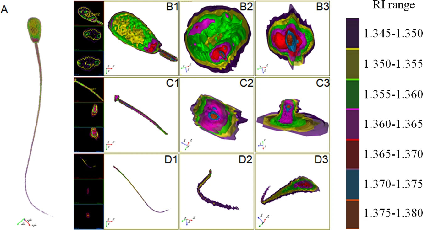
Now, a Korean team of scientists led by Dr Nam-Hyung Kim has used the Tomocube holotomography microscope to develop a 3D label-free imaging technique. For the first time, individual spermatozoon can be non-invasively imaged in high resolution and quantitative analysis of its morphological and biophysical properties performed (1).
Without altering the physiological state of the spermatozoa, the team’s quantitative phase imaging (QPI) approach of refractive index (RI) tomography reveals the structure, volume, surface area, concentration, and dry matter mass of the individual spermatozoon. As part of the study, Holstein cows and Korean native cattle spermatozoa were systematically analysed, revealing significant differences in anatomical structure between the two breeds, including spermatozoa head length, head width, midpiece length, and tail length.
Furthermore, the QPI techniques not only acquired the full 3D structure of the spermatozoa but also revealed their 3D translational head motion and the angular velocity of their head spin as well as the 3D flagellar motion which influences the spermatozoa’s swimming trajectory. In addition, the biophysical parameters retrieved from the RI tomograms, such as dry mass and cell volume, allowed determination of the concentration of the spermatozoa because the RI value of the cell cytoplasm is linearly proportional to its concentration.
According to Aubrey Lambert, Tomocube’s chief marketing officer, “despite more than 200 years’ experience with artificial insemination in humans and animals, this label-free imaging approach provides a new technique for understanding the physiology of spermatozoa.
“The results show the benefits of this new technique for further in-depth structural analysis, quality monitoring, and spermatozoa selection in a rapid and label-free manner. Whether the internal RI distribution of spermatozoa can be used as a diagnostic parameter to evaluate the state of a spermatozoa and fertilization ability remains an open question; however, it is now accessible to direct experimental study.”
The HT-2 from Tomocube is the world’s first microscope to combine both holotomography and 3D fluorescence imaging into one unit. Capable of simultaneously capturing high resolution 3D optical diffraction tomography and 3D fluorescence images, the new microscope enables long-term tracking of specific targets in live cells while minimising stress. The capability to easily deliver holotomography and fluorescence correlative analysis in 2D, 3D and 4D will enable researchers and clinicians to open new frontiers in bioscience and better understand, diagnose, and treat disease.
Reference
Hao Jiang, Jeong-woo Kwon, Sumin Lee, Yu-Jin Jo, Suk Namgoong, Xue-rui Yao, Bao Yuan, Jia-bao Zhang, Yong-Keun Park & Nam-Hyung Kim. Reconstruction of bovine spermatozoa substances distribution and morphological differences between Holstein and Korean native cattle using three-dimensional refractive index tomography. Nature Scientific Reports volume 9, Article number: 8774 (2019)
About Tomocube, Inc.
Tomocube is dedicated to delivering products that can enhance biological and medical research via novel optical solutions that can assist in understanding, diagnosing, and treating human diseases. Our microscope platform enables researchers to measure nanoscale, real-time, dynamic images of individual living cells without the need for sample preparation through the measurement of 3D refractive index tomograms. This enables researchers and clinicians to work with primary cells and non-invasively observe label-free 3D dynamics of live cells and tissues, make quantitative measurements and retrieve unique cell properties such as cell volume, cytoplasmic density, and surface area.
Media Partners


