CytoSMART Technologies has announced the launch of a new automated live-cell imaging system designed for long-term experiments, comparison studies, and large laboratory teams. The CytoSMART Multi Lux is a cost-effective solution for researchers who want to carry out side-by-side comparisons between cell cultures, run long-term experiments, and monitor cells from their home’s comfort....
HORIBA, a global leader in measurement and analysis solutions for advanced research and industry, and the world leader in Raman microscopy and nanoscopy, is thrilled to announce a strategic collaboration with Qnami, leader and pioneer in scanning NV magnetometry based on diamond quantum sensing. After a series of joint research projects, both companies have agreed to accelerate their developments to address the growing needs of cutting-edge technology for the quantitative and non-perturbative analysis...
NanoImaging Services, Inc. (NIS), specialists in transmission electron microscopy (TEM), has announced they have partnered with Thermo Fisher Scientific to increase accessibility to cryoelectron microscopy (cryoEM) technology for the global pharmaceutical and biotechnology community. Having pioneered cryoEM for use in drug discovery and vaccine development applications, NIS brings its proven expertise and experience into this partnership, supported by its growing network of state-of-the-art facilities....
The FRITSCH ANALYSETTE 28 ImageSizer for dry and wet measurement is the ideal Particle Sizer for all applications that require accurate and reproducible measuring results for both particle shape and size. The optical process of Dynamic Image Analysis delivers accurate measuring results, which are available immediately in less than 5 minutes. Your advantage: Great flexibility for different measurement tasks for particle sizes of 20 µm – 20 mm in quality control, research and laboratory....
More than 305,000 high-resolution, multi-channel COVID-19 cellular images captured in less than four weeks with Molecular Devices ImageXpress® Micro Confocal High-Content Imaging System are now available to the scientific community. With the COVID-19 pandemic impacting millions around the globe, researchers are racing to develop a vaccine or drug treatment. In an effort to better understand the cellular responses to COVID-19, the digital biology company Recursion has publicly released the world’s largest imaging dataset....
ZEISS introduces the Advanced Reconstruction Toolbox for its industry-leading ZEISS Xradia 3D X-ray microscope and computed tomography systems. With the Toolbox, two modules are announced: an upgraded ZEISS OptiRecon for iterative reconstruction, and ZEISS DeepRecon, microscopy’s first commercially available deep learning reconstruction technology....
CytoSMART Technologies has announced the launch of an automated organoid counter. The software application detects organoids using bright-field image analysis and runs on the Corning® Cell Counter, a CytoSMART product. Said Joffry Maltha, CEO at CytoSMART Technologies "The 3D cellular structures that are formed in organoids have great potential in drug discovery and developmental biology. This has fueled the ever-growing interest in culturing new sub-types of organoids and optimization of current protocols...
Olympus, a leading manufacturer of high-end research microscopes, and Cytosurge, a precision manufacturer of cell manipulation technologies, have entered a co-marketing agreement to become a complete system provider to the scientific community’s growing need for next-generation single-cell and CRISPR genetic manipulation solutions....
NanoImaging Services, Inc., specialists in transmission electron microscopy (TEM) with a vision to make cryoEM workflows accessible to all, today announced the opening of a new facility close to their San Diego headquarters. The new data collection center is the second facility opening in as many months and serves as further evidence of the company’s investment in growth and ongoing service expansion....
Researchers have developed a family of engineered nanobodies which neutralise the SARS-CoV-2 virus, targeting the viral spike protein in a novel way. The research team from The Rosalind Franklin Institute, working with colleagues at Diamond Light Source, Oxford University and Public Health England, have hailed the breakthrough as a potential therapy....
The ZEISS Innovation Hub on the campus of the Karlsruhe Institute of Technology (KIT) has seen a number of successful collaborations and projects since it opened in early 2020. ZEISS wants the hub to house high-tech and digital start-ups, as well as its own innovation and new business activities. KIT will thus join forces with ZEISS experts to pave the way for the technologies of the future....
The launch of a new JEOL Field Emission Scanning Electron Microscope during the summer of 2020 includes virtual demonstrations of its powerful performance directly to those in the market for an analytical ultrahigh resolution SEM. JEOL’s new JSM-IT800 is the company’s top-of-the-line microscope with ultrahigh spatial resolution imaging and analysis at the nanoscale....
BioTek Instruments announces the availability of variable bandwidth monochromators on their modular and versatile Synergy™ H1 Hybrid Multi-Mode Reader. This allows researchers to achieve even greater levels of assay sensitivity and specificity compared to fixed bandwidth systems. The microplate reader includes BioTek’s patented Hybrid Technology™...
A radical new way of thinking about soil has finally solved the mystery of why adding organic material like manure improves flood and drought resilience, climate control and crop yields - universal ‘ecosystem services’ that are widely recognised as worth billions to the global economy. Founded on more than 50 years’ worth of data from a unique field experiment, researchers have demonstrated...
The new SCHOTT® VisiLED UV Ring Light for stereo microscopes combines classic bright-field illumination with UV illumination. It is the only segment ring light on the market in which white-light and UV-LEDs are alternately installed in eight segments. This LED arrangement allows objects to be examined from the same illumination angle, which significantly improves the ability to compare and reproduce resulting images....
NanoImaging Services, Inc. (NIS), specialists in transmission electron microscopy (TEM) with a vision to make cryoEM workflows accessible to all, has announced the opening of a new facility in Boston. NIS is expanding with new services and locations to support its growing customer base requiring cryoEM for drug discovery and vaccine development applications. COVID-19 projects are receiving prioritization....
3D label-free imaging identifies real-time cholesterol sorting mechanism in Plasmodium falciparum-infected human erythrocytes. The first study to elucidate the sequential dynamics of membrane cholesterol transport in erythrocytes infected with live Plasmodium falciparum parasites has been successfully concluded using the 3D label-free imaging capability of holotomography microscopy....
Rutherford has entered into a £55m development framework agreement with Equitix Limited. Under of the terms of the agreement, Rutherford and Equitix will establish up to five new diagnostic facilities in the UK, to provide diagnostic services to the NHS and to private patients. Each centre will provide a variety of diagnostics services including Positron Emission Tomography–Computed Tomography, Magnetic Resonance Imaging, Computed Tomography, Ultrasound, Endoscopy, X-Ray and other relevant diagnostic services...
CytoSMART Technologies has announced the launch of a new live-cell imaging system. The CytoSMART Lux2 Duo Kit offers a straightforward, cost-effective solution for researchers carrying out immediate side-by-side comparisons between cell cultures. Said Jan-Willem van Bree, CTO at CytoSMART Technologies "Employing this two camera mini live cell imaging system is especially useful for stem cell research....
Having launched a unique AI-aided drug discovery platform last month, this company grows quickly with concerted support from many talented researchers and scientists. It offers drug R & D solutions from the perspective of AI for medical institutions and pharmaceutical enterprises worldwide...
Oxford Instruments and Digital Surf, creator of the industry-standard Mountains® surface and image analysis software platform, has announced the release of Relate software for users of Oxford Instruments' leading-edge tools for materials characterization. This software will bring great value to Oxford Instruments' users working in R&D across a wide range of academic and industrial applications including semiconductors, renewable energy, mining, metallurgy, and forensics....
In this new 10-page Technology Note, learn how different image processing methods have the potential to make widefield microscope systems more powerful and versatile. Understand each methods limitations and pitfalls, as well as suitability for your specific applications. For decades, fluorescence microscopy has been an invaluable tool in life sciences research, with new variations and implementations emerging almost every year....

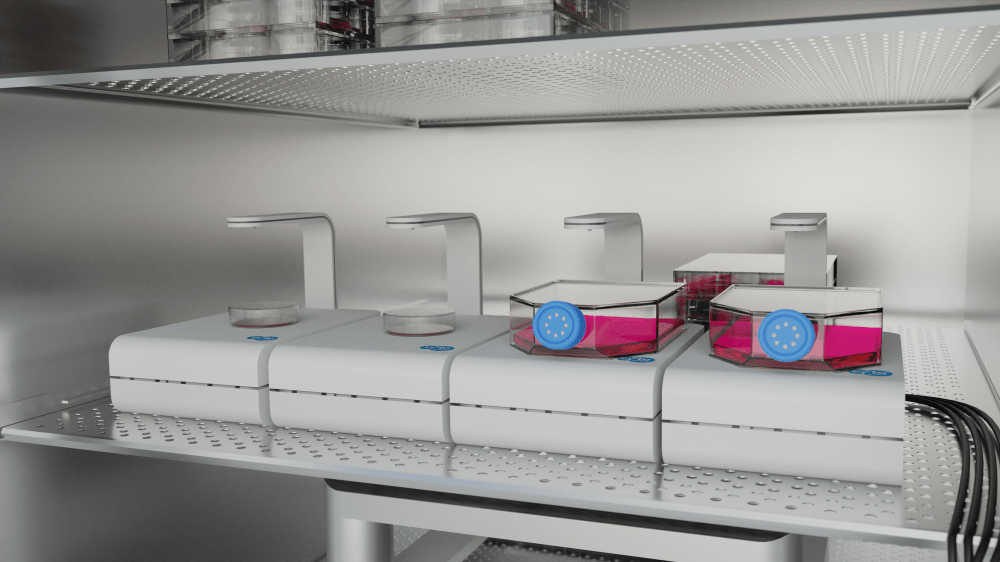 CytoSMART Technologies has announced the launch of a new automated live-cell imaging system designed for long-term experiments, comparison studies, and large laboratory teams. The CytoSMART Multi Lux is a cost-effective solution for researchers who want to carry out side-by-side comparisons between cell cultures, run long-term experiments, and monitor cells from their home’s comfort....
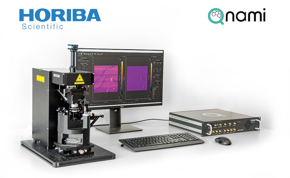 HORIBA, a global leader in measurement and analysis solutions for advanced research and industry, and the world leader in Raman microscopy and nanoscopy, is thrilled to announce a strategic collaboration with Qnami, leader and pioneer in scanning NV magnetometry based on diamond quantum sensing. After a series of joint research projects, both companies have agreed to accelerate their developments to address the growing needs of cutting-edge technology for the quantitative and non-perturbative analysis...
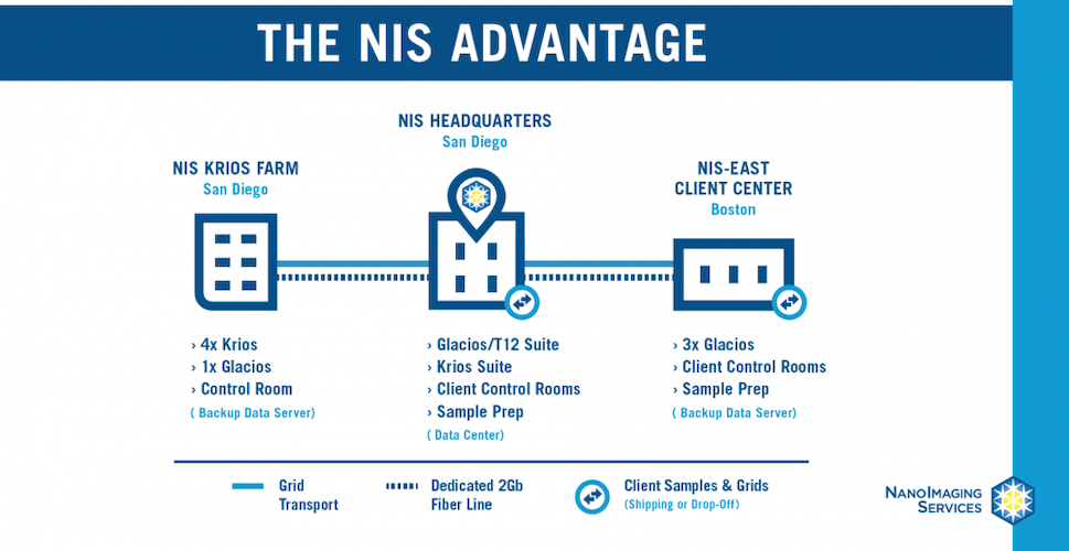 NanoImaging Services, Inc. (NIS), specialists in transmission electron microscopy (TEM), has announced they have partnered with Thermo Fisher Scientific to increase accessibility to cryoelectron microscopy (cryoEM) technology for the global pharmaceutical and biotechnology community. Having pioneered cryoEM for use in drug discovery and vaccine development applications, NIS brings its proven expertise and experience into this partnership, supported by its growing network of state-of-the-art facilities....
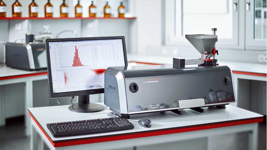 The FRITSCH ANALYSETTE 28 ImageSizer for dry and wet measurement is the ideal Particle Sizer for all applications that require accurate and reproducible measuring results for both particle shape and size. The optical process of Dynamic Image Analysis delivers accurate measuring results, which are available immediately in less than 5 minutes. Your advantage: Great flexibility for different measurement tasks for particle sizes of 20 µm – 20 mm in quality control, research and laboratory....
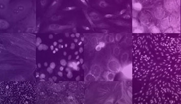
 ZEISS introduces the Advanced Reconstruction Toolbox for its industry-leading ZEISS Xradia 3D X-ray microscope and computed tomography systems. With the Toolbox, two modules are announced: an upgraded ZEISS OptiRecon for iterative reconstruction, and ZEISS DeepRecon, microscopy’s first commercially available deep learning reconstruction technology....
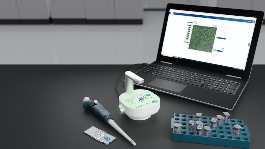 CytoSMART Technologies has announced the launch of an automated organoid counter. The software application detects organoids using bright-field image analysis and runs on the Corning® Cell Counter, a CytoSMART product. Said Joffry Maltha, CEO at CytoSMART Technologies "The 3D cellular structures that are formed in organoids have great potential in drug discovery and developmental biology. This has fueled the ever-growing interest in culturing new sub-types of organoids and optimization of current protocols...
 Olympus, a leading manufacturer of high-end research microscopes, and Cytosurge, a precision manufacturer of cell manipulation technologies, have entered a co-marketing agreement to become a complete system provider to the scientific community’s growing need for next-generation single-cell and CRISPR genetic manipulation solutions....
 NanoImaging Services, Inc., specialists in transmission electron microscopy (TEM) with a vision to make cryoEM workflows accessible to all, today announced the opening of a new facility close to their San Diego headquarters. The new data collection center is the second facility opening in as many months and serves as further evidence of the company’s investment in growth and ongoing service expansion....
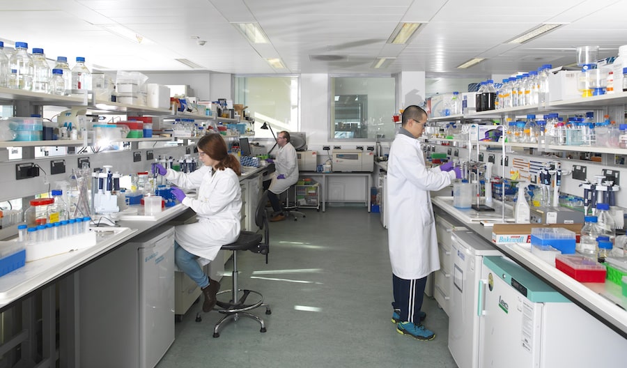 Researchers have developed a family of engineered nanobodies which neutralise the SARS-CoV-2 virus, targeting the viral spike protein in a novel way. The research team from The Rosalind Franklin Institute, working with colleagues at Diamond Light Source, Oxford University and Public Health England, have hailed the breakthrough as a potential therapy....
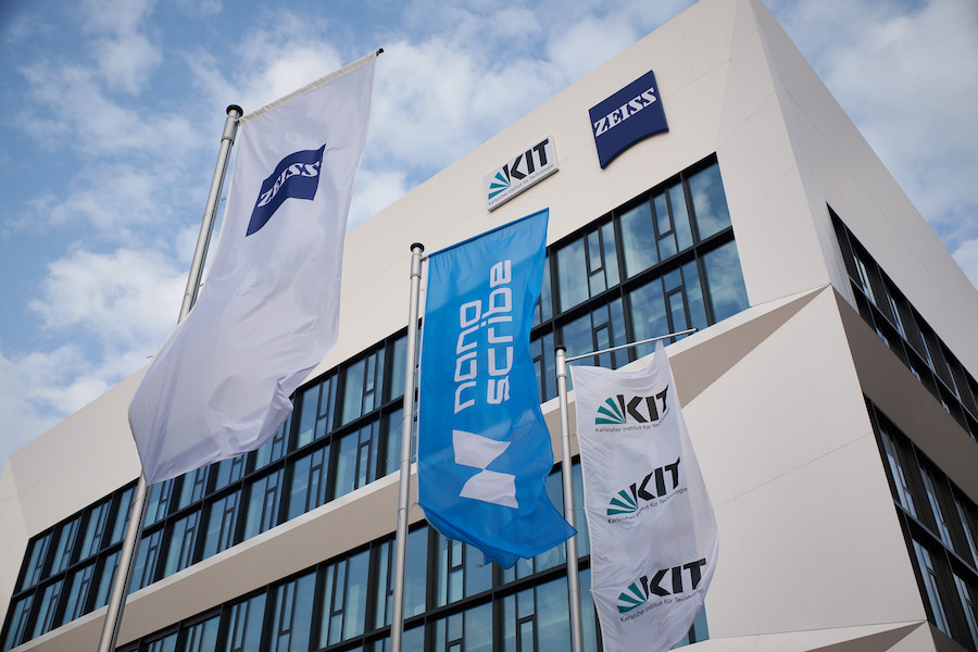 The ZEISS Innovation Hub on the campus of the Karlsruhe Institute of Technology (KIT) has seen a number of successful collaborations and projects since it opened in early 2020. ZEISS wants the hub to house high-tech and digital start-ups, as well as its own innovation and new business activities. KIT will thus join forces with ZEISS experts to pave the way for the technologies of the future....
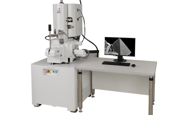 The launch of a new JEOL Field Emission Scanning Electron Microscope during the summer of 2020 includes virtual demonstrations of its powerful performance directly to those in the market for an analytical ultrahigh resolution SEM. JEOL’s new JSM-IT800 is the company’s top-of-the-line microscope with ultrahigh spatial resolution imaging and analysis at the nanoscale....
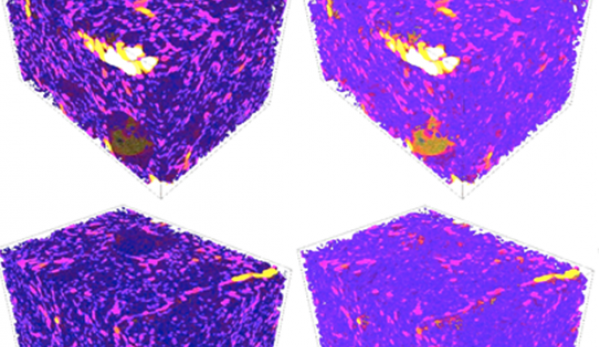 A radical new way of thinking about soil has finally solved the mystery of why adding organic material like manure improves flood and drought resilience, climate control and crop yields - universal ‘ecosystem services’ that are widely recognised as worth billions to the global economy. Founded on more than 50 years’ worth of data from a unique field experiment, researchers have demonstrated...
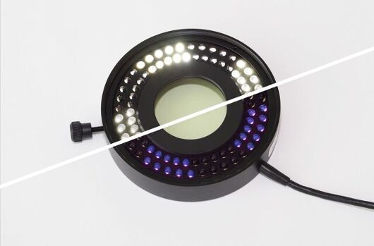 The new SCHOTT® VisiLED UV Ring Light for stereo microscopes combines classic bright-field illumination with UV illumination. It is the only segment ring light on the market in which white-light and UV-LEDs are alternately installed in eight segments. This LED arrangement allows objects to be examined from the same illumination angle, which significantly improves the ability to compare and reproduce resulting images....
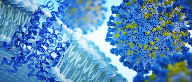 NanoImaging Services, Inc. (NIS), specialists in transmission electron microscopy (TEM) with a vision to make cryoEM workflows accessible to all, has announced the opening of a new facility in Boston. NIS is expanding with new services and locations to support its growing customer base requiring cryoEM for drug discovery and vaccine development applications. COVID-19 projects are receiving prioritization....
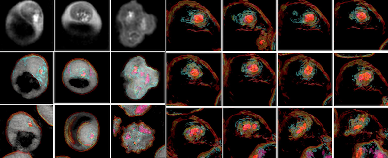 3D label-free imaging identifies real-time cholesterol sorting mechanism in Plasmodium falciparum-infected human erythrocytes. The first study to elucidate the sequential dynamics of membrane cholesterol transport in erythrocytes infected with live Plasmodium falciparum parasites has been successfully concluded using the 3D label-free imaging capability of holotomography microscopy....
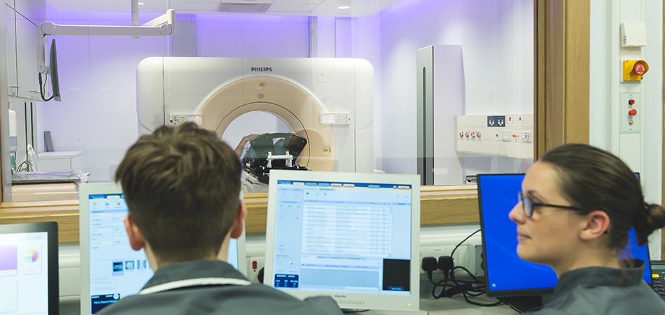 Rutherford has entered into a £55m development framework agreement with Equitix Limited. Under of the terms of the agreement, Rutherford and Equitix will establish up to five new diagnostic facilities in the UK, to provide diagnostic services to the NHS and to private patients. Each centre will provide a variety of diagnostics services including Positron Emission Tomography–Computed Tomography, Magnetic Resonance Imaging, Computed Tomography, Ultrasound, Endoscopy, X-Ray and other relevant diagnostic services...
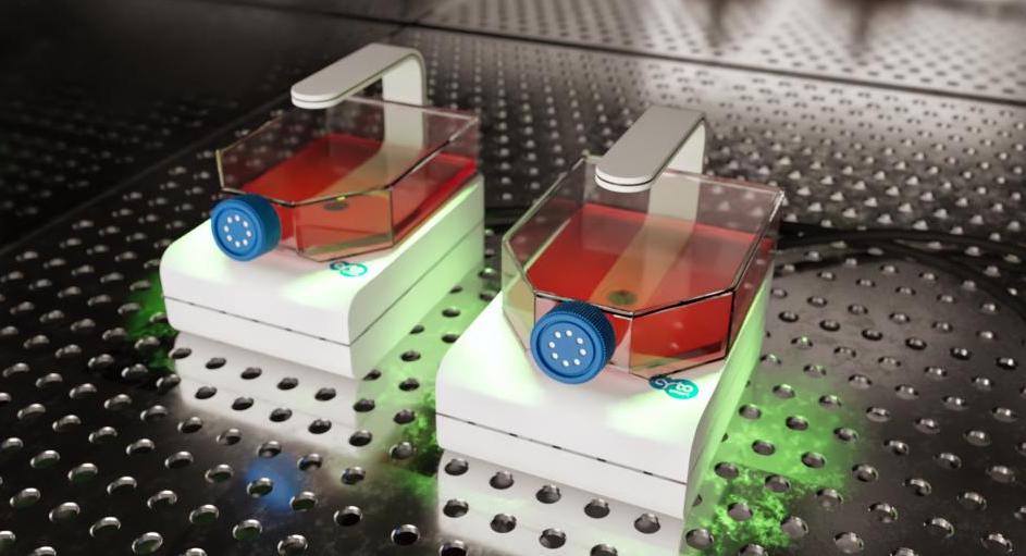 CytoSMART Technologies has announced the launch of a new live-cell imaging system. The CytoSMART Lux2 Duo Kit offers a straightforward, cost-effective solution for researchers carrying out immediate side-by-side comparisons between cell cultures. Said Jan-Willem van Bree, CTO at CytoSMART Technologies "Employing this two camera mini live cell imaging system is especially useful for stem cell research....
 Having launched a unique AI-aided drug discovery platform last month, this company grows quickly with concerted support from many talented researchers and scientists. It offers drug R & D solutions from the perspective of AI for medical institutions and pharmaceutical enterprises worldwide...
 Oxford Instruments and Digital Surf, creator of the industry-standard Mountains® surface and image analysis software platform, has announced the release of Relate software for users of Oxford Instruments' leading-edge tools for materials characterization. This software will bring great value to Oxford Instruments' users working in R&D across a wide range of academic and industrial applications including semiconductors, renewable energy, mining, metallurgy, and forensics....
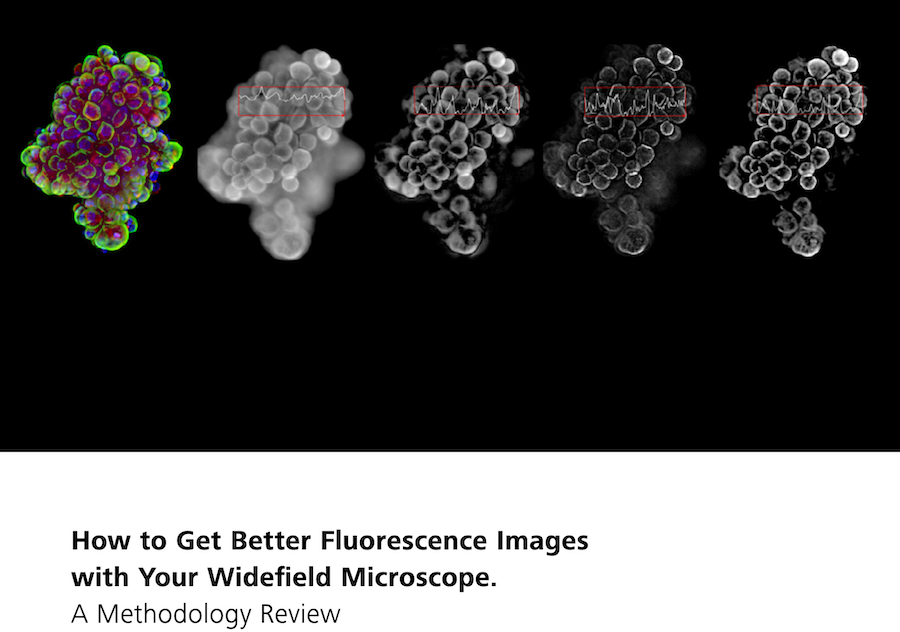 In this new 10-page Technology Note, learn how different image processing methods have the potential to make widefield microscope systems more powerful and versatile. Understand each methods limitations and pitfalls, as well as suitability for your specific applications. For decades, fluorescence microscopy has been an invaluable tool in life sciences research, with new variations and implementations emerging almost every year....