
Channels
Special Offers & Promotions
Next-Generation Microscopy Image Analysis with Deep-Learning Technology
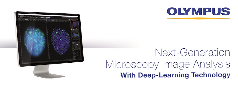
Olympus cellSens imaging software improves the efficiency of your research with accurate object detection and segmentation.
Leveraging the power of deep learning, Olympus cellSens imaging software for microscopy offers significantly improved segmentation analysis, such as label-free nucleus detection and cell counting, for more accurate data and efficient experiments.
Image analysis is a critical part of many life science applications. Analyses that rely on segmentation to extract targets, such as cells and organelles, from the rest of the image are commonplace. However, conventional thresholding methods that depend on brightness and color can miss critical information or may not be able to detect the targets at all. cellSens software’s deep-learning technology enables users to quickly train the system to automatically capture this information, improving the speed and accuracy of label-free object detection, quantitative analysis of fluorescent-labeled cells and segmentation based on morphological features.
Improve Experiment Efficiency with Label-Free Nuclei Detection
The fluorescent staining and UV excitation required for conventional nucleus detection is time consuming and can damage the cells. However, cellSens software can identify and segment nuclei from simple transmission images so that fluorescent labeling is not required.
Reducing Phototoxicity During Fluorescence Imaging to Support Accurate Data Acquisition
With cellSens software’s deep-learning technology, users can get accurate analysis data from low signal-to-noise ratio images. The technology produces outstanding accuracy while significantly reducing the amount of excitation light the cells are exposed to. This enables high-resolution segmentation while helping keep the cells healthy.
Save Time by Automating Cell Counting and Measuring
Deep-learning technology saves time by identifying and counting mitotic cells automatically. This technology is also useful for segmenting images of tissue specimens, such as kidney glomeruli, which is challenging when using conventional methods.
learn more about cellSens imaging software
News Channels
- Latest News
- New Laboratory Products
- Industry News
- Laboratory Automation | IT Solutions
- Microscopy | Image Analysis
- Separation Science
- Research | Case Studies
- Video Presentations
- Events | Conferences
Subscribe to any of our newsletters for the latest on new laboratory products, industry news, case studies and much more!
Popular this Month
Top 10 most popular articles this month
Today's Picks
Looking for a Supplier?
Search by company or by product
Please note Lab Bulletin does not sell, supply any of the products featured on this website. If you have an enquiry, please use the contact form below the article or company profile and we will send your request to the supplier so that they can contact you directly.
Lab Bulletin is published by newleaf marketing communications ltd.
Media Partners