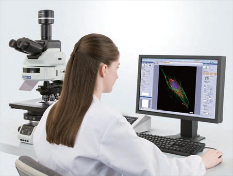
cellSens 1.7 offers multi-well imaging, HDR, 2D deconvolution, real-time control and more
Olympus has announced a new release of its powerful and flexible cellSens software (now version 1.7), offering a range of new benefits that allow any user to generate expert results, regardless of experience level. These include the brand new ‘Well Navigator’ Solution, which opens up a new world of flexible, multi-well imaging experiments. cellSens 1.7 also offers High Dynamic Range (HDR) imaging for the perfect exposure of challenging samples, 2D deconvolution for maximising image clarity and a new database model for streamlining the management of large data repositories with many users...
Read MoreRutgers Selects Albira Preclinical Imaging System from Carestream Molecular Imaging, Signs Collaborative AgreementAug 21, 2012Rutgers University has added an Albira PET/CT system from Carestream Molecular Imaging to its new Molecular Imaging Center in Piscataway, N.J.
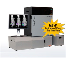
Located in a specially renovated facility near Rutgers' Livingston campus, the Molecular Imaging Center now houses the Albira PET/CT, a Carestream In-Vivo MS FX PRO optical imaging system, an MRI system, 3-D displays and other technologies, plus a federally registered holding facility for small animals. The center will support research in basic sciences, cardiovascular and neurodegenerative diseases, cancer, and many other fields...
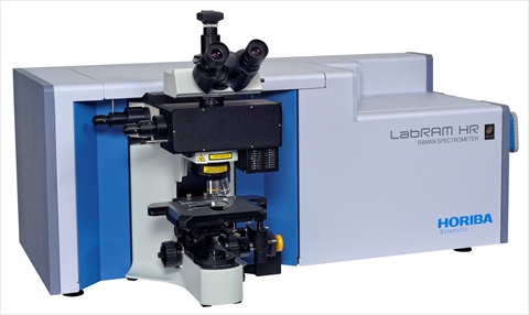
Read MoreHORIBA Scientific announces their new LabRAM HR Evolution spectrometerAug 17, 2012
Fully automated extended wavelength range capability and new generation of software offer versatility without compromise
HORIBA Scientific, global leader in Raman and Fluorescence spectroscopy systems, announces their new LabRAM HR Evolution, the latest spectrometer to join the LabRAM series of research Raman spectrometers. The LabRAM HR Evolution is highly versatile. It offers a fully automated, extended wavelength range capability. It also has an unsurpassed achromatic optical design with a high focal length. In fact, it provides high spectroscopic resolution and a unique wavelength range capability from 200nm to 2000nm with an access to frequencies as low as 10cm-1 thanks to the Ultra Low Frequency module, providing high content spectroscopic information for chemical and structural identification as well as an accurate spatial resolution at sub-micron scale...
Carl Zeiss Microscopy, a company of the Carl Zeiss Group and leading provider of light, laser-scanning and electron and ion beam microscopes, announces a new program which allows users to trade in their current Zeiss systems for credit towards a new 7-series laser scanning, spectral confocal system. The old products that are being accepted for trade in are the LSM 510, LSM 5 LIVE, LSM 5 Pascal or Bio-Rad confocal. The credit from trading in your old systems can be applied to Zeiss' LSM 780, LSM 710 or LSM 700 systems.
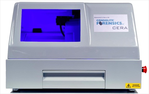
Read MoreNovel Lens Enhances Forensic Fingerprinting TechniqueAug 8, 2012Resolve Optics Ltd. has supplied a novel custom lens to Consolite Forensics Ltd. (Zeals, UK) to optimise the performance of their Cartridge Electrostatic Recovery and Analysis (CERA) system
The revolutionary CERA technology is now able to extract fingerprints from discharged cartridge cases, not visible by any other means. The scenes of many crimes involving firearms, where spent cartridge cases are the only evidence recovered, has traditionally posed a particular challenge for forensic scientists. Using fingerprints, caused by corrosive sweat, on brass metal cartridge cases has enjoyed some recent success in producing identifiable fingerprints* where conventional techniques have failed...
Read MoreCatalogue features extensive range of Ceti MicroscopesAug 8, 2012Laboratory equipment specialists MEDLINE SCIENTIFIC have recently published a new dedicated CETI MICROPSCOPE catalogue which was launched at ACHEMA 2012.
This new 44 page catalogue features the extensive range of CETI microscopes which are designed to suit all budgets and most popular applications. The range includes models for simple educational applications through to more sophisticated models for research and clinical tasks. Important new models include the LED powered Stereo microscopes, such as the STAR24LED, Steddy LED and the Varizoom. These offer variable top and bottom LED lighting as standard to ensure that the sample can be viewed without suffering any thermal damage over a prolonged viewing time...
Read MoreCambridge Research Biochemicals Signs Deal with LI-COR Biosciences for Infrared Dye Labelled PeptidesAug 7, 2012Leading research peptide manufacturer, Cambridge Research Biochemicals (CRB) announced today that it has signed a non-exclusive license with LI-COR Biosciences (LI-COR) to develop, manufacture and sell IRDye® and IRDye® QC-1 near-infrared dye-labelled peptides for research use only.
CRB is a world leader in providing dye labelled products to researchers. The partnership with LI-COR will enable the company to provide dye labelled peptides, which are unique in the field of fluorescence imaging applications...
Read MoreTecMicroscopes announce sales of their Microtec light microscopes into multiple education facilities across the UKAug 7, 2012TecMicroscopes in conjunction with Mazurek Optical Services, suppliers of the Microtec range of light microscopes and accessories, is pleased to announce multiple sales into academic institutions across the UK.
TecMicroscopes and its principle sales partner, Mazurek Optical Services are very pleased to announce the installation of three new suites of microscopes to UK universities and schools. Mazurek's excellent service and support record as noted as doing "an excellent job, so promptly and so efficiently" by Marta Salamonowicz, Research Administrator at the UCL Prostate Cancer Research Centre, backed by the high level of optical performance at an economical price of Microtec has been the basis of these sales...
Read MoreJPK reports on the use of their NanoWizard systems in the Microscopy & Imaging Facility at the University of CalgaryAug 7, 2012JPK Instruments, a world-leading manufacturer of nanoanalytic instrumentation for research in life sciences and soft matter, reports on the use of their NanoWizard® AFM systems at the Microscopy & Imaging Facility (MIF) at the University of Calgary in Canada.
The Calgary Microscopy and Imaging Facility (MIF) is a world-class university-wide facility housing transmission electron microscopy (TEM), scanning electron microscopy (SEM), advanced light microscopy, atomic force microscopy (AFM), including single cell force spectroscopy (SCFS), and advanced image processing for three-dimensional electron and light microscopy, directed by Professor Matthias Amrein.
Read MoreNew Olympus NA 1.7 APON100Aug 7, 2012High quality single molecule imaging and analysis in cells
Olympus, a leading manufacturer of professional opto-digital products, has launched the NA 1.7 APON100×HOTIRF objective for the imaging of intracellular molecules. With a world record-breaking numerical aperture (NA) of 1.7, the objective achieves the highest Z-resolution ever recorded for a Total Internal Reflection Fluorescence (TIRF) microscope. The benefits for research using TIRF microscopy include extremely thin penetration depth and high signal-to-noise ratio, making the lens ideal for single molecule dynamic studies and super resolution applications... Read MoreCarl Zeiss reveals High Definition FE-SEM SIGMA HDAug 6, 2012The High Definition Field Emission Scanning Electron Microscope (FE-SEM) SIGMA HD is revealed at the Microscopy & Microanalysis conference 2012 in Phoenix, Arizona by Carl Zeiss Microscopy.
SIGMA HD offers customers high resolution, fast imaging and easy sample navigation for nanoscale analytics in addition to the performance of the established SIGMA series...
Leica SCN400 2.2 - Fast, Flexible Whole Slide Capture for Digital Pathology
Leica Microsystems announces the release of the SCN400 2.2 scanning platform. Batch processing in both brightfield and multi-channel fluorescence, coupled with user-friendly workflows, makes the SCN400 2.2 an all-round high performance solution for digital pathology scanning...
Customers can now choose between the systems MERLIN Compact, MERLIN VP Compact and MERLIN and configure them individually. The Plug-and-Play feature allows the customer to add and change detectors with minimum effort to handle tasks ranging from simple image capture to extensive material analysis. A large frame store of 32k x 24k pixels allows imaging of very large areas. New features include in-situ 3D surface reconstruction and calculation of 3D data from 2D data...
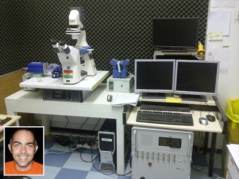
Read MoreJPK reports on the research in the Marseille INSERM/CNRS laboratories where the NanoWizard AFM system is being used for cell studiesAug 1, 2012JPK Instruments, a world-leading manufacturer of nanoanalytic instrumentation for research in life sciences and soft matter,reports on the use of the NanoWizard® AFM system at the INSERM/CNRS Laboratories in Marseille, France.
The Laboratory of Cell Adhesion and Inflammation is located in Marseille as part of the INSERM/CNRS complex on the Luminy campus. It is a multidisciplinary research unit comprised of investigators with backgrounds in physics, biology and medicine. Their aim is to adapt physical concepts and methodologies to the understanding of cell function, notably cell adhesion in the immune system, with the goal of applying the results of this fundamental research to relevant clinical problems...
Read MoreSteady Funding Initiatives are Instrumental in Channeling Optical Imaging into New Application Areas, States Frost & SullivanAug 1, 2012Application scope set to expand rapidly from current base of ophthalmology, cardiology, neurology and gastroenterology
Photometrics introduces the CoolSNAP™ MYO and the CoolSNAP™ KINO CCD cameras, the newest members of the popular CoolSNAP camera line
Designed to discern finer details in biological samples under lower light levels, the MYO and KINO enable scientists to achieve higher quality, higher resolution images than previous CCD technology...
Read MoreDefiniens Expands Large Data Analytics Functionality to Accelerate Image-based Research and DevelopmentJul 16, 2012New Definiens Image MinerTM 2 tightly integrates data mining with image analysis to streamline the knowledge-generation process
Definiens®, the leading provider of image analysis and data mining solutions for quantitative digital pathology, today announced the release of Definiens Image MinerTM 2. The new product provides researchers in the life sciences with deeper insights into underlying biology by integrating image with data analysis. Image Miner 2 makes the wealth of information in biomedical images accessible, accelerating life sciences research and allowing for successful biomarker development...
Read MoreEuropean launch of new microscope systems to be held at the EMC 2012 in ManchesterJul 16, 2012Olympus has announced that it will be launching a totally re-engineered line of highly customisable inverted microscope systems for life science research at the European Microscopy Congress (EMC) 2012 in Manchester, UK
As well as hosting a booth at the show, Olympus plan to demo some of its systems in the RMS learning zone at the meeting. This area is specifically designed to provide scientists and technicians with the opportunity to learn from commercial application specialists, as well as their academic peers. The company will also be hosting several breakout workshops at EMC 2012, where Olympus experts will be discussing the range of exciting applications the new systems will offer - seating will be allocated on a first-come-first-serve basis, so please arrive promptly to avoid disappointment...
Read MoreSectioning of bone as a specialist histology specimenJul 13, 2012Thermo Scientific HM355S and Finesse ME+ Automated Microtomes deliver quality sections from bone tissue
The significance of histological examination in the classification and diagnosis of clinical conditions is reliant on the expertise of the histology laboratory in managing the wide spectrum of specimen types submitted for analysis. From receipt of the tissue sample to presentation of a slide for microscopic examination, histologists must consider the composition of the specimen to effectively determine how it should be handled...
Read MoreCarl Zeiss Licenses Digital Pathology Patents from OlympusJul 13, 2012Carl Zeiss Microscopy GmbH and Olympus America Inc. CENTER VALLEY, Pa. have signed a nonexclusive worldwide licensing agreement allowing Carl Zeiss to access an extensive portfolio of patents held by Olympus in the field of digital pathology and virtual microscopy.
The patents included in the licensing deal cover methods and equipment for creating, storing and delivering virtual microscopy slides. The technology enables individuals to view and share high-resolution virtual microscopy images over the Internet...
Read MoreXEI Scientific announces the first sales of their Evactron TEM Wand localized plasma cleaning system for TEM applicationsJul 13, 2012XEI Scientific Inc, maker of the popular EVACTRON® De-Contaminator Plasma Cleaning System for electron microscopes and other vacuum chambers, announce that they have begun shipments of the Evactron TEM WandTM systems.
Specifically designed to deliver the proven downstream plasma cleaning technique efficiently to critical areas of the Transmission Electron Microscope (TEM), the Evactron TEM Wand allows microscopists to easily clean the sample introduction, goniometer and pole piece areas of their microscopes. Removal of the unwanted carbon contamination provides clearer images and data without artifacts...
Read MoreR&D 100 Award for Super-Resolution Microscope from Leica MicrosystemsJul 10, 2012Leica SR GSD Convinces Jury with Unprecedented Performance in Fluorescence Microscopy
The Leica SR GSD is a winner of the R&D100 Award, granted by R&D Magazine for outstanding technical developments. It is the 50th anniversary of this prestigeous award. Leica Microsystems was honored together with the Max Planck Institute for Biophysical Chemistry in Göttingen for the development of this ground-breaking widefield super-resolution microscope...
Read More

 Olympus has announced a new release of its powerful and flexible cellSens software (now version 1.7), offering a range of new benefits that allow any user to generate expert results, regardless of experience level. These include the brand new ‘Well Navigator’ Solution, which opens up a new world of flexible, multi-well imaging experiments. cellSens 1.7 also offers High Dynamic Range (HDR) imaging for the perfect exposure of challenging samples, 2D deconvolution for maximising image clarity and a new database model for streamlining the management of large data repositories with many users...
 Located in a specially renovated facility near Rutgers' Livingston campus, the Molecular Imaging Center now houses the Albira PET/CT, a Carestream In-Vivo MS FX PRO optical imaging system, an MRI system, 3-D displays and other technologies, plus a federally registered holding facility for small animals. The center will support research in basic sciences, cardiovascular and neurodegenerative diseases, cancer, and many other fields...
 HORIBA Scientific, global leader in Raman and Fluorescence spectroscopy systems, announces their new LabRAM HR Evolution, the latest spectrometer to join the LabRAM series of research Raman spectrometers. The LabRAM HR Evolution is highly versatile. It offers a fully automated, extended wavelength range capability. It also has an unsurpassed achromatic optical design with a high focal length. In fact, it provides high spectroscopic resolution and a unique wavelength range capability from 200nm to 2000nm with an access to frequencies as low as 10cm-1 thanks to the Ultra Low Frequency module, providing high content spectroscopic information for chemical and structural identification as well as an accurate spatial resolution at sub-micron scale...
 Carl Zeiss Microscopy, a company of the Carl Zeiss Group and leading provider of light, laser-scanning and electron and ion beam microscopes, announces a new program which allows users to trade in their current Zeiss systems for credit towards a new 7-series laser scanning, spectral confocal system. The old products that are being accepted for trade in are the LSM 510, LSM 5 LIVE, LSM 5 Pascal or Bio-Rad confocal. The credit from trading in your old systems can be applied to Zeiss' LSM 780, LSM 710 or LSM 700 systems.
 The revolutionary CERA technology is now able to extract fingerprints from discharged cartridge cases, not visible by any other means. The scenes of many crimes involving firearms, where spent cartridge cases are the only evidence recovered, has traditionally posed a particular challenge for forensic scientists. Using fingerprints, caused by corrosive sweat, on brass metal cartridge cases has enjoyed some recent success in producing identifiable fingerprints* where conventional techniques have failed...
 This new 44 page catalogue features the extensive range of CETI microscopes which are designed to suit all budgets and most popular applications. The range includes models for simple educational applications through to more sophisticated models for research and clinical tasks. Important new models include the LED powered Stereo microscopes, such as the STAR24LED, Steddy LED and the Varizoom. These offer variable top and bottom LED lighting as standard to ensure that the sample can be viewed without suffering any thermal damage over a prolonged viewing time...
 CRB is a world leader in providing dye labelled products to researchers. The partnership with LI-COR will enable the company to provide dye labelled peptides, which are unique in the field of fluorescence imaging applications...
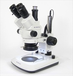 TecMicroscopes and its principle sales partner, Mazurek Optical Services are very pleased to announce the installation of three new suites of microscopes to UK universities and schools. Mazurek's excellent service and support record as noted as doing "an excellent job, so promptly and so efficiently" by Marta Salamonowicz, Research Administrator at the UCL Prostate Cancer Research Centre, backed by the high level of optical performance at an economical price of Microtec has been the basis of these sales...
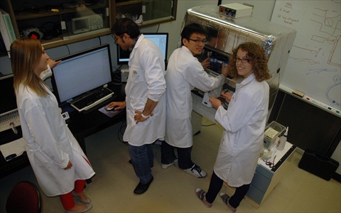 The Calgary Microscopy and Imaging Facility (MIF) is a world-class university-wide facility housing transmission electron microscopy (TEM), scanning electron microscopy (SEM), advanced light microscopy, atomic force microscopy (AFM), including single cell force spectroscopy (SCFS), and advanced image processing for three-dimensional electron and light microscopy, directed by Professor Matthias Amrein.
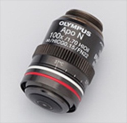 Olympus, a leading manufacturer of professional opto-digital products, has launched the NA 1.7 APON100×HOTIRF objective for the imaging of intracellular molecules. With a world record-breaking numerical aperture (NA) of 1.7, the objective achieves the highest Z-resolution ever recorded for a Total Internal Reflection Fluorescence (TIRF) microscope. The benefits for research using TIRF microscopy include extremely thin penetration depth and high signal-to-noise ratio, making the lens ideal for single molecule dynamic studies and super resolution applications...
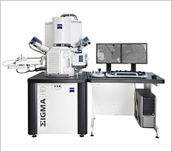 SIGMA HD offers customers high resolution, fast imaging and easy sample navigation for nanoscale analytics in addition to the performance of the established SIGMA series...
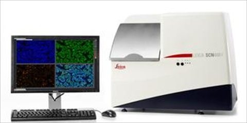 Leica Microsystems announces the release of the SCN400 2.2 scanning platform. Batch processing in both brightfield and multi-channel fluorescence, coupled with user-friendly workflows, makes the SCN400 2.2 an all-round high performance solution for digital pathology scanning...
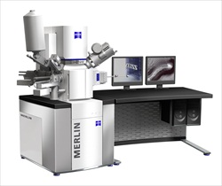 Customers can now choose between the systems MERLIN Compact, MERLIN VP Compact and MERLIN and configure them individually. The Plug-and-Play feature allows the customer to add and change detectors with minimum effort to handle tasks ranging from simple image capture to extensive material analysis. A large frame store of 32k x 24k pixels allows imaging of very large areas. New features include in-situ 3D surface reconstruction and calculation of 3D data from 2D data...
 The Laboratory of Cell Adhesion and Inflammation is located in Marseille as part of the INSERM/CNRS complex on the Luminy campus. It is a multidisciplinary research unit comprised of investigators with backgrounds in physics, biology and medicine. Their aim is to adapt physical concepts and methodologies to the understanding of cell function, notably cell adhesion in the immune system, with the goal of applying the results of this fundamental research to relevant clinical problems...
 Definiens®, the leading provider of image analysis and data mining solutions for quantitative digital pathology, today announced the release of Definiens Image MinerTM 2. The new product provides researchers in the life sciences with deeper insights into underlying biology by integrating image with data analysis. Image Miner 2 makes the wealth of information in biomedical images accessible, accelerating life sciences research and allowing for successful biomarker development...
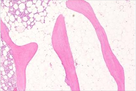 The significance of histological examination in the classification and diagnosis of clinical conditions is reliant on the expertise of the histology laboratory in managing the wide spectrum of specimen types submitted for analysis. From receipt of the tissue sample to presentation of a slide for microscopic examination, histologists must consider the composition of the specimen to effectively determine how it should be handled...
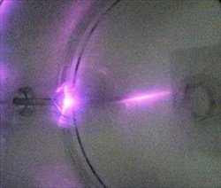 Specifically designed to deliver the proven downstream plasma cleaning technique efficiently to critical areas of the Transmission Electron Microscope (TEM), the Evactron TEM Wand allows microscopists to easily clean the sample introduction, goniometer and pole piece areas of their microscopes. Removal of the unwanted carbon contamination provides clearer images and data without artifacts...
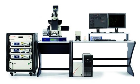 The Leica SR GSD is a winner of the R&D100 Award, granted by R&D Magazine for outstanding technical developments. It is the 50th anniversary of this prestigeous award. Leica Microsystems was honored together with the Max Planck Institute for Biophysical Chemistry in Göttingen for the development of this ground-breaking widefield super-resolution microscope...