The Thermo Scientific Cool Cut specimen clamp maintains block temperature for more consistent, better quality sections
Histological examination is considered the gold standard for detection, diagnosis and characterization of many clinical conditions, and producing suitable sections for microscopic examination constitutes one of the primary skills of the histologist...
Olympus has released the new FluoView FV1200 confocal laser scanning microscope, optimised for live cell imaging
The new system uniquely combines the accuracy of the newly engineered IX83 frame with enhanced fluorescence sensitivity and simultaneous laser stimulation of cells, making it ideal for advanced life science applications such as FRAP, FLIP and photo-activation. In particular, the new highly-reflective, silver-coated galvanometer scanning mirrors and dual channel GaAsP FluoView PMT module both act to maximise light transfer and detection. This allows for reduced laser power, protecting against the effects of photobleaching and phototoxicity...
Read MoreFLIR Launch Range of Portable R&D CamerasDec 7, 2012
FLIR Systems announces the launch of SC650 and SC450 thermal imaging camera packs for scientific research and development
Delivering the highest sensitivity and most advanced features available on a portable thermal imaging camera - the FLIR SC650 / SC450 packs offer users the no-compromise flexibility of an R&D grade camera they can take from experiment to experiment. Included as standard in all FLIR SC650 / SC450 thermal imaging camera packs is ResearchIR software which allows high speed recording, advanced thermal pattern analysis and users to make the most of their new thermal imaging camera...
Read MoreNew Masterflex Tygon E-LFL Tubing designed to respond to changing environmental concernsDec 6, 2012
Tubing formulation offers performance with minimal environmental impact
The new Masterflex® Tygon® E-LFL Tubing from Cole-Parmer is now available for use in Masterflex® peristaltic pumps. This new tubing formulation is replacing the discontinued Masterflex® LFL tubing. The transparent tubing offers similar chemical resistance, purity, and long pumping life to its predecessor and is intended for use in a variety of biological, pharmaceutical, and chemical processes....
Read MoreResearchers from the University of East Anglia, UK, use nanoscale thermal analysis techniques to improve drug delivery systemsDec 5, 2012
Anasys Instruments report on the use of their award-winning nanoscale characterization instrumentation to advance developments in the understanding of drug delivery systems
The Drug Delivery & Materials Characterization Group at the University of East Anglia, UK, is internationally recognized for work involving the development of novel thermal, dielectric, rheological and microscopic techniques as analytical tools within the pharmaceutical sciences. There is particular emphasis on the study of the physical properties of drugs and dosage forms in relation to performance...
Read MoreLI-COR Now Accepting Applications for New Undergraduate Research GrantNov 30, 2012
LI-COR is now accepting applications for its Science Undergraduate Research Grant (SURG) program. This program is designed for faculty researchers and their students to gain access to cutting edge life science technology and incorporate it into the classroom
LI-COR Biosciences is awarding a limited number of matching fund grants (value up to $18,400) to eligible academic institutions within the United States and Puerto Rico to be used toward the purchase of a LI-COR Odyssey® Fc Imaging System, including the instrument, software, and reagents. Also included with the system are Western blot and DNA gel imaging sample curriculum, installation, and training...
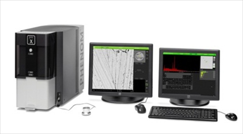
Read MoreCollect more than just SEM images with Phenom proX Desktop SEM!Nov 26, 2012
The Phenom proX is a fast and intuitive desktop electron microscope with fully integrated EDS elemental analysis. For pharmaceutical, chemical industries, failure analysis purposes and metallurgical applications as well as biological applications and forensics, the Phenom will give your whole team access to affordable and reliable imaging and material analysis
Adding an X-ray detector to an SEM is an obvious upgrade to implement elemental identification of microstructures on a sample. The Phenom proX solution takes it one step further and extends the ease of use, speed and one system solution into the x-ray analysis field. The 15kV electron beam in the Phenom proX creates back scatter electrons for fast and reliable SEM imaging and generates large numbers of X-rays for fast data collection...
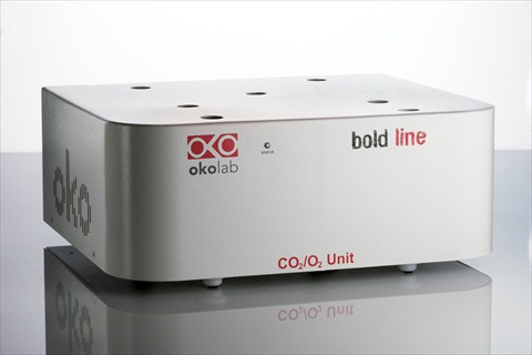
Read MoreCO2/O2 Controller Ideal for Hypoxia and Hyperoxia Live-Cell Imaging ExperimentsNov 23, 2012
Warner Instruments is pleased to introduce the Bold Line CO2 - O2 Gas Controllers, part of the complete line of gas controllers from Okolab. Warner Instruments is the Authorized Distributor for Okolab products in the US
The Bold Line combination CO2 - O2 gas controllers are ideal for hypoxia and hyperoxia experiments. Two versions are available, CO2; 0-10% and O2; 1-18%, and CO2; 0-20% and O2; 1-95%. Gas flow rates are digitally controlled. A long life zirconium oxide sensor precisely measures the O2 concentration, and a non dispersive InfraRed (NDIR) dual wave length detector sets the CO2 concentration...
The Olympus VS120 Slide Scanning System has earned three first place awards at the prestigious second International Scanner Contest (ISC), which took place in Berlin earlier this year
The advanced technology of the VS120 creates a “virtual slide”, a high resolution image of the complete specimen that can be electronically stored on a central server for simultaneous viewing anywhere in the world, at a range of magnifications. Awards were obtained in the fields of Colour and Precision, Fluorescence 20X, and Fluorescence 40X. Exhibiting unsurpassed performance in fluorescence microscopy, combined with...
Enhanced speed and efficiency for recording, processing and displaying large amounts of data - including 3D visualization in real-time
Leica Microsystems has launched the new version of its powerful research microscopy software LAS AF 3 (Leica Application Suite Advanced Fluorescence). LAS AF 3 sets new standards for intuitive operation. It covers the whole spectrum of fluorescence applications from routine work to sophisticated tasks in biomedical research, such as deep imaging of thicker tissue or interactive time lapse experiments... Read MoreOcean Thin Films Announces Multispectral Imaging Research Grant Program Nov 9, 2012
Academic, industry professionals can use free or subsidized SpectroCam for data collection
Ocean Thin Films is calling for grant proposals featuring multispectral imaging applications that would utilize the SpectroCam™ platform. SpectroCam is an imaging system that integrates scientific-grade sensors with eight filters to produce multispectral images. This grant gives participants the unique opportunity to add a high-end instrument to their lab at little or no cost...
Read MoreA Whole-Microscope Enclosure for Maintaining Cell Culture ConditionsNov 8, 2012
Warner Instruments is pleased to introduce the Okolab Bold Line Cage Incubator that provides a controlled environment all around the microscope
Obtain a fully integrated workflow with OLYMPUS Stream 1.8
Olympus today launched the latest version of its highly successful OLYMPUS Stream materials science microscopy imaging software family. OLYMPUS Stream 1.8 provides new and improved features, enhancing the overall capabilities of the software to help users fully integrate and automate their workflows. Users will benefit from the advanced automation of a multiple stage location engine, extended data management, new measurement options and new additions to the optional Materials Solutions: Particle Distribution, Porosity, Throwing Power and Phase Analysis. These Materials Solutions enable users to build a fully guided, or even automated, system to precisely match the needs of their materialographic analysis...
Read MoreNIST research highlights promise of AFM-IR for quantitative nanoscale chemical analysisNov 5, 2012
Anasys Instruments announces a new paper authored by Dr Andrea Centrone and his colleagues at NIST published recently in Small, a leading publication which focuses on the nano and micro worlds
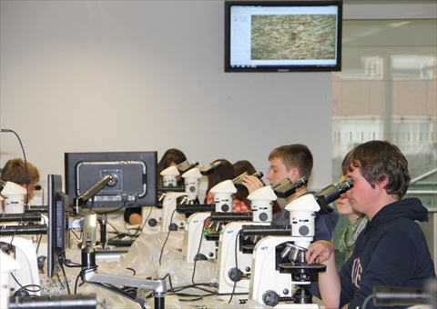
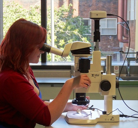
Meiji Techno in association with Mazurek Optical Services are pleased to announce they have installed a suite of 365 light microscopes to the new Central Teaching Laboratory at the University of Liverpool
The new laboratory complex was officially opened on Monday, October 22nd but has already been extensively used by students returning for the new academic year at the beginning of the month. The University of Liverpool's new Central Teaching Laboratories for the Faculty of Science and Engineering are set to transform the way in which Physical Sciences (Physics, Chemistry, Archaeology, Geography, and Earth and Ocean Science) are taught at the University...
The qualities of particles can now be determined using Axio Zoom.V16 and Correlative Microscopy
The Carl Zeiss Microscopy business group is presenting two new options for particle analysis at this year's parts2clean trade fair in Stuttgart, Germany. Using Axio Zoom.V16, testers can investigate a component's cleanliness quickly and efficiently, while correlative microscopy provides chemical information and material classifications on individual particles...
Read MoreMazurek Optical Services announces a new microscopy award for students at the University of LiverpoolOct 25, 2012
Mazurek Optical Services is pleased to announce the first awarding of an annual prize given to the most promising first year student demonstrating outstanding work in microscopy while studying at new the Central Teaching Laboratory of the University of Liverpool
Continuing their support of the development of young microscopists, Mazurek Optical Services (MOS) have endowed a prize to be presented annually to the most promising first year student who demonstrates outstanding work in light microscopy at the University of Liverpool's newly opened Central Teaching Laboratories...
Read MoreAndor Neo sCMOS cameras at core of Light Sheet MicroscopyOct 17, 2012
European and US labs announce simultaneous adoption of Andor Neo sCMOS camera to power high-speed 4-axis live imaging of biological specimens
Laboratories in Europe and the USA have independently announced in the same issue of Nature Methods the development of new microscopes capable of imaging rapid biological processes in thick samples in unprecedented detail and ideally suited to the long-term study of live biological specimens... Read MoreCarl Zeiss Introduces Lightsheet Z.1 Light Sheet Microscope SystemOct 16, 2012
3D fluorescence imaging of large living specimens with low phototoxicity
The Microscopy business group at Carl Zeiss is presenting a new microscopy technology at the Society for Neuroscience Annual Meeting in New Orleans, Louisiana. Lightsheet Z.1 provides biologists with a new method of imaging dynamic processes in living organisms...
Read MoreScientifica presented with Queen's Award for EnterpriseOct 15, 2012
Scientifica was presented with its prestigious Queen's Award for Enterprise: International Trade 2012 at a ceremony held at its headquarters in Uckfield last Friday (5 October). The Award was given to Mark Johnson, Managing Director of Scientifica, by Peter Field, Lord Lieutenant for East Sussex
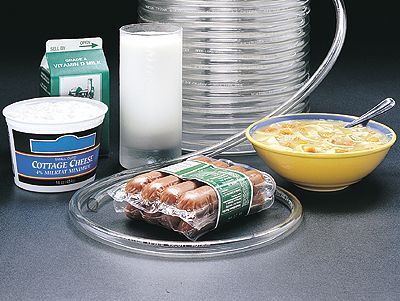
Market leaders in temperature controlled microscopy, Linkam Scientific Instruments report on the use of their Linksys32-DV software to visualise and measure the in-situ growth of fat crystals on the surface of chocolate
Reformulation is a common area of study for food scientists, as worldwide, many universities and private organisations are investing heavily in research to create healthier versions of popular foods. Reformulation is the process of adding or taking away ingredients from an established recipe. This is challenging as many of the chemical processes that occur within the foods are unknown and changing a specific constituent can have consequences on health and safety, cooking, storage and on manufacturing processes... Read More

 Histological examination is considered the gold standard for detection, diagnosis and characterization of many clinical conditions, and producing suitable sections for microscopic examination constitutes one of the primary skills of the histologist...
 The new system uniquely combines the accuracy of the newly engineered IX83 frame with enhanced fluorescence sensitivity and simultaneous laser stimulation of cells, making it ideal for advanced life science applications such as FRAP, FLIP and photo-activation. In particular, the new highly-reflective, silver-coated galvanometer scanning mirrors and dual channel GaAsP FluoView PMT module both act to maximise light transfer and detection. This allows for reduced laser power, protecting against the effects of photobleaching and phototoxicity...
 The new Masterflex® Tygon® E-LFL Tubing from Cole-Parmer is now available for use in Masterflex® peristaltic pumps. This new tubing formulation is replacing the discontinued Masterflex® LFL tubing. The transparent tubing offers similar chemical resistance, purity, and long pumping life to its predecessor and is intended for use in a variety of biological, pharmaceutical, and chemical processes....

 LI-COR Biosciences is awarding a limited number of matching fund grants (value up to $18,400) to eligible academic institutions within the United States and Puerto Rico to be used toward the purchase of a LI-COR Odyssey® Fc Imaging System, including the instrument, software, and reagents. Also included with the system are Western blot and DNA gel imaging sample curriculum, installation, and training...
 Adding an X-ray detector to an SEM is an obvious upgrade to implement elemental identification of microstructures on a sample. The Phenom proX solution takes it one step further and extends the ease of use, speed and one system solution into the x-ray analysis field. The 15kV electron beam in the Phenom proX creates back scatter electrons for fast and reliable SEM imaging and generates large numbers of X-rays for fast data collection...
 The Bold Line combination CO2 - O2 gas controllers are ideal for hypoxia and hyperoxia experiments. Two versions are available, CO2; 0-10% and O2; 1-18%, and CO2; 0-20% and O2; 1-95%. Gas flow rates are digitally controlled. A long life zirconium oxide sensor precisely measures the O2 concentration, and a non dispersive InfraRed (NDIR) dual wave length detector sets the CO2 concentration...
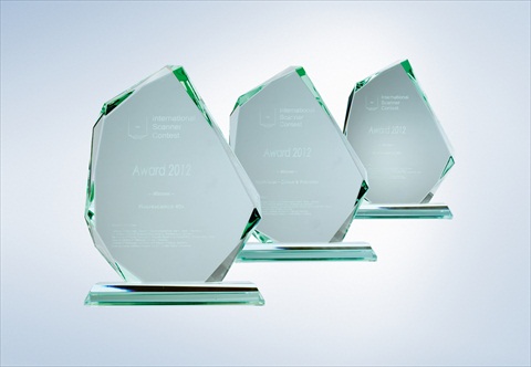 The advanced technology of the VS120 creates a “virtual slide”, a high resolution image of the complete specimen that can be electronically stored on a central server for simultaneous viewing anywhere in the world, at a range of magnifications. Awards were obtained in the fields of Colour and Precision, Fluorescence 20X, and Fluorescence 40X. Exhibiting unsurpassed performance in fluorescence microscopy, combined with...
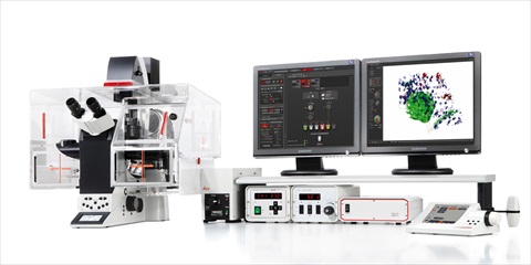 Leica Microsystems has launched the new version of its powerful research microscopy software LAS AF 3 (Leica Application Suite Advanced Fluorescence). LAS AF 3 sets new standards for intuitive operation. It covers the whole spectrum of fluorescence applications from routine work to sophisticated tasks in biomedical research, such as deep imaging of thicker tissue or interactive time lapse experiments...
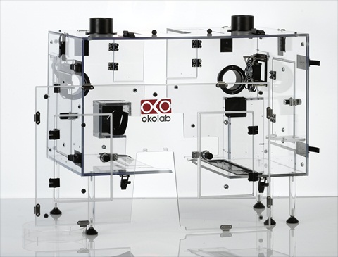 Cells proliferate as well as bench top incubators
  Olympus today launched the latest version of its highly successful OLYMPUS Stream materials science microscopy imaging software family. OLYMPUS Stream 1.8 provides new and improved features, enhancing the overall capabilities of the software to help users fully integrate and automate their workflows. Users will benefit from the advanced automation of a multiple stage location engine, extended data management, new measurement options and new additions to the optional Materials Solutions: Particle Distribution, Porosity, Throwing Power and Phase Analysis. These Materials Solutions enable users to build a fully guided, or even automated, system to precisely match the needs of their materialographic analysis...
 The new laboratory complex was officially opened on Monday, October 22nd but has already been extensively used by students returning for the new academic year at the beginning of the month. The University of Liverpool's new Central Teaching Laboratories for the Faculty of Science and Engineering are set to transform the way in which Physical Sciences (Physics, Chemistry, Archaeology, Geography, and Earth and Ocean Science) are taught at the University...
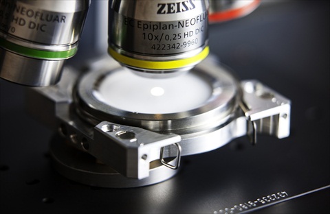 The Carl Zeiss Microscopy business group is presenting two new options for particle analysis at this year's parts2clean trade fair in Stuttgart, Germany. Using Axio Zoom.V16, testers can investigate a component's cleanliness quickly and efficiently, while correlative microscopy provides chemical information and material classifications on individual particles...
 Continuing their support of the development of young microscopists, Mazurek Optical Services (MOS) have endowed a prize to be presented annually to the most promising first year student who demonstrates outstanding work in light microscopy at the University of Liverpool's newly opened Central Teaching Laboratories...
 Laboratories in Europe and the USA have independently announced in the same issue of Nature Methods the development of new microscopes capable of imaging rapid biological processes in thick samples in unprecedented detail and ideally suited to the long-term study of live biological specimens...
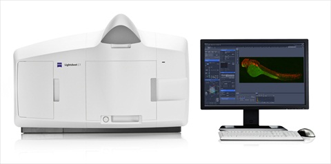 The Microscopy business group at Carl Zeiss is presenting a new microscopy technology at the Society for Neuroscience Annual Meeting in New Orleans, Louisiana. Lightsheet Z.1 provides biologists with a new method of imaging dynamic processes in living organisms...
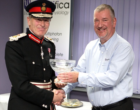 The event was also attended by local MP, Charles Hendry, along with Scientifica's team from its Uckfield and Maidenhead offices and specially invited guests. These included Councillors Chris Dowling, Jonica Fox and John Carvey, Mayor of Uckfield, and David Marshall, Chairman of The Uckfield Chamber of Commerce.
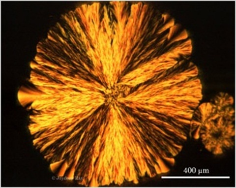 Reformulation is a common area of study for food scientists, as worldwide, many universities and private organisations are investing heavily in research to create healthier versions of popular foods. Reformulation is the process of adding or taking away ingredients from an established recipe. This is challenging as many of the chemical processes that occur within the foods are unknown and changing a specific constituent can have consequences on health and safety, cooking, storage and on manufacturing processes...