Channels
Special Offers & Promotions
Andor Neo sCMOS cameras at core of Light Sheet Microscopy
European and US labs announce simultaneous adoption of Andor Neo sCMOS camera to power high-speed 4-axis live imaging of biological specimens
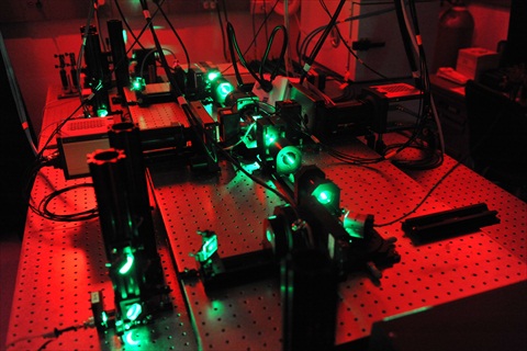
Laboratories in Europe and the USA have independently announced in the same issue of Nature Methods the development of new microscopes capable of imaging rapid biological processes in thick samples in unprecedented detail and ideally suited to the long-term study of live biological specimens.Philipp J Keller designed the microscope used by the group from the Howard Hughes Medical Institute in Virginia, USA, while Lars Hufnagel led the team from the European Molecular Biology Laboratory (EMBL) in Heidelberg, Germany. Called SiMView by Keller and Multi-View SPIM (MuVi-SPIM) by Hufnagel, the new 4-axis light-sheet microscopes build upon the SPIM (Selective-Plane Illumination Microscopy) technology developed by Keller and Stelzer at EMBL and feature Andor's Neo sCMOS camera at their cores.
Like SPIM, the new microscopes shine a thin sheet of light on the sample, illuminating one layer at a time to obtain an image of the whole sample with minimal light-induced damage. However, SiMView/MuVi-SPIM uses two diagonally-opposed light sources and two cameras to capture four images from different angles, eliminating the need to rotate the sample. This speeds up the imaging and enables the four images to be merged into a single high-quality, three-dimensional image for the first time.
Hufnagel and Keller agree that every aspect of the microscope must be optimised for speed and light sensitivity - optics, mechanics, cameras, and software. Both teams independently chose the 5.5 megapixel Andor Neo sCMOS camera, which offers a higher sensitivity than any CCD, rapid frame rates of up to 100fps, and vacuum thermoelectric cooling to -40?C to minimise dark current and maintain low noise.
"For global measurements of the dynamic behaviour and structural changes of all cells in a developing organism, data acquisition must occur at speeds that match the timescales of the fastest processes of interest and with minimal time shifts between complementary views," says Keller. "SiMView/MuVi-SPIM overcomes the limitations of sequential multiview strategies and enables quantitative systems-level imaging of fast dynamic events in large living specimens under physiological conditions."
Both teams used the Andor Neo sCMOS cameras to capture a complete view of an embryo of Drosophila melanogaster within a few seconds. Lars Hufnagel focused on the biomechanics of development using one-photon microscopy and recorded the movements of every nucleus in the embryo throughout the first three hours of life when nuclei divide very rapidly as well as producing a 20-hour video showing the fruit fly embryo from approx. two-and-a-half hours to the stage it walked away from the microscope as a larva. Philipp Keller used both one- and two-photon microscopy, penetrating deeper inside the embryo with multiphoton microscopy to look at subcellular events happening throughout the whole embryo development until eventual hatching.
"Understanding the development and function of complex bio¬logical systems relies on capturing and quantifying fast spatiotemporal dynamics on a microscopic scale," says Orla Hanrahan, Application Specialist at Andor. "The innovative approach of both teams in developing their four-axis SPIM microscopes allows them to take full advantage of the large field of view, high sensitivity, and fast frame rate capabilities of Andor's Neo sCMOS detector and unleashes a host of possibilities for the future of imaging".
Andor's Neo sCMOS camera can be used for a broad range of microscopy applications, including wide field fluorescence microscopy, calcium ratio imaging, FRET, and TIRF microscopy. To learn more about the Neo sCMOS camera and its use in microscopy, please visit the Andor website
Media Partners


