Channels
Special Offers & Promotions
Pioneering publication locates origin of Autophagosomal Membranes
Trio of Andor iXon EMCCD cameras on Revolution XD microscope make possible the simultaneous tracking of key autophagosome markers to deliver new insights into organelle biogenesis
 The location of the site where autophagosomal membranes are created within the cell has been the subject of two competing models for many years. Now, a group of researchers from Osaka University led by Prof. Maho Hamasaki has located the origin of autophagosomes within the cell and shown that neither model is correct.
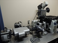
Writing in Nature, they show that autophagosomes form at the contact site of the endoplasmic reticulum (ER) with mitochondria. Live cell imaging studies, using an Andor Revolution XD spinning disk confocal microscope equipped with three Andor iXon 3 EMCCD cameras, captured previously hidden detail. The research revealed the re-localisation of the pre-autophagosome/autophagosome marker ATG14 to the ER-mitochondria contact site after starvation, along with the complementary localisation of the autophagosome-formation marker ATG5 until formation is complete.
"Following highly dynamic events within the live cell requires a fast, high-resolution system and this led us to select the Andor Revolution XD spinning disk confocal microscope as the heart of our set-up," says Prof. Maho Hamasaki. "But, we also needed to observe three markers simultaneously and using a single camera with three filters would have caused unacceptable delays. Instead, we deployed a trio of Andor iXon 3 EMCCD cameras to capture the three colours and Z-axis data for the Andor IQ software. The iXon camera was chosen for its combination of very high levels of speed, sensitivity and resolution."
According to Geraint Wilde, Product Specialist at Andor, "This is a very important technical and scientific achievement. Although autophagy was discovered fifty years ago and autophagy-related proteins were identified in the 1990s, the argument over whether autophagosome membrane formation takes place in the ER or the mitochondria has raged ever since. Prof. Hamasaki's team has shown that results previously thought to exclusively support either model are finally explained by their new model in which the autophagosome is formed at the ER-mitochondria contact site.
"Speed and sensitivity were key to tracking the three markers necessary for their discovery. The Andor iXon 3 camera offers the highest sensitivity from a quantitative scientific digital camera, particularly at fast frame rates, and proved the ideal partner for the spinning disk technology of the Andor Revolution XD fluorescence microscope system."
Andor's microscopy systems and scientific cameras address a broad range of optical microscopy techniques including laser spinning disk confocal microscopy, photo-bleaching, activation, conversion and ablation, TIRF, white light spinning disk confocal, calcium ratio imaging, comet assay and bioluminescence.
About Andor
Andor is a world leader in Scientific Imaging, Spectroscopy Solutions and Microscopy Systems. Established in 1989 from Queen's University in Belfast, Northern Ireland, Andor Technology now employs over 340 people in 16 offices worldwide, distributing its portfolio of over 70 products to 10,000 customers in 55 countries.
Andor’s digital cameras, designed and manufactured using pioneering techniques developed in-house, allow scientists around the world to measure light down to a single photon and capture events occurring within 1 billionth of a second. This unique capability is helping them push back the boundaries of knowledge in fields as diverse as drug discovery, toxicology analysis, medical diagnosis, food quality testing and solar energy research.
Media Partners


