
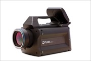
 The versatile FLIR X6580sc camera uniquely ombines high speed and high resolution with ease of use and the flexibility to be configured for just about any scientific or research application. The X6580sc features a high-speed 640x512 digital InSb detector with broadband (1.5-5.5µm) spectral sensitivity and F/3 aperture. The X6580sc provides images up to 350Hz in full frame and up to 4500 Hz in a 320x8 sub-windowing mode....

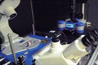 The CBS includes a research group focused on single molecule physics. Dr Pierre-Emmanuel Milhiet runs a team which applies AFM and advanced fluorescence microscopies (single molecule tracking and single-molecule localization microscopy or SMLM) in the study of both structure and dynamics of biological membranes. Speaking about his work, Dr Milhiet says "One of our aims is to decipher the molecular mechanisms involved in the lateral segregation of membrane components using artificial bilayers and intact cell membranes. Part of our activities is also to develop new methodologies and we have recently mounted a new setup combining a JPK AFM and home-made SMLM (especially PALM and STORM)...
 These new versions provide a cost effective way of displaying HD images directly on a monitor without the need for a computer. Cameras are now available in 1920 x 1080 (CVC AN-1080) or 1280 x 720 (CVC AN-072) pixel resolution, both providing 60 frames/s output. The CVC AN-1080 offers the additional benefit of being able to capture images to an SD memory card inserted in the camera back using a wired remote control. The cameras are compatible with C-mount lenses and offer an excellent high definition alternative to standard video cameras...
 École Polytechnique Federale de Lausanne, better known as EPFL, has recently reported on how a group of its scientists have used powerful imaging techniques including nanoIR to support a study which sheds light on photosynthesis. All plants use a form of photosynthesis to produce energy, though not all rely exclusively on it. In higher plants, capturing light takes place in specialized compartments called thylakoids. These are found in cell organelles called chloroplasts, which are the equivalent of a power station for the plant....
 The IRIS spectrograph has begun to observe with unprecedented detail the lowest parts of the sun's atmosphere, known as the interface region. “The quality of images and spectra we are receiving from IRIS is amazing.” said Dr. Alan Title, IRIS principal investigator. IRIS data will allow scientists to study and better understand the energy transport on the sun. The diffraction gratings for the IRIS spectrograph have been produced by HORIBA Jobin Yvon S.A.S.Longjumeau – France...
 The first 3D surgical microscopes from Leica Microsystems with TrueVision 3D technology inside are available to customers. By incorporating the digital smart 3D system inside select models of Leica Microsystems' surgical microscopes, the two companies have eliminated the need for a separate 3D cart. Surgeons can control the 3D recording functions, without interrupting their workflow, via the microscope hand and foot controls, and OR staff will benefit from easier, faster setup and more space to maneuver in the operating room....
 Solentim, the developer of tools for shortening the upstream cell line development workflow, has announced the release of Cell Metric™ rapid, a high speed imaging and analysis system for cell line development. The new product can be used to quickly and reliably screen hundreds of plates from limited dilutions via automated image capture and analysis....
 Over forty international scientists gathered for the 5th Annual Light Sheet Fluorescence Microscopy Workshop in September in Thornwood, New York. The meeting was held at the U.S. headquarters of Carl Zeiss Microscopy, LLC. Researchers presented data and shared ideas regarding instrument development, standards, applications and specific applications of light sheet fluorescence microscopy (LSFM) and related technologies to various biological model organisms such as zebrafish, drosophila, mouse and others...
 Recent advances in nanotechnology have created a novel breed of optically active materials fabricated from highly-structured two-dimensional arrays of noble metal nanoparticles. Now, a team from Rice University in Houston, Texas, has developed a method to manufacture stable, three-dimensional (3-D) nanoclusters that could be used to impart metamaterial optical properties into unstructured substrates such as liquids, glasses and plastics....
 Leica Microsystems continues to extend the capabilities of confocal super-resolution with its new STED platform Leica TCS SP8 STED 3X. The first presentation to the public will be at the annual meetings of the Society for Neuroscience in San Diego, USA, and of the American Society for Cell Biology in New Orleans, USA, during November and December 2013 respectively....

Early detection of breast cancer is one of the best methods to improve survival rate. This can be done in two ways - by hand and by using mammography screening. A mammogram is an X-ray exam of the breasts to look for abnormal changes. The results can go directly into a computer and become a digital image ready to be read and analyzed by a radiologist. ISO has developed a number of International Standards to support digital imaging, in particular ISO 12052:2006, Health informatics – Digital imaging and communication in medicine (DICOM) including workflow and data management...

 from -196°C to 500°C.")
 TalkingPoint allows a Biomedical Scientist (BMS) to efficiently generate a cut-up report using speech recognition (SR) unassisted by a scribe. TalkingPoint also automates LIMS entry, saving a further hour per day.Dean Harmse, Consultant Histopathologist, Derriford Hospital, said:” The TalkingPoint implementation is one part of a complete process change here. SR has been a vital cog in the wheel, complemented by process and procedural developments throughout the pathology department....
 Anasys Instruments is proud to announce the
  XEI is pleased to announce that the company has been granted a new US patent # US8507879B2 which describes a new oxidative cleaning method and system for electron microscopes and other high vacuum instrumentation using UV excitation in an oxygen radical source. The new method and apparatus comprises a vacuum ultraviolet light source housed in an irradiation chamber where it photo dissociates oxygen to be used downstream to clean the chamber or samples in the absence of the UV light....