Quorum Technologies has recently relocated to a new, purpose-built factory and offices in the village of Laughton in East Sussex – an attractive, rural location on the edge of the South Downs National Park, yet with ready access to London, Gatwick airport and continental Europe.
The move has united the specimen preparation division (previously Emitech) and cryo preparation division in a single modern facility shared with Quorum’s sister company UHV Design, although both companies will continue to operate as distinct and separate businesses. “This superb new state-of-the-art facility has been designed and built to meet the specific needs of both companies” explained David Barnbrook, Managing Director of Quorum Technologies. “Built to the very highest design standards....
JPK Instruments, a world-leading manufacturer of nanoanalytic instrumentation for research in life sciences and soft matter, reports on how the Laboratory for Mechanobiology and Bioengineering at Nagoya Institute of Technology in Japan use the NanoWizard®3 AFM to investigate biomaterials.
Professor Shinji Deguchi leads a research group at Nagoya Institute of Technology (NITech). The Laboratory for Mechanobiology & Bioengineering has a multi-disciplinary approach to the growing field of Mechano-Biology. This brings together the areas of cell biology, bioengineering and biophysics. Describing his work, Professor Deguchi said "Every living cell keeps their tension constantly always and pulls surrounding cells each other. It is known as "Tensional Homeostasis." We believe that tension homeostasis is one of the important factors used to control cell function such as proliferation, differentiation and apoptosis. We study important protein complexes which play a part of the tension homeostasis process."..
Porvair Sciences new generation Krystal™ glass bottom plates are precision manufactured using a proprietary process resulting in excellent light transmission and an ultra-flat optical plane (+/- 15µm) making them perfect for imaging growing cells.
The complete range of 24-, 96- and 384-well Krystal glass bottom microplates is available sterilised for tissue culture to optimise cell growth or non-sterile for assay development. Deep black plates for fluorescent studies are available, together with brilliant white plates which increase sensitivity for luminescence and absorbance work. The high optical clarity base of each plate in the range offers the advantage of easy positioning and crisp images when used with confocal microscopes....
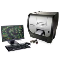
Cytation 3 is a cell imaging multi-mode microplate reader that combines automated digital microscopy and conventional microplate detection.
This unique patent pending design provides rich phenotypic cellular information with well-based quantitative data. The multi-mode microplate detection system features patented Hybrid Technology™, incorporating both high sensitivity filter-based and flexible monochromator based optics for unmatched versatility and performance....
Read MoreVision Source appointed as authorised UK specialist dealer for Sony Medical Division's productsOct 30, 2013
Leading UK digital clinical imaging specialists, Vision Source, announce their appointment as Sony Medical's authorized specialist dealer for their cameras, recorders, monitors & printers.
Vision Source is very pleased to announce their appointment as UK authorized specialist dealer for Sony Medical's range of cameras, recorders, monitors and printers. Further to acquiring assets and the hiring of staff from former dealers, Optivision, Vision Source are committed to a seamless integration of Sony's products into their portfolio offering users old and new continued excellent service and support...
Linkam Scientific Instruments report on the use of their popular THMS600 stage for polymer research at the University of A Coruña (A UDC), Spain.
Founded in 1989, the University of A Coruña (A UDC) promotes the generation, management and dissemination of cultural, scientific, technological and professional development through research and teaching. The University aims to raise levels of welfare for the whole society through the pursuit of social progress, science and technology. Dr. Birgit Bittmann and her team at the University are using the Linkam THMS600 stage to help understand polymer materials....
Leica SR GSD 3D - Highly Precise and Reproducible Localization of Single Molecules and Cellular Structures in Three Dimensions
Super-resolution is a major trend in light microscopy. Extending its portfolio of super-resolution microscopy products, Leica has now launched the Leica SR GSD 3D, a widefield system that not only offers 2D, but now also 3D super-resolution imaging of molecules and cellular structures. Based on GSD (Ground State Depletion) or dSTORM technology, the widefield fluorescence microscope attains resolutions of down to 20 nm in the lateral and 50 nm in the axial direction. The Leica SR GSD 3D also scores with its unique precision for localizing single molecules, its system stability and its optical performance. These attributes are essential for reproducible, high-quality results in a very short time. The benefits of the Leica SR GSD 3D are rounded off by the easy operation of the fully automated system and software...
Cleaver Scientific and Biocenter Collaboration is Appreciated at the University Of Pécs in Hungary
Gel Electrophoresis specialists CLEAVER SCIENTIFIC Ltd (CSL) and their Hungarian distributor Biocenter recently exhibited at and sponsored the 11th International Symposium on Vasoactive Intestinal Peptide (VIP) and Pituitary Adenylate Cyclase-Activating Peptide (PACAP) and related peptides. The event, staged at the University of Pécs in Hungary at the end of August, was an excellent example of the successful collaboration between CSL and their Hungarian distributor...
Andor Zyla high-speed cameras power novel four-lens light-sheet microscope to deliver whole embryo images in under ten seconds.
Understanding embryo development is still one of the most intriguing questions facing today's biologists. Light-sheet microscopy is rapidly gaining recognition for whole organism imaging, allowing cell growth, differentiation and morphogenesis to be studied in detail. However, compared to confocal microscopy, the amount of data generated by this new technique is approximately three thousand times greater and poses new storage and image post-processing challenges...
Leading UK digital clinical imaging specialists, Vision Source, announce their appointment as Panasonic's authorized specialist dealer for imaging products for use in the medical and life science environments.
Vision Source is very pleased to announce their appointment as UK authorized specialist dealer for Panasonic's imaging products from Industrial Medical Vision division. Further to acquiring assets and the hiring of staff from former dealers, Optivision, Vision Source are committed to a seamless integration of Panasonic's products into their portfolio offering users old and new continued excellent service and support....
BioTek’s Cytation™3 Cell Imaging Multi-Mode Reader was recently honored with the first annual New Product Innovation Award, given by MipTec organizers at the 2013 MipTec Conference & Exhibition in Basel, Switzerland. Cytation3 was one of three winning products announced at the exhibition.
Cytation3 combines automated digital microscopy and conventional microplate multi-mode detection to provide both rich phenotypic and well-based quantitative analyses in a streamlined workflow. This unique, patent pending design is ideal for both cell-based and biochemical assays, and its modular architecture allows users to select the modes they need now, and easily upgrade as their needs evolve...
Agar Scientific, a leading supplier of microscopy accessories and consumables, has increased the production of their world-leading filaments for electron microscopy.
As a leading supplier of accessories for microscopy, Agar Scientific has a world-renowned reputation for the manufacture and supply of replacement filaments for many of the world's leading makes of electron microscopes. Now back in production are new and re-filamented, JEOL K Type filaments. Agar filaments are specifically designed for electron microscopes for the majority of the leading manufacturers including Cambridge/LEO/Zeiss, FEI, JEOL and Camscan. The filaments are made with specially designed jigs which ensure accuracy and reproducibility in production...
iXon EMCCD camera's exceptional low light performance opens the door to real-time implementation of atomtronics
Since the first demonstrations of Bose-Einstein condensation in 1995, ultra-cold matter has become a fertile and lively field of study. Research around the world is establishing a high level of understanding of the underlying physics for applications, such as inertial guidance systems, atomic clocks and quantum computing. Teams are also beginning to design and build compact, low-power instrumentation for handling ultra-cold atoms...
The new Olympus FluoView FVMPE-RS, a dedicated multiphoton microscope system, enables high-precision, ultra-fast scanning and stimulation, allowing researchers to see deep within specimens, take measurements at the highest speeds, and capture images, even when working under the most demanding conditions.
With its high speed and precision performance, the FVMPE-RS is designed for electrophysiology and optogenetics studies. It is also a good match for applications such as high-speed calcium and in vivo imaging, peristalsis and blood flow studies, mosaic imaging, connectomics and functional brain imaging, stem cell research, and any field that requires precise colocalisation, uncaging, simultaneous imaging/stimulation, extensive real-time signal processing, or multipoint mapping....
ZEISS has installed 13 systems worth over two million euros at Istanbul Medipol University as well as trained the staff there.
Various light microscopes, as well as stereo and confocal microscopes are in operation at the university's Regenerative and Restorative Medical Research Center now. At the same time, the university agreed to sign a cooperation agreement with ZEISS to hold joint workshops and training, as well as to introduce the new light sheet fluorescence microscope system Lightsheet Z.1 to Turkey...
IDRaman micro optimizes Raman microscopy data collection
Now available from Ocean Optics is the IDRaman micro, a compact microscope designed for Raman measurements in research, quality control and quality assurance environments. The IDRaman micro is a versatile, high-performance analytical tool for applications where sampling requires careful focus and high spatial resolution to optimize the Raman signal. The new instrument debuted September 29, 2013 at SciX 2013, an industry event that covers analytical chemistry, with an emphasis on emerging technologies. SciX, which was held in Milwaukee, Wisconsin, attracts a mix of scientific and engineering experts from around the world...
The software platform for advanced life science research, Leica Appplication Suite Advanced Fluorescence (LAS AF), originally launched in 2005 for Leica Microsystems' widefield and confocal microscope systems, has been released in its 3.2 version. This version focuses on 3D analysis, 3D measurements and 3D visualization. The 3D functionalities are especially interesting for users of Leica Microsystems' widefield super-resolution system Leica SR GSD 3D: They make GSD (Ground State Depletion) data visible as volume rendered images and therefore add a third dimension to super-resolution images. Further features include...
The global reaching partnership aims to provide clinical researchers with new biomarkers and validated image analysis protocols, ensuring a continuum from preclinical to clinical phase in the development of anticancer therapies
Oncodesign,a contract research laboratory dedicated to the discovery of new anticancer therapies, and Banook Central Imaging, a company specializing in the centralized reading of medical images, today announce the start of a partnership to expand the use of new approaches in pharmaco-imaging. The partners expect to share expertise, co-promote joint activities and jointly engage in scientific collaboration to standardize image acquisition and analysis protocols in early clinical phases. The financial details of the agreement were not disclosed...
Artemis CCD Limited, a leading manufacturer of cooled CCD cameras is delighted to announce the supply of OEM grade cooled CCD cameras to NeutronOptics for the application of Neutron imaging. Neutron imaging compliments x-ray radiography, especially for materials opaque to x-rays or where damage may occur. In both cases a "scintillator" film is used to convert the radiation to visible light that can then be imaged with a camera. But unlike x-rays, neutrons penetrate heavy metals, and even lead, to reveal structural details that are otherwise invisible, yet they are strongly scattered by light atoms such as hydrogen...
New Stain-Free Gel Visualisation Application of Syngene’s G:BOX System Helps Save Time and Make Protein Analysis Safer
Scientific Digital Imaging’s [SDI’s] Syngene Division, a world-leading manufacturer of image analysis solutions is delighted to announce that the lighting and filter conditions in the G:BOX image analysis system have been optimised to allow faster, safer visualisation of proteins on stain-free gels. Technical specialists at Syngene tested filter and lighting conditions in a G:BOX XR5 image analysis system to accurately image a range of proteins (2-300ng) run on a Criterion TGX Stain-Free gel. For optimum imaging performance, the gel was exposed to 5 minutes of mid wave UV (302nm) on a UV transilluminator and the gel was imaged using a UV filter and an exposure time of 2 seconds. The gel was then stained with ProtoBlue Safe and imaged again.
As part of the annual EMBO and EMBL events, Olympus will be showcasing the open source microscopy concept, led by the IX3 inverted microscope frames, optimised for life science research.
Demonstrating the very latest in live cell imaging technologies, Olympus will exhibit its range of IX3 inverted microscope frames with associated software and accessories at the upcoming European Molecular Biology Organisation (EMBO) Meeting 2013, taking place in Amsterdam on 21-24 September. Delegates of the “Seeing is Believing –Imaging the Processes of Life” Symposium hosted by EMBO in collaboration with the European Molecular Biology Laboratory (EMBL), which takes place on the 3-6 October in Heidelberg (Germany), will also have the chance to discover how the Olympus open source microscopy concept is advancing live cell imaging...
Scientific Digital Imaging’s [AIM-SDI] Synoptics Health Division, a manufacturer of innovative digital imaging systems for healthcare applications.
Dr Julian Huppert, Member of Parliament for Cambridge, visited Synoptics Health at 2.00 p.m. on Friday 30th August to see the company’s ProReveal Sensitive Protein Detection Test and to discuss how implementing this technology could help save money by reducing the numbers of post-operative hospital acquired infections (HAIs) from dirty surgical instruments...

 The move has united the specimen preparation division (previously Emitech) and cryo preparation division in a single modern facility shared with Quorum’s sister company UHV Design, although both companies will continue to operate as distinct and separate businesses. “This superb new state-of-the-art facility has been designed and built to meet the specific needs of both companies” explained David Barnbrook, Managing Director of Quorum Technologies. “Built to the very highest design standards....

 This unique patent pending design provides rich phenotypic cellular information with well-based quantitative data. The multi-mode microplate detection system features patented Hybrid Technology™, incorporating both high sensitivity filter-based and flexible monochromator based optics for unmatched versatility and performance....


 Gel Electrophoresis specialists CLEAVER SCIENTIFIC Ltd (CSL) and their Hungarian distributor Biocenter recently exhibited at and sponsored the 11th International Symposium on Vasoactive Intestinal Peptide (VIP) and Pituitary Adenylate Cyclase-Activating Peptide (PACAP) and related peptides. The event, staged at the University of Pécs in Hungary at the end of August, was an excellent example of the successful collaboration between CSL and their Hungarian distributor...


 Since the first demonstrations of Bose-Einstein condensation in 1995, ultra-cold matter has become a fertile and lively field of study. Research around the world is establishing a high level of understanding of the underlying physics for applications, such as inertial guidance systems, atomic clocks and quantum computing. Teams are also beginning to design and build compact, low-power instrumentation for handling ultra-cold atoms...
 With its high speed and precision performance, the FVMPE-RS is designed for electrophysiology and optogenetics studies. It is also a good match for applications such as high-speed calcium and in vivo imaging, peristalsis and blood flow studies, mosaic imaging, connectomics and functional brain imaging, stem cell research, and any field that requires precise colocalisation, uncaging, simultaneous imaging/stimulation, extensive real-time signal processing, or multipoint mapping....

 3.2") The software platform for advanced life science research, Leica Appplication Suite Advanced Fluorescence (LAS AF), originally launched in 2005 for Leica Microsystems' widefield and confocal microscope systems, has been released in its 3.2 version. This version focuses on 3D analysis, 3D measurements and 3D visualization. The 3D functionalities are especially interesting for users of Leica Microsystems' widefield super-resolution system Leica SR GSD 3D: They make GSD (Ground State Depletion) data visible as volume rendered images and therefore add a third dimension to super-resolution images. Further features include...
The software platform for advanced life science research, Leica Appplication Suite Advanced Fluorescence (LAS AF), originally launched in 2005 for Leica Microsystems' widefield and confocal microscope systems, has been released in its 3.2 version. This version focuses on 3D analysis, 3D measurements and 3D visualization. The 3D functionalities are especially interesting for users of Leica Microsystems' widefield super-resolution system Leica SR GSD 3D: They make GSD (Ground State Depletion) data visible as volume rendered images and therefore add a third dimension to super-resolution images. Further features include...

 Scientific Digital Imaging’s [SDI’s] Syngene Division, a world-leading manufacturer of image analysis solutions is delighted to announce that the lighting and filter conditions in the G:BOX image analysis system have been optimised to allow faster, safer visualisation of proteins on stain-free gels. Technical specialists at Syngene tested filter and lighting conditions in a G:BOX XR5 image analysis system to accurately image a range of proteins (2-300ng) run on a Criterion TGX Stain-Free gel. For optimum imaging performance, the gel was exposed to 5 minutes of mid wave UV (302nm) on a UV transilluminator and the gel was imaged using a UV filter and an exposure time of 2 seconds. The gel was then stained with ProtoBlue Safe and imaged again.
 Demonstrating the very latest in live cell imaging technologies, Olympus will exhibit its range of IX3 inverted microscope frames with associated software and accessories at the upcoming European Molecular Biology Organisation (EMBO) Meeting 2013, taking place in Amsterdam on 21-24 September. Delegates of the “Seeing is Believing –Imaging the Processes of Life” Symposium hosted by EMBO in collaboration with the European Molecular Biology Laboratory (EMBL), which takes place on the 3-6 October in Heidelberg (Germany), will also have the chance to discover how the Olympus open source microscopy concept is advancing live cell imaging...