Members Login

Channels
Special Offers & Promotions
Pioneering publication reports first ratiometric, single-protein red fluorescent pH sensor in live neurons
 The importance that intracellular pH plays in cell
biology is highlighted in a new paper published by the American Chemical
Society. Written by a group of researchers from Harvard Medical School led by
Professor Gary Yellen, it reports the engineering of the first ratiometric,
single-protein red fluorescent sensor of pH, called pHRed, and its use to image
intracellular pH in live neuronal cells.
The importance that intracellular pH plays in cell
biology is highlighted in a new paper published by the American Chemical
Society. Written by a group of researchers from Harvard Medical School led by
Professor Gary Yellen, it reports the engineering of the first ratiometric,
single-protein red fluorescent sensor of pH, called pHRed, and its use to image
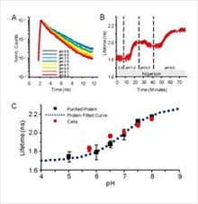
intracellular pH in live neuronal cells.Using Andor's Revolution DSD spinning disk confocal technology, the team also established the unique capability of pHRed to be used in simultaneous measurements of intracellular ATP and pH because of its spectral compatibility with a second ratiometric ATP sensor, GFP-based Perceval. Further, the team showed that pHRed provides a new tool for imaging pH in thick samples and in vivo when imaged by two-photon FLIM (fluorescence lifetime imaging microscopy).
"Sensitivity to pH is a critical problem for GFP-based sensors and can cause severe artefacts if not taken into account," says Dr Yellen. "Our multicolour imaging experiment illustrates the value of pHRed in providing a simultaneous pH signal. This can be used to correct the pH sensitivity of a green sensor and optical sectioning with excitation ratio-imaging enables access to intracellular compartments and organelles.
"We chose the Andor Revolution DSD structured illumination system because of its speed, flexibility and price. This spinning disk system is compatible with a range of non-laser light and filters are user-selectable to suit individual applications. Compared to expensive, maintenance-heavy, laser-based instrumentation, the DSD is ideal for individual laboratories. In eliminating the need to share with other research groups, the DSD allowed us to optimise the experimental set-up and experimental scheduling," he adds.
"This is an important scientific achievement and builds on the increasing interest in genetically encoded fluorescent indicators (GEFIs) for live cell microscopy," according to Dr Mark Browne, Director of Systems at Andor. "The use of the Revolution DSD for simultaneous two parameter ratio excitation imaging in live neurons really highlights the instrument's flexibility. We are delighted that Professor Yellen's group chose Andor as its partner in this work".
DSD offers simple and flexible confocal imaging in a cost-effective add-on to existing upright or inverted microscopes. Capable of delivering high contrast, low background images of fixed and live specimens, it is provides a flexible alternative to laser scanning confocal for individual researchers and small laboratories.
Cellular pH, though often ignored, is a key parameter that affects not only most cellular processes but also the sensing properties of most of the available GEFIs. Because of its red emission colour, pHRed may be used simultaneously with any of the many green or yellow fluorescing GEFIs to correct for their pH sensitivity as well as to give intrinsically interesting information about pH changes.
Andor's microscopy systems address a broad range of optical microscopy techniques including laser spinning disk confocal microscopy, photo-bleaching, activation, conversion and ablation, TIRF, white light spinning disk confocal, calcium ratio imaging, comet assay and bioluminescence. To learn more about the Revolution Differential Spinning Disk (DSD) system and its use in live cell imaging, please visit the microscopy section of the Andor website
Media Partners


