
Channels
Special Offers & Promotions
Application Note: Gemstone Identification Using Raman Microscopy
Introduction
Gemstones are pieces of mineral crystal cut and polished for use in the gem and jewellery industry. The term gemstone covers a wide variety of gems, roughly over 200 types of natural gemstones exist. Gemstones can be separated into two classifications: precious stones, such as sapphires, and semi-precious, such as garnet. The value of these stones depends on their colour, size, quality, and rarity. Gemstones are generally further classified into species based on their chemical composition, and these species can have several varieties. For example, quartz, with the elemental configuration SiO2, has a large list of varieties, depending on impurities such as citrine and amethyst.
The market frequently suffers from imitation gemstones being sold claiming to be more valuable gems. Several techniques can be used to make gemstones of lesser quality appear like their more expensive counterpart. For example, dyes can be added to add the correct colouring to stones and heating can be used to increase clarity. Even with analysis by an experienced jeweller, one cannot always distinguish between real and “fake” gems and therefore additional analytical techniques are required for accurately identifying gemstones and determining quality.
Raman microscopy is an ideal method for analysing gemstones and other geological samples. Its non-destructive nature and lack of sample preparation make it particularly advantageous for analysing all gemstones with no concerns about damage or reducing their value. Additionally, the technique of Raman scattering is highly sensitive to crystalline structures and the presence of minor components within a sample. In this application note an RM5 Raman Microscope is used to identify several gemstones and through Raman mapping highlight how Raman microscopy can be used to study the different components of a sample.
Materials and Methods
An RM5 Raman Microscope equipped with 532 nm and 785 nm lasers for excitation was used. Two laser lines were needed because some samples gave large fluorescence backgrounds at one excitation. Generally, when selecting a laser excitation moving from the 532 nm laser to the 785 nm laser will reduce fluorescence but also reduce Raman scatter.
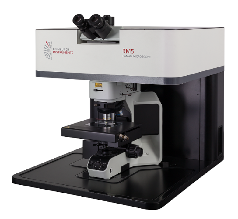
Figure 1: RM5 Raman Microscope
Results and Discussion
Spectral identification
Three popular gemstones: citrine, peridot, and carnelian were analysed as known gemstones. Citrine is a type of quartz and is the most valuable quartz-gem. Citrine is a yellow-coloured gemstone and is associated with November, being commonly used in birthstone jewellery. Although officially the birthstone is topaz, historically citrine was known as gold topaz, when it is actually a different class of gemstone entirely. Citrine is still used as a November birthstone and is a cheaper alternative to topaz. Peridot is a green gemstone, the gem form of olivine, it is a member forsterite-fayalite family, and its green colour can be credited to the amount of iron it contains. Peridot is the birthstone for August and is one of the few gems that forms in only one colour. Carnelian is an orangey gemstone variety of chalcedony. The chalcedony group represents microcrystalline quartz. Carnelian’s colour varies from light pink to a rust brown and is commonly used as a bead in jewellery.
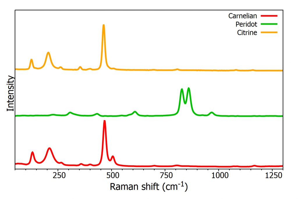
Figure 2: Raman spectra from 3 known gemstones: carnelian, peridot, and citrine
The spectra from each gemstone are shown in Figure 2, their identification confirmed by using the KnowItAll® Raman database. The database contains over 25,000 spectra to aid in sample identification and also allows the user to create their own databases, all Edinburgh Instruments Raman Microscopes come with a one-year free subscription to the package. When a Raman spectrum is entered into the software the result will be a list of potential matches and the percentage match between the measured spectrum and the database spectrum. When the samples spectra were put through the spectral database each one returned a percentage match, with citrine being successfully identified at 97.47%, peridot at 97.78%, and carnelian at 97.23%. These results highlight how Raman spectroscopy combined with databases can be used to confirm the identification of gemstones.
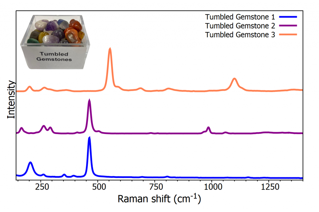
Figure 3: Raman spectra from 3 unknown gemstones
Next, 3 samples were selected from a box of unidentified gemstones for analysis, Figure 3 presents the spectrum from these 3 randomly selected samples. The first sample was determined to be tiger’s eye, with a spectral match of 98.89%. Tiger’s eye is a brown coloured chalcedony gem. Gemstone 2 was identified as nepheline, with a match of 96.07%. Nepheline is a member of the feldspathoid group and is commonly used in glass manufacturing and ceramics. The final tumbled gemstone could be detected as lapis lazuli at 98.27%. Lapis lazuli is a deep blue semi-precious stone and used in jewellery and ornaments for its intense colouring.
Analysing Ornaments and Jewellery
The samples analysed up to this point were purchased in a polished, isolated gemstone form and not truly representative of real samples. Two samples, Figure 4, were analysed by Raman microscopy to test gem identification under more realistic conditions.
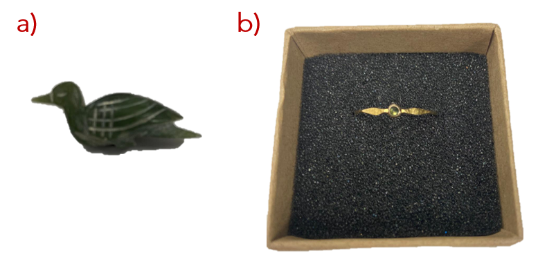
Figure 4: a) Small bird ornament b) ring
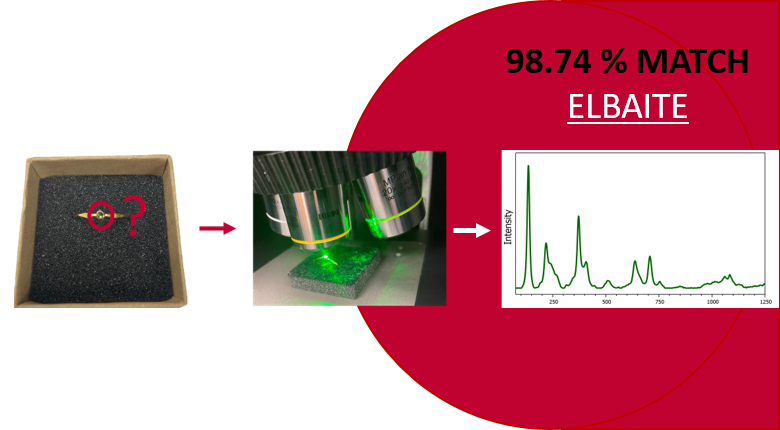
Firstly, a small bird-shaped ornament was analysed and identified, Figure 5a), as edenite, a silicate mineral ranging in colours from white and grey to shades of green. As mentioned, the main use for gemstones is the jewellery industry so a ring containing a small gemstone was placed under the microscope, Figure 5b). The gemstone in the ring was identified as elbaite, a member of the tourmaline group with varying colours and quality.
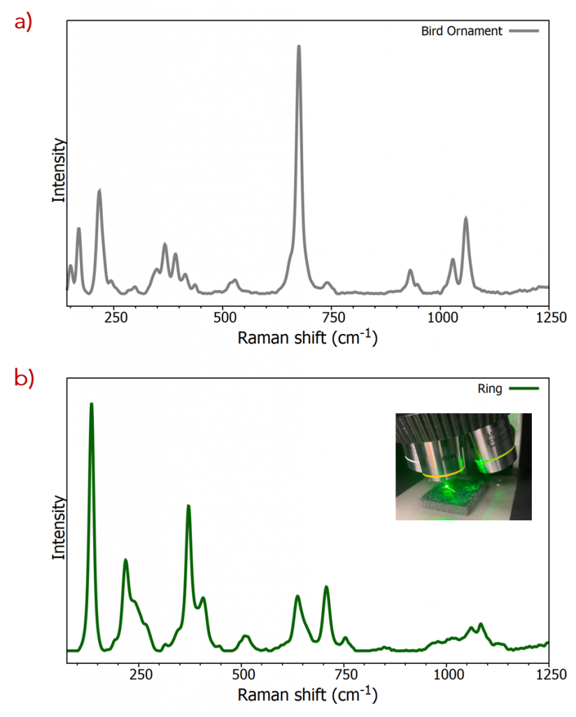
Figure 5: Raman spectrum from a) a small ornament and b) a ring
Imitation
One of the biggest challenges in the gemstone industry is the imitation of expensive gemstones by cheaper counterparts. An example of this is turquoise, turquoise is a blue-green hydrated phosphate of aluminium and copper. It is the birthstone for December and used commonly in both jewellery and ornaments. Turquoise is a rare gemstone due to the precise conditions needed for its formation; dry regions with acidic groundwater rich in cooper which reacts with minerals containing aluminium and phosphorus. The highest quality turquoise is more expensive than diamond, and most turquoise in jewellers will have been treated to enhance the colouring and strength. Due to the demand for the gem and the few areas it can be mined turquoise frequently finds itself a victim of imitation. Mangesite and howlite have been discovered dyed to imitate turquoise, fortunately, their Raman spectra vary significantly, meaning fakes can easily and rapidly be identified.
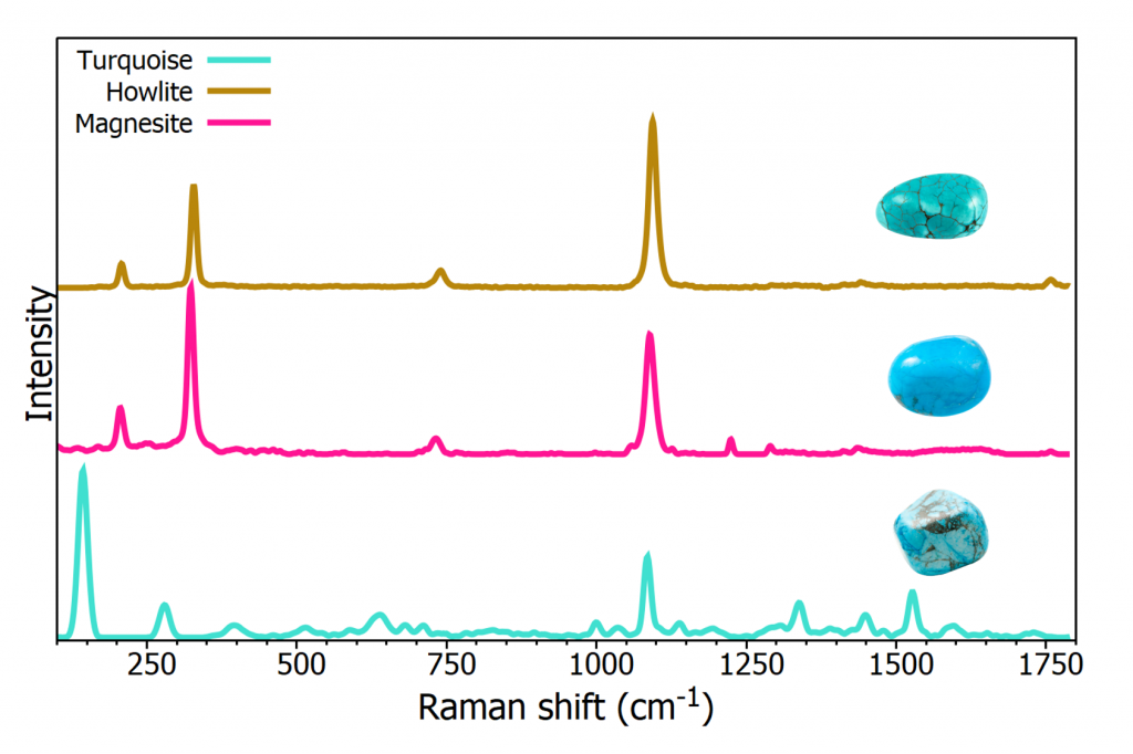
Figure 6: Raman spectrum from known turquoise and 2 imitations
Figure 6 shows the different Raman spectrum from each gemstone along with an image of each gem claiming to be turquoise. Whilst turquoise is a phosphate, magnesite is a magnesium carbonate material and howlite a calcium borosilicate hydroxide, both significantly different in chemical composition to turquoise. This characteristic results in notably different Raman spectra between the imposters and turquoise. This example shows how easily Raman spectroscopy can be used to aid the gemstone industry for identifying imitations.
Raman Mapping
So far, in this application note, Raman microscopy has been used in single point analysis. However, samples will not always simply be one component. In these cases, the entire sample can be Raman mapped (also referred to as Raman imaging). A white light visible image can be taken across the gemstone being analysed, and subsequently the Raman map acquired. The sample analysed here, Figure 7, was described as sodalite. Sodalite is strongly blue coloured tectosilicate mineral largely found in Canada and Greenland. When mining for any mineral there will typically be a mix of minerals and the processing of the sample will determine its purity and quality. In this example Raman mapping identified 3 constituents in the sample.
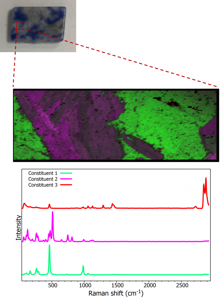
Figure 7: Top – reflected brightfield image from a sample of Sodalite, Middle – Raman image from sample, Bottom – Raman spectrum from the 3 constituents found by Raman imaging
The Raman map is shown in three colours to represent the three constituents that were identified. The green areas represent the sodalite that the gemstone the sample was labelled as. The pink/purple area is orthoclase which is a potassium feldspar material and represents one of the most abundant minerals on Earth. Finally, a small amount of the sample (top right of the Raman map), shown in red, was identified as nepheline.
Conclusion
In this application note the capabilities of the RM5 Raman Microscope for gemstone analysis were demonstrated. Spectral analysis was used to identify unknown minerals and detect the presence of forgeries. Thanks to its non-destructive qualities Raman can be used on fine jewellery to ensure identification without risking damage to the item. Finally, Raman mapping was used to show how one can identify all constituents in a gem sample.
The RM5 Confocal Raman Microscope
The RM5 is a compact and fully automated Raman microscope for analytical and research purposes. The truly confocal design of the RM5 is unique to the market and offers uncompromised spectral resolution, spatial resolution, and sensitivity.
more news from Edinburgh Instruments
News Channels
- Latest News
- New Laboratory Products
- Industry News
- Laboratory Automation | IT Solutions
- Microscopy | Image Analysis
- Separation Science
- Research | Case Studies
- Video Presentations
- Events | Conferences
Subscribe to any of our newsletters for the latest on new laboratory products, industry news, case studies and much more!

Popular this Month
Top 10 most popular articles this month
Today's Picks
Looking for a Supplier?
Search by company or by product
Please note Lab Bulletin does not sell, supply any of the products featured on this website. If you have an enquiry, please use the contact form below the article or company profile and we will send your request to the supplier so that they can contact you directly.
Lab Bulletin is published by newleaf marketing communications ltd.
Media Partners