Channels
Special Offers & Promotions
Anasys
Anasys Instruments reports on EPFL's publication in Plant Cell on the use of nanoIR to look into the process of photosynthesis to shed more light on how plants produce energy
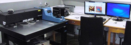
École Polytechnique Federale de Lausanne, better known as EPFL, has recently reported on how a group of its scientists have used powerful imaging techniques including nanoIR to support a study which sheds light on photosynthesis.
All plants use a form of photosynthesis to produce energy, though not all rely exclusively on it. In higher plants, capturing light takes place in specialized compartments called thylakoids. These are found in cell organelles called chloroplasts, which are the equivalent of a power station for the plant. Despite being well-defined from a biochemical perspective, photosynthesis is still a mystery when we consider what happens at the level of the cell. Collaborating in a study published in Plant Cell, EPFL scientists have used a range of microscopy and visualization techniques to understand how the largest photosynthetic pigment-protein antenna complex, known as light-harvesting complex II (LHCII) behave to capture light.
Andrzej Kulik from Giovanni Dietler's group at EPFL, collaborating with Wies?aw Gruszecki at the Maria Curie-Sklodowska University and with researchers at the University of Warsaw compared LHCII-membrane complexes isolated from spinach leaves. The difference lay in the amount of light the complexes had received: One group came from leaves adapted to the dark and the other from leaves previously exposed to high-intensity light. Using X-ray diffraction, nanoscale infrared imaging microscopy*, confocal laser scanning microscopy, and transmission electron microscopy, the researchers found that the dark-adapted LHCII-membranes complexes assembled into rivet-like stacks of bilayers (like a typical chloroplast membranes), while the pre-illuminated complexes formed 3-D forms that were considerably less structured.
The authors conclude that the formation of bilayer, rivet-like structures is crucial in determining how the thylakoid membrane structures itself in response to light exposure. Depending on how much light they receive, the membranes can either stack up on each other or unstack in order to better utilize the energy captured.
* Dr Kulik describes nanoIR as "one of the most important breakthroughs in the AFM technique since it adds chemical composition information to nanoscale morphology. Its ease of use will ensure its wide adoption given the crucial importance of nanoscale chemical composition in most research applications.'
Media Partners


