
 ZEISS presents a new generation of focused ion beam scanning electron microscopes (FIB-SEMs) for high-end applications in research and industry. ZEISS Crossbeam 550 features a significant increase in resolution for imaging and material characterization and a speed gain in sample preparation....
 These awards are announced on the Queen’s birthday, 21April and are one of the UK’s highest accolades recognising business achievement. Last year MR Solutions won the Queen’s Award for Innovation. This has been achieved in recognition of MR Solutions’ outstanding business success with strong export sales growth of 119% over the last three years....
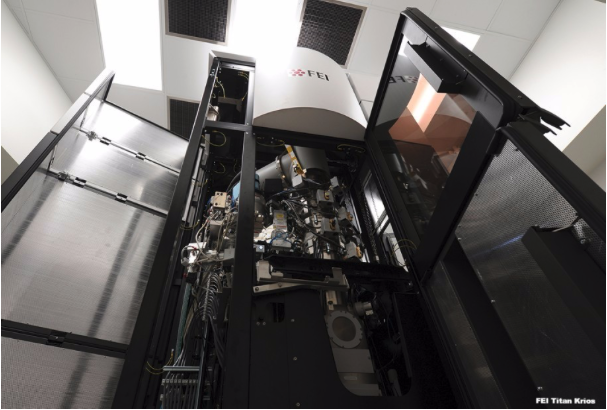 Cryo-electron microscopy (cryo-EM) is a revolutionary technique that reveals in unprecedented detail the molecular and atomic interactions at the foundation of life, allowing scientists to observe exactly how viruses enter cells, how DNA replicates, how chemical compounds assemble into functional biological components, and much more....
 These versatile cameras are available as a single board camera without a lens holder, as a single board camera with an S mount or CS/C mount, or as a housed version with a C/CS mount. With its miniature dimensions – the single board solution measures just 36 x 36 mm – it can be integrated in the tightest of spaces, with applications in instrument manufacturing and microscopy....
 The unique medical research facility, which was built in collaboration with NHS Greater Glasgow and Clyde (NHSGGC) and with £16m funding from the Medical Research Council and Glasgow City Region City Deal, was opened by the Chief Executive Designate of UK Research and Innovation (UKRI), Professor Sir Mark Walport....
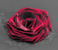 Italian Institute of Technology’s Andrea Jacassi is the grand prize winner of the Sixth Annual 2016 Thermo Fisher Scientific Electron Microscopy image contest for his “Cysteine Rose” image. The image, acquired using the FEI Helios NanoLab 650 DualBeam, focused ion beam/scanning electron microscope (FIB/SEM) and was selected by a vote of Thermo Fisher employees from more than 270 entries...
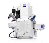 ZEISS presents a new generation of focused ion beam scanning electron microscopes (FIB-SEMs) for high-end applications in research and industry. ZEISS Crossbeam 550 features a significant increase in resolution for imaging and material characterization and a speed gain in sample preparation....
 Comprising a leading conference for biomedical sciences, a significant exhibition and other networking opportunities the Photonex Roadshow, which will be held on the 14th June, brings scientists from across Scotland together for a technology-crammed one-day extravaganza....
 As well as being extremely powerful this new scanner can be multi-modality, incorporating PET and/or SPECT capabilities. MR Solutions’ helium free scanners offer a compact scanner with a small stray field at a very competitive price. SCMR has worldwide attendance amongst top healthcare professionals who are committed to cardiovascular development and their clinical application....
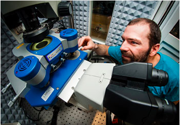 They use a co-located SEM-Raman system from Renishaw to provide comprehensive in situ sample characterisation. Dr Guillaume Wille works in the Mineral Physicochemical and Textural Characterization Unit. In describing the analysis methods used at BRGM, Dr Wille said, “Numerous techniques are used, like infrared spectroscopy, X-ray diffraction, electron microscopy and microanalyses....
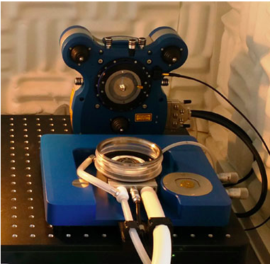 Linkam has been developing cryo stages for correlative microscopies for many years while JPK Instruments is regarded as a leading supplier of nanoscale resolution SPM & Optical Tweezer systems. Bringing their technologies together has enabled JPK to offer cryo-stage capability for their NanoWizard® AFM systems. This means AFM users may now study surface property changes as a function of temperature over the range of -120 °C to +220 °C. Head of Applications at JPK...
 Dr Nic Mullin is a senior experimental officer in the SPM Group of Professor Jamie Hobbs in the Department of Physics & Astronomy at the University of Sheffield. His research is based around instrument development for scanning probe microscopy for the study of soft matter and biological systems at the molecular scale....
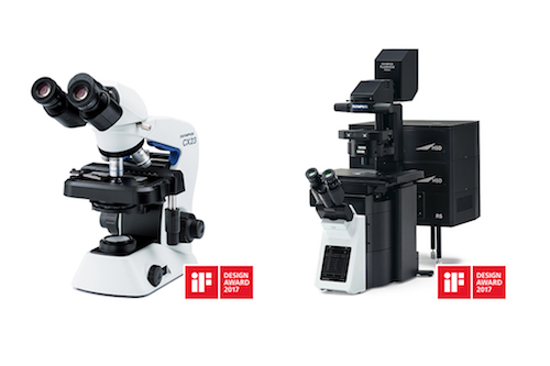 Globally renowned as a symbol of design excellence, the iF design awards celebrate the best in user-focused, ergonomic and efficient design. With over 5,000 submissions from 70 countries, Olympus Scientific Solutions Division has received two of these prestigious awards for its CX23 upright microscope and FLUOVIEW FV3000 confocal laser scanning...
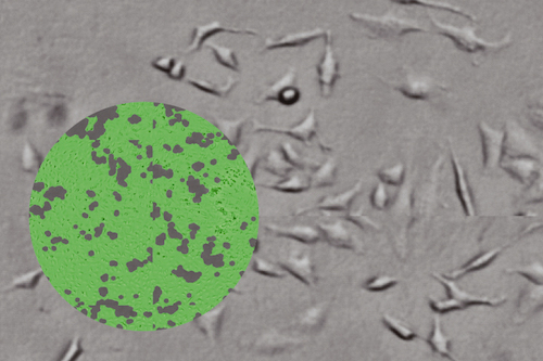 Unlike other multimode readers, Spark lets you actually see what’s happening to your cells, offering automated cell imaging, confluence measurements, cell counting and viability assessments to simplify cell biology protocols and enable long-term, walkaway experiments. Spark has been developed specifically to address the needs of cell-based workflows...
 The ioLight microscope is the first professional quality pocket digital microscope. It is a laboratory grade microscope that fits in a jacket pocket, is simple to use and robust. It unfolds quickly to record and share 5MP still images and real time HD video on a tablet or phone....
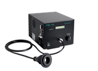 Mercury vapour lamps and bulbs suffer from a number of limiting disadvantages including an operational lifetime rarely exceeding 200 hours and the need for time consuming alignment procedures during installation. In addition Mercury vapour bulbs are susceptible to flickering, limiting the use of these light sources in quantitative fluorescent microscopy....
 Dr Torsten Mueller is a member of the development team with German nanoscience instrument makers, JPK Instruments. Dr Mueller and his colleagues are based in Berlin supporting a worldwide user network which is continuously testing JPK with requests for new capabilities for their life science and materials nanoscience systems....
 One of the research goals of the Molecular Nanoscience Group at ISMO is to work towards circuits and devices in which surface plasmons (not electrons or photons) are used to transfer and manipulate information. However, why develop plasmonics when there are already solutions using electronics and photonics?....