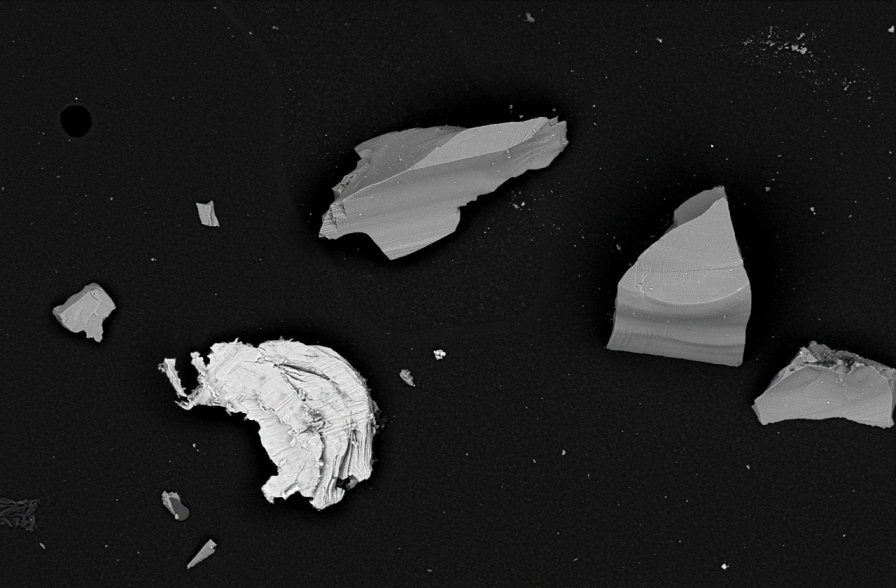
Manufacturers are constantly seeking faster, more effective methods of producing parts. But to achieve the best quality final products, it’s crucial not to rush the technical cleanliness process. Here, Salomé Larmier, electron microscopy specialist at Thermo Fisher Scientific, explains why technical cleanliness is critical and how to achieve it using microscopy techniques...
Biotium, a leading innovator in life science reagents, is proud to support the advancement of super-resolution imaging with the release of their MiniMab™ single-domain antibody (SdAb) series. The MiniMab™ SdAb series, like Nanobodies®, are high-affinity recombinant alpaca VHHs against Glial Fibrillary Acidic Protein (GFAP), Synaptotagmin 1 (SYT1) and Vesicular Glutamate Transporter 1 (VGLUT1)...
In early July, Carl Zeiss Microscopy GmbH has acquired all equity shares of Pi Imaging Technology SA, based in Lausanne, Switzerland. Pi Imaging Technology SA now operates as "Pi Imaging Technology SA – a ZEISS company". The Lausanne location with all employees will be retained. Pi Imaging Technology SA has been a trusted partner of ZEISS Research Microscopy Solutions for many years...
Leica Microsystems, a leading provider of microscopy and scientific instrumentation, has expanded its online shopping experience to the United Kingdom. UK customers will be the first in Europe to enjoy the convenience and personalisation of this digital store. Initially launched in the United States last year, UK customers now have a straightforward way to find the best microscope for their specific needs...
Evident, a global leader in microscopy solutions, announces the launch of the SLIDEVIEW™ DX VS200 universal whole slide imaging scanner, a CE-marked device under the EU IVDR (Regulation (EU) 2017/746) for use in clinical diagnostics in Europe. The SLIDEVIEW DX VS200 scanner delivers high-resolution, high-throughput whole slide scanning for a wide range of pathology applications, accommodating versatile slide types, magnifications and imaging modes...
Leica Biosystems proudly presents the Aperio FL Digital System– versatile digital pathology system designed for both brightfield and fluorescent whole slide imaging. This system combines speed, flexibility, and ease of use, delivering crisp, highresolution images with intuitive one-click operation. Whether you’re managing highthroughput studies or focused research, The Aperio FL Digital System offers a unified solution built for precision and performance...
In case you missed it, Prior Scientific's Product Manager, Simon Bush, recently delivered an informative online presentation titled “Modular Microscopy for Prototyping and Discovery” at the BioPhotonics Microscopy Summit. This session showcased how Prior's innovative OpenStand microscopy platform and our adaptable automation components can play a vital role in today's microscopy landscape...
Thermo Fisher Scientific Inc., the world leader in serving science, has launched the spectral-enabled Invitrogen™ Attune™ Xenith™ Flow Cytometer, allowing immunology and immuno-oncology researchers to automate and streamline workflows to obtain more detailed and accurate insights from critical cellular samples. By leveraging Thermo Fisher’s legacy core acoustic focusing technology, this new solution offers improved time to results for scientists researching cellular behaviors and mechanisms and discovering targeted therapies..
TESCAN Group, a global manufacturer of electron microscopes and advanced scientific instruments, announces a new collaboration with The University of British Columbia (UBC). UBC’s partnership with TESCAN USA will provide researchers with cutting-edge MIRA, TENSOR, and AMBER electron microscopes, bringing together world-class research and innovative technology to drive innovation in microscopy and materials characterization...
Leica Microsystems, a leading provider of microscopy and scientific instrumentation, has released the latest version of its AI-driven image analysis software. Aivia 15 empowers scientists to set up quickly, deploy intuitive AI-powered analysis for accurate detection, and then easily batch process their analyses...
Evident announces the release of two new spinning disk confocal microscopes, the IXplore™ IX85 SpinXL and the IXplore™ IX85 SpinSR. Built on Evident’s new IXplore IX85 automated inverted microscope system, the IX85 SpinXL and IX85 SpinSR add powerful new technology to the platform, expanding the possibilities of what researchers can capture and discover in live-cell imaging...
TESCAN GROUP introduces the MIRA XR, an ultra-high-resolution SEM-EDS solution designed for fast, precise materials analysis in academic research and quality control environments. Designed to bridge the gap between conventional FEG SEMs and ultra-high-resolution (UHR) systems, MIRA XR offers analytical flexibility, ease of use, and streamlined workflows for both industrial and research applications...
Leica Microsystems, a leading provider of microscopy and scientific instrumentation, has launched the Visoria series of upright microscopes, to help enhance the efficiency and comfort of routine microscopy work for pathologists, quality control managers, and researchers. There are three distinct microscope models in the Visoria series, each designed to meet the needs of users for different applications in various fields..
ZEISS and Alpenglow Biosciences have announced a new partnership to jointly develop an inverted light-sheet microscope and bioinformatics pipeline tailored specifically for clinical applications. Alpenglow, a pioneering 3D spatial biology company specializing in open-top light-sheet microscopy, data processing, and AI-driven analysis for biopharma and clinical applications, has partnered with ZEISS, a global leader in light-sheet imaging technologies and visualization software – including ZEISS Lattice Lightsheet 7 and arivis software...
Zeiss simplify high-throughput multiplex IF imaging and analysis, making it robust, scalable and accessible. Our seamless tissue multiplexing workflow aims to empower routine histopathology labs, including those with no prior spatial biology expertise, to generate comprehensive, reproducible biomarker data across large sample cohorts. By combining automation, optimized imaging, and AI-powered analysis...
Revvity Inc. has unveiled the VivoJect™ Image-Guided Injection System as part of its distinguished cancer research and discovery portfolio at the AACR Annual Meeting 2025 in Chicago. Paired with the Vega™ automated preclinical ultrasound system, the VivoJect system allows for real-time imaging and precise, nimble operation for researchers at a higher throughput compared to traditional techniques...
Bruker Corporation has announced the launch of the nVista 2P miniature, two-photon microscope, a groundbreaking addition to the Inscopix product line for functional imaging of freely behaving animals. It leverages advanced two-photon fluorescence microscopy technology to enable high-resolution imaging at greater depths with 3D data reconstruction, providing researchers with unprecedented insights into neuronal activity...
Onboard the International Space Station (ISS), the Extant Life Volumetric Imaging System, dubbed ELVIS, is not about resurrecting rock-n-roll legends but pioneering scientific discovery. Using innovative holographic technology to deliver detailed 3D views of cells and microbes, the system allows scientists to study the adaptability and resilience of life under extreme conditions. Knowledge gained could reveal how life might persist on distant moons and planets, significantly enhancing our search for life outside Earth...
The NanoScan OP200 piezo objective positioner has been added to Queensgate's already successful NanoScan OP series product line. The NanoScan OP200 provides class-leading step settle performance over a range of up to 200 µm. With user-configurable parameters to accommodate varying objective sizes and weights as well as performance requirements, it works with the majority of microscope types and objective lenses...
In the rapidly evolving field of structural biology, single particle analysis (SPA) and cryo-electron tomography (cryo-ET) are powerful techniques that allow scientists to better understand the intricacies of biology, providing atomic-level insights that reveal how viruses, proteins and cells work. The Krios 5 Cryo-TEM optimizes productivity and performance to enhance these techniques...
ZEISS announces the introduction of its highly flexible and efficient software suite, ZEN core, for operating all ZEISS scanning electron microscopes (SEMs), including focused ion beam scanning electron microscopes (FIB-SEMs). ZEN stands for ZEISS Efficient Navigation and lets microscopists obtain more meaningful information. Users across various fields – including materials research, natural resources, electronics, and life sciences...
New alliance for advanced biological imaging aimed at enhancing some of the world's most innovative structural cell biology capabilities to help scientists answer pressing research questions. Thermo Fisher Scientific Inc., the world leader in serving science, has announced a Technology Alliance Agreement with the Chan Zuckerberg Institute for Advanced Biological Imaging (CZ Imaging Institute). This agreement aims to develop new technologies to enable researchers to better visualize human cells, significantly advancing scientific research and discovery...

 Manufacturers are constantly seeking faster, more effective methods of producing parts. But to achieve the best quality final products, it’s crucial not to rush the technical cleanliness process. Here, Salomé Larmier, electron microscopy specialist at Thermo Fisher Scientific, explains why technical cleanliness is critical and how to achieve it using microscopy techniques...
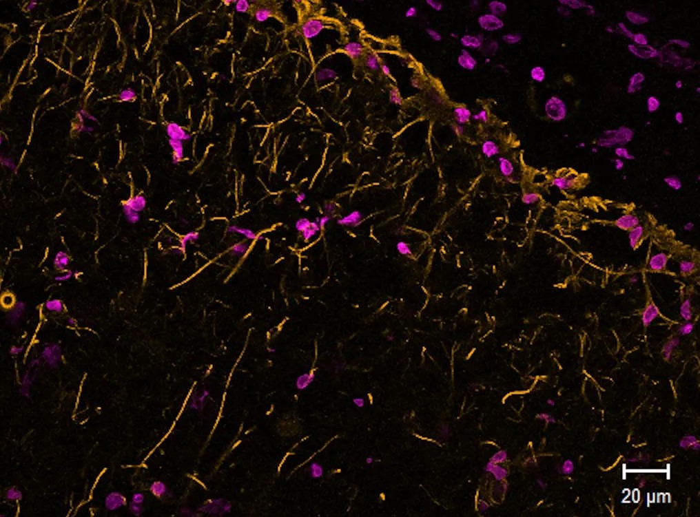 Biotium, a leading innovator in life science reagents, is proud to support the advancement of super-resolution imaging with the release of their MiniMab™ single-domain antibody (SdAb) series. The MiniMab™ SdAb series, like Nanobodies®, are high-affinity recombinant alpaca VHHs against Glial Fibrillary Acidic Protein (GFAP), Synaptotagmin 1 (SYT1) and Vesicular Glutamate Transporter 1 (VGLUT1)...
 In early July, Carl Zeiss Microscopy GmbH has acquired all equity shares of Pi Imaging Technology SA, based in Lausanne, Switzerland. Pi Imaging Technology SA now operates as "Pi Imaging Technology SA – a ZEISS company". The Lausanne location with all employees will be retained. Pi Imaging Technology SA has been a trusted partner of ZEISS Research Microscopy Solutions for many years...
 Leica Microsystems, a leading provider of microscopy and scientific instrumentation, has expanded its online shopping experience to the United Kingdom. UK customers will be the first in Europe to enjoy the convenience and personalisation of this digital store. Initially launched in the United States last year, UK customers now have a straightforward way to find the best microscope for their specific needs...
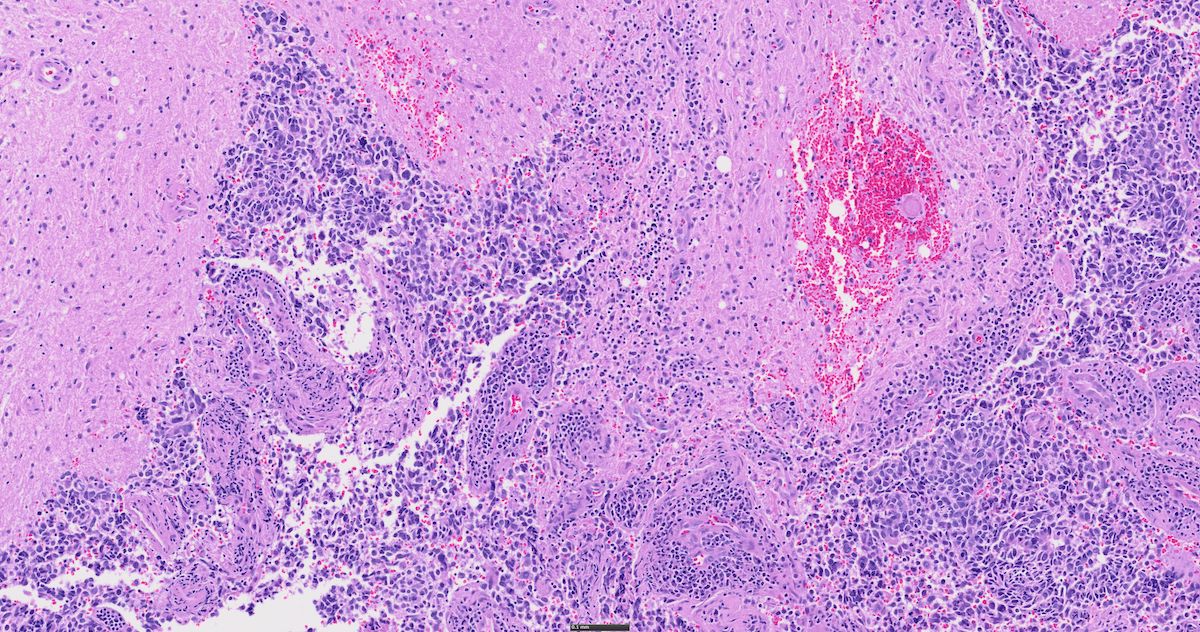 Evident, a global leader in microscopy solutions, announces the launch of the SLIDEVIEW™ DX VS200 universal whole slide imaging scanner, a CE-marked device under the EU IVDR (Regulation (EU) 2017/746) for use in clinical diagnostics in Europe. The SLIDEVIEW DX VS200 scanner delivers high-resolution, high-throughput whole slide scanning for a wide range of pathology applications, accommodating versatile slide types, magnifications and imaging modes...
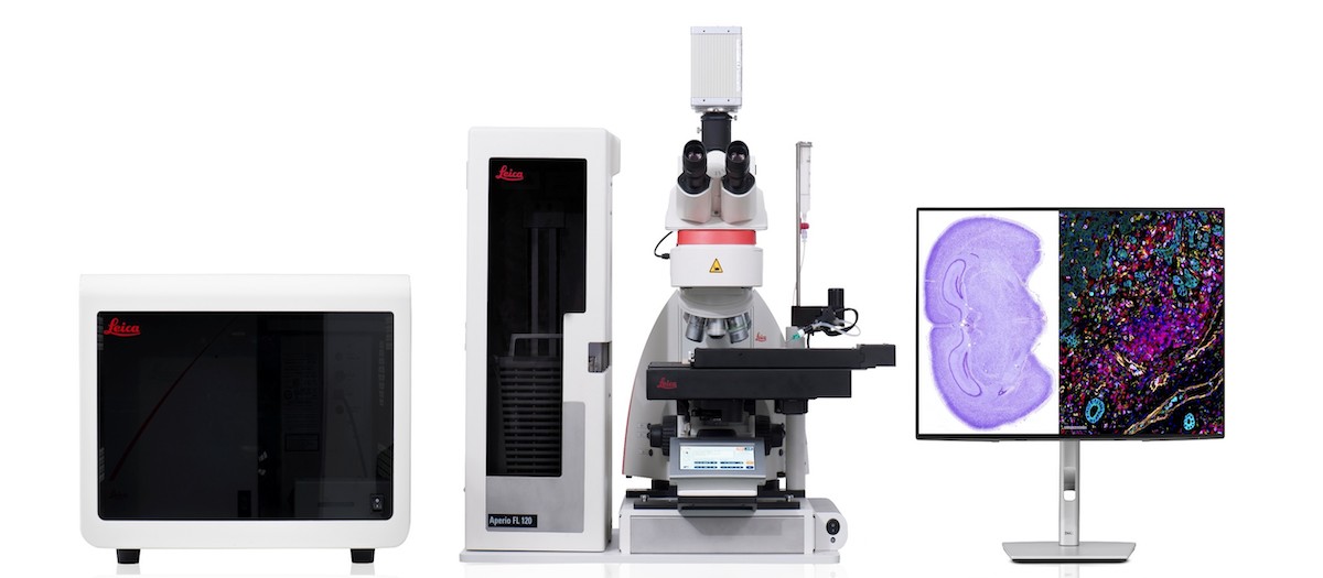 Leica Biosystems proudly presents the Aperio FL Digital System– versatile digital pathology system designed for both brightfield and fluorescent whole slide imaging. This system combines speed, flexibility, and ease of use, delivering crisp, highresolution images with intuitive one-click operation. Whether you’re managing highthroughput studies or focused research, The Aperio FL Digital System offers a unified solution built for precision and performance...
 In case you missed it, Prior Scientific's Product Manager, Simon Bush, recently delivered an informative online presentation titled “Modular Microscopy for Prototyping and Discovery” at the BioPhotonics Microscopy Summit. This session showcased how Prior's innovative OpenStand microscopy platform and our adaptable automation components can play a vital role in today's microscopy landscape...
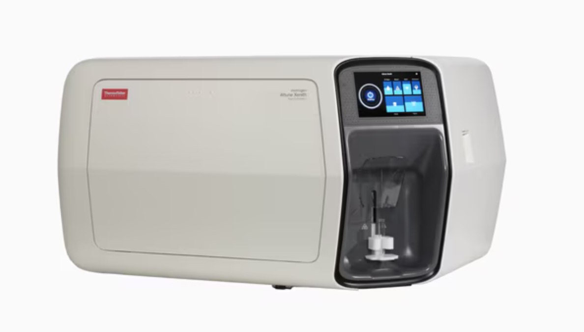 Thermo Fisher Scientific Inc., the world leader in serving science, has launched the spectral-enabled Invitrogen™ Attune™ Xenith™ Flow Cytometer, allowing immunology and immuno-oncology researchers to automate and streamline workflows to obtain more detailed and accurate insights from critical cellular samples. By leveraging Thermo Fisher’s legacy core acoustic focusing technology, this new solution offers improved time to results for scientists researching cellular behaviors and mechanisms and discovering targeted therapies..
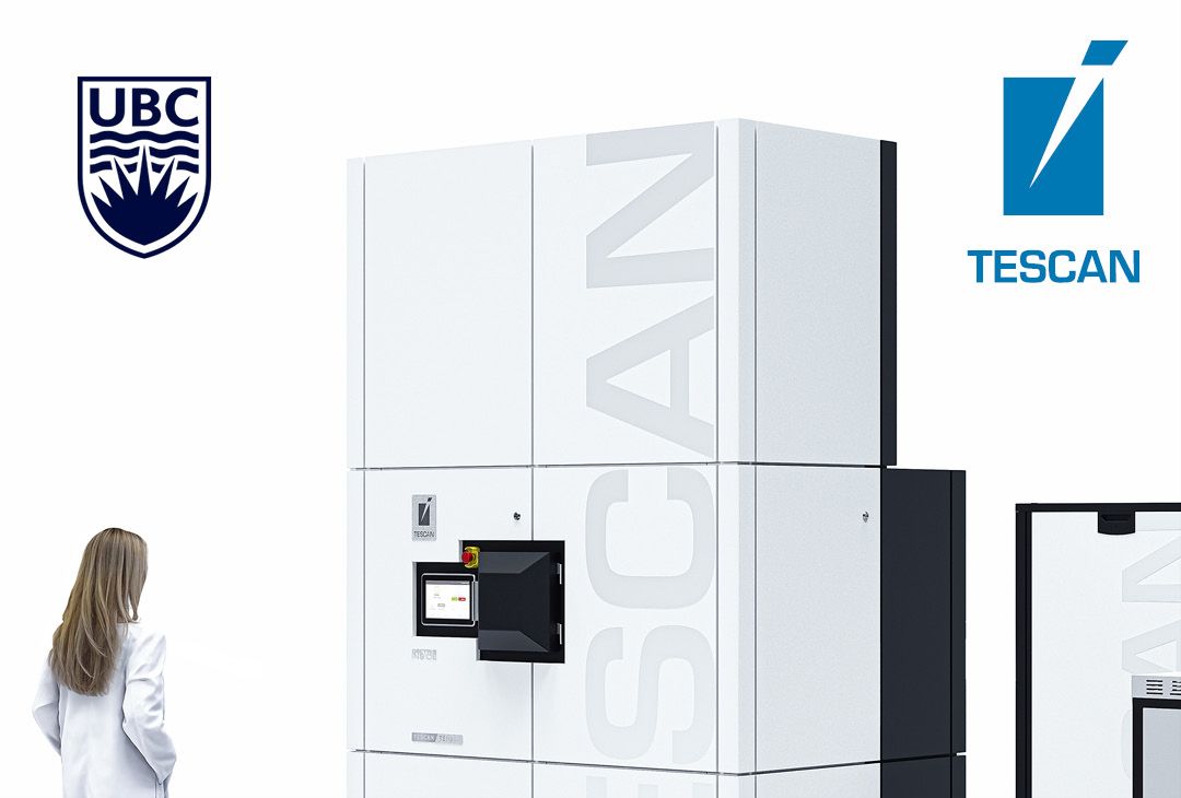 TESCAN Group, a global manufacturer of electron microscopes and advanced scientific instruments, announces a new collaboration with The University of British Columbia (UBC). UBC’s partnership with TESCAN USA will provide researchers with cutting-edge MIRA, TENSOR, and AMBER electron microscopes, bringing together world-class research and innovative technology to drive innovation in microscopy and materials characterization...
 Leica Microsystems, a leading provider of microscopy and scientific instrumentation, has released the latest version of its AI-driven image analysis software. Aivia 15 empowers scientists to set up quickly, deploy intuitive AI-powered analysis for accurate detection, and then easily batch process their analyses...
 Evident announces the release of two new spinning disk confocal microscopes, the IXplore™ IX85 SpinXL and the IXplore™ IX85 SpinSR. Built on Evident’s new IXplore IX85 automated inverted microscope system, the IX85 SpinXL and IX85 SpinSR add powerful new technology to the platform, expanding the possibilities of what researchers can capture and discover in live-cell imaging...
 TESCAN GROUP introduces the MIRA XR, an ultra-high-resolution SEM-EDS solution designed for fast, precise materials analysis in academic research and quality control environments. Designed to bridge the gap between conventional FEG SEMs and ultra-high-resolution (UHR) systems, MIRA XR offers analytical flexibility, ease of use, and streamlined workflows for both industrial and research applications...
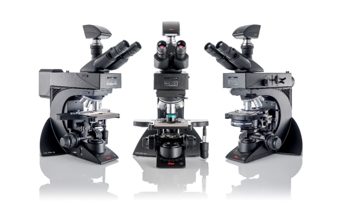 Leica Microsystems, a leading provider of microscopy and scientific instrumentation, has launched the Visoria series of upright microscopes, to help enhance the efficiency and comfort of routine microscopy work for pathologists, quality control managers, and researchers. There are three distinct microscope models in the Visoria series, each designed to meet the needs of users for different applications in various fields..
 ZEISS and Alpenglow Biosciences have announced a new partnership to jointly develop an inverted light-sheet microscope and bioinformatics pipeline tailored specifically for clinical applications. Alpenglow, a pioneering 3D spatial biology company specializing in open-top light-sheet microscopy, data processing, and AI-driven analysis for biopharma and clinical applications, has partnered with ZEISS, a global leader in light-sheet imaging technologies and visualization software – including ZEISS Lattice Lightsheet 7 and arivis software...
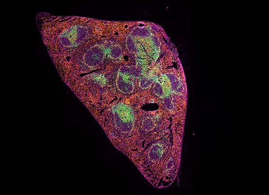 Zeiss simplify high-throughput multiplex IF imaging and analysis, making it robust, scalable and accessible. Our seamless tissue multiplexing workflow aims to empower routine histopathology labs, including those with no prior spatial biology expertise, to generate comprehensive, reproducible biomarker data across large sample cohorts. By combining automation, optimized imaging, and AI-powered analysis...
 Revvity Inc. has unveiled the VivoJect™ Image-Guided Injection System as part of its distinguished cancer research and discovery portfolio at the AACR Annual Meeting 2025 in Chicago. Paired with the Vega™ automated preclinical ultrasound system, the VivoJect system allows for real-time imaging and precise, nimble operation for researchers at a higher throughput compared to traditional techniques...
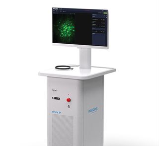 Bruker Corporation has announced the launch of the nVista 2P miniature, two-photon microscope, a groundbreaking addition to the Inscopix product line for functional imaging of freely behaving animals. It leverages advanced two-photon fluorescence microscopy technology to enable high-resolution imaging at greater depths with 3D data reconstruction, providing researchers with unprecedented insights into neuronal activity...
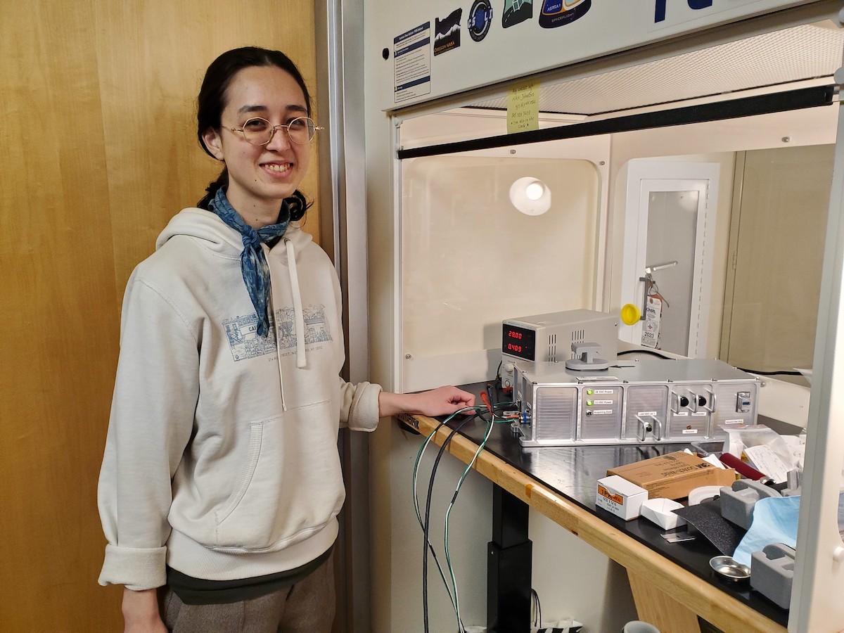 Onboard the International Space Station (ISS), the Extant Life Volumetric Imaging System, dubbed ELVIS, is not about resurrecting rock-n-roll legends but pioneering scientific discovery. Using innovative holographic technology to deliver detailed 3D views of cells and microbes, the system allows scientists to study the adaptability and resilience of life under extreme conditions. Knowledge gained could reveal how life might persist on distant moons and planets, significantly enhancing our search for life outside Earth...
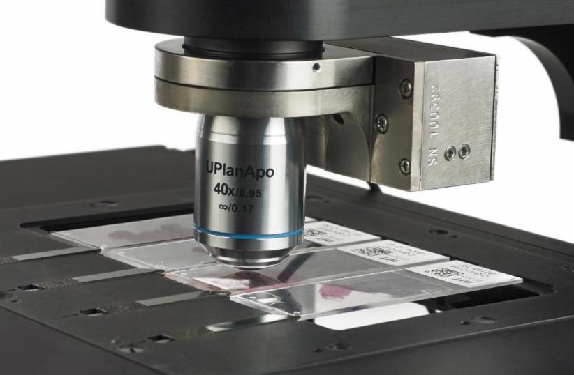 The NanoScan OP200 piezo objective positioner has been added to Queensgate's already successful NanoScan OP series product line. The NanoScan OP200 provides class-leading step settle performance over a range of up to 200 µm. With user-configurable parameters to accommodate varying objective sizes and weights as well as performance requirements, it works with the majority of microscope types and objective lenses...
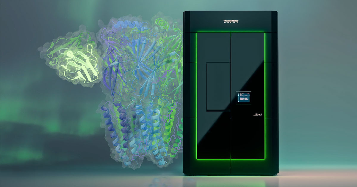 In the rapidly evolving field of structural biology, single particle analysis (SPA) and cryo-electron tomography (cryo-ET) are powerful techniques that allow scientists to better understand the intricacies of biology, providing atomic-level insights that reveal how viruses, proteins and cells work. The Krios 5 Cryo-TEM optimizes productivity and performance to enhance these techniques...
 ZEISS announces the introduction of its highly flexible and efficient software suite, ZEN core, for operating all ZEISS scanning electron microscopes (SEMs), including focused ion beam scanning electron microscopes (FIB-SEMs). ZEN stands for ZEISS Efficient Navigation and lets microscopists obtain more meaningful information. Users across various fields – including materials research, natural resources, electronics, and life sciences...