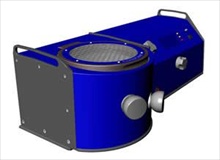
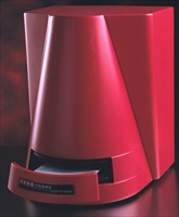
Carestream Introduces In-Vivo Xtreme - a Powerful New Multimodal Optical and X-ray Small Animal Imaging SystemSep 9, 2011Carestream Molecular Imaging is pleased to announce the In-Vivo Xtreme, the newest addition to their award-winning* family of multimodal imaging products dedicated to preclinical research. Xtreme will be introduced at the World Molecular Imaging Congress in San Diego, CA, September 7-10...
Read MoreAgar Scientific announces new products from the SIMPore range of precision membrane accessories for electron microscopistsSep 8, 2011Agar Scientific, a leading supplier of microscopy accessories and consumables, announces new products from the SIMPore range of precision membranes for electron microscopists. Agar Scientific is a market leader in the supply of high quality accessories to assist with sample preparation for electron microscopy. As applications become more challenging, the sample preparation process has to be rapid, accurate, consistent and reproducible...
Read MoreCCD System is up to 10 Times Faster than Laser Scanner for IR Image Analysis New Study Shows G:BOX Chemi IR6 Rapidly Captures Western Blot ImagesSep 8, 2011Syngene, a world-leading manufacturer of image analysis solutions, today announced the results of a head-to-head study carried out at the University of Cambridge to demonstrate that the G:BOX Chemi IR6 can produce images of IR labelled Western blots up to 10 times faster than laser based scanning...
Read MoreFast, fluid and natural microscopy using your computer monitorSep 8, 2011Olympus has released the new DP26, a five megapixel colour digital camera optimised for viewing, documentation, reporting and analysis using your microscope. The new camera is ideal for browsing a sample using the monitor, utilising a rapid progressive scan readout that provides fluid, natural panning and focusing, and avoids distracting artefacts. This removes the physical strain imposed by using the eyepieces, improving user comfort and efficiency...
Read MoreXEI Scientific launches Evactron CombiClean at M&M 2011Sep 7, 2011XEI Scientific Inc, manufacturers of more than 1,100 EVACTRON® De-Contaminator Plasma Cleaning Systems for electron microscopes and other vacuum chambers, announces the release of their new Evactron® CombiCleanTM system which simplifies the control and operation of plasma radical sources for both column and desktop cleaning of specimens for electron columns used in SEMs, TEMs and FIBs...
Read MoreNew Widefield Super-Resolution System for Applications in Biomedical ResearchSep 7, 2011With the new Leica SR GSD from Leica Microsystems, scientists can now achieve resolutions far below the limit of diffraction that have never been attained before in widefield fluorescence microscopy. The system is capable of resolving details as small as 20 nanometers. This enables research of structures of single proteins and other biomolecules in cells and observation of molecular processes to gain new insights into fundamental processes of life...
Read MoreAnasys Instruments receives Microscopy TodaySep 6, 2011Anasys Instruments' AFM-IR system has been recognized by Microscopy Today in the receipt of the 2011 Innovation Award. It was presented to CEO, Roshan Shetty, at the 2011 M&M Annual conference held this year in Nashville, TN. The AFM-IR technique was developed by Dr. Alexandre Dazzi at the University of Paris-Sud. It uses an AFM probe as the IR absorbance detector and hence obtains IR spectroscopy at up to 2 orders of magnitude better than traditional IR spectroscopy...
Read MoreJPK announces the 10th annual International Meeting on the application of SPM & Optical Tweezers for Life SciencesSep 6, 2011JPK Instruments are happy to announce that registration is now open for the tenth annual international symposium on the applications of scanning probe microscopy (SPM) and optical tweezers. The symposia will be held on the 5-6th October 2011 in Berlin focusing on applications developments in life sciences. These meetings continue to be highly regarded on the international life sciences meetings calendar. More than 100 scientists from around the world are expected to come to Berlin to discuss their results and share scientific knowledge in a relaxed and informal atmosphere...
Read MoreThe solution to high quality fluorescent image deconvolutionSep 5, 2011Olympus' new BX3 microscope systems are designed with the user in mind to maximise the quality of fluorescence images captured as part of life science research projects. The high-end BX63 system comfortably handles day-to-day applications, while boasting a range of advanced features to satisfy the most complex of needs, including rapid deconvolution of fluorescent Z-stack images...
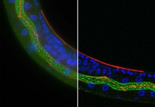
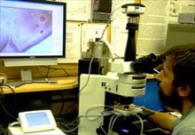
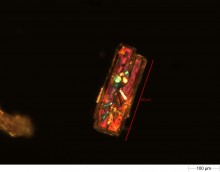
Read MoreDetecting a Mouse Pancreatic Cancer Cell Line Using iBox Explorer Imaging MicroscopeAug 24, 2011In vivo imaging of small animals for pre-clinical research using fluorescent markers is a novel approach to visualize cellular processes occurring at the micron level. There is growing interest in understanding what occurs at the level of tumor metastasis through direct visualization...
Read MoreThermal Imaging of High Speed EventsAug 19, 2011The FLIR SC7000 Series of Infrared Cameras from FLIR Advanced Thermal Solutions (ATS) provide the high performance and facilities for thermal imaging of high speed events. These high-speed, high-resolution research grade cameras provide Gigabit Ethernet, Camera Link and USB interfaces for maximum flexibility and performance. They also feature simultaneous digital and analogue outputs, and are available in multiple wavebands, detector resolutions, and lens configurations...
Read MoreJPK Instruments extends the European sales team with two new members in GermanyAug 19, 2011JPK Instruments, a world-leading manufacturer of nanoanalytic instrumentation for research in life sciences and soft matter is pleased to announce the appointment of Thomas Neicke and Dr. Elmar Hartmann as new members of the sales team...
Read MoreThe Olympus VS120 virtual slide system for digitising slides at high magnification Aug 9, 2011Olympus has released the latest version of its virtual slide (VS) product range, the VS120. The system can rapidly scan a slide in less than two minutes, creating a digital ‘virtual slide' that is an exact copy of the original. This is easily created and navigated using an intuitive graphical interface. The new unit provides semi-automatic and manual focusing modes for working with challenging samples, enhanced image clarity using the new Sample Detection Mask for automatic slide scanning, and increased optimisation of user workflow....
Read MoreCarl Zeiss Obtains License from University of California for Illumination TechniqueAug 9, 2011Carl Zeiss has received a license from the University of California in San Francisco (UCSF) for the commercialization of "Multidirectional Selective Plane Illumination Microscopy" (mSPIM), an advanced illumination technique for light sheet fluorescence microscopy...
Read MoreChina Medical Technologies and Leica Microsystems Announce CollaborationAug 9, 2011China Medical Technologies, Inc. (CMED) (Nasdaq: CMED), a leading China-based advanced in-vitro diagnostic ("IVD") company, and Leica Biosystems, a division of Leica Microsystems, a world leader in microscopes and scientific instruments, today announced that they have established a sales, research and development collaboration to co-develop and market automated FISH kits to be used on the Leica BOND system. CMED will sell the Automated FISH Kits in China and Leica will have an option to sell the FISH kits in the rest of the world...
Read MoreCambridge Technology Company ionscope helps researchers make heart discoveryAug 9, 2011SICM, a high resolution microscopy technique, has been used by a team of researchers to identify a drug that could one day be used to prevent abnormal heart rhythms, or arrhythmia. The study, published in Hepatology[1] this week, was enabled by SICM, which is the result of 7 years of research and development by ionscope Ltd. The global market for SICM equipment is supplied using patented technology from ionscope's offices in Cambridge, UK...
Read MoreZ Polarizer: a Unique Converter Presents New AdvantagesAug 8, 2011A direct laser writing technique for production of radial polarization converter also called as Z polarizer was developed by Workshop of Photonnics in cooperation with prof. Peter G. Kazansky.
Read MoreNEW ChemiFast Substrate from Syngene Offers Higher Sensitivity and Long Signal Life with Chemiluminescent BlotsAug 8, 2011Syngene, a world-leading manufacturer of image analysis solutions is pleased to introduce ChemiFast, a new chemiluminescence substrate specifically designed for use with all Syngene's chemiluminescence imaging systems. ChemiFast is highly sensitive and produces an enhanced signal that is stable for up to 24 hours, making it ideal for detecting even faint protein bands on Western blots...
Read MoreLinkam TS1400 high temperature stage selected by the Bodnar Group at Virginia Tech to study geological specimensAug 8, 2011Market leaders in temperature controlled microscopy, Linkam Scientific Instruments report the use of their 1400°C high temperature stage to study silicate melt inclusions in the Fluids Research Group of the Geosciences Department at Virginia Tech. Researchers in the Fluids Research Group at Virginia Tech are concerned with the distribution, properties and role of fluids in and on the Earth, from its surface (shallow Earth's crust) to its deep interior (Earth's mantle)...
Read MoreNew software for MalvernAug 3, 2011Malvern Instruments has released a new version of software for the company's Morphologi G3 particle characterization system. This fully automated image analysis-based system delivers particle size and shape information for wet and dry samples, and for the analysis of foreign particulate matter collected on filters. Among the new applications in the version 7.40 software are facilities for user-defined light settings and automated dark-field measurements...
Read MoreDigital Cameras Ideal for All Contrast Methods in Light MicroscopyAug 2, 2011Warner Instruments is pleased to introduce its NEW line of ProgRes® Digital CMOS and CCD Microscope Cameras. These cameras are the result of more than two decades experience in the development and production of high-end solutions for digital imaging. The highly versatile and cost effective ProgRes® CMOS Camera range allows for quick and precise setting of both specimen and microscope...
Read MoreGlass Bottom Microplates for Imaging ApplicationsJul 29, 2011Precision manufactured to be extremely flat (+/- 15 microns across base) - KrystalTM glass bottom plates from Porvair Sciences provide unmatched performance for whole-plate CCD imaging and laser detection applications. Available in a choice of 24-, 96- and 384-well formats - Krystal glass bottom plates combine the advantageous optical properties of glass, low background and low birefringence, with the versatility of a microplate...
Read More

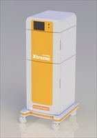 Carestream Molecular Imaging is pleased to announce the In-Vivo Xtreme, the newest addition to their award-winning* family of multimodal imaging products dedicated to preclinical research. Xtreme will be introduced at the World Molecular Imaging Congress in San Diego, CA, September 7-10...
 Agar Scientific, a leading supplier of microscopy accessories and consumables, announces new products from the SIMPore range of precision membranes for electron microscopists. Agar Scientific is a market leader in the supply of high quality accessories to assist with sample preparation for electron microscopy. As applications become more challenging, the sample preparation process has to be rapid, accurate, consistent and reproducible...
 Syngene, a world-leading manufacturer of image analysis solutions, today announced the results of a head-to-head study carried out at the University of Cambridge to demonstrate that the G:BOX Chemi IR6 can produce images of IR labelled Western blots up to 10 times faster than laser based scanning...
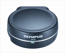 Olympus has released the new DP26, a five megapixel colour digital camera optimised for viewing, documentation, reporting and analysis using your microscope. The new camera is ideal for browsing a sample using the monitor, utilising a rapid progressive scan readout that provides fluid, natural panning and focusing, and avoids distracting artefacts. This removes the physical strain imposed by using the eyepieces, improving user comfort and efficiency...
 XEI Scientific Inc, manufacturers of more than 1,100 EVACTRON® De-Contaminator Plasma Cleaning Systems for electron microscopes and other vacuum chambers, announces the release of their new Evactron® CombiCleanTM system which simplifies the control and operation of plasma radical sources for both column and desktop cleaning of specimens for electron columns used in SEMs, TEMs and FIBs...
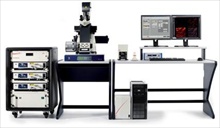 With the new Leica SR GSD from Leica Microsystems, scientists can now achieve resolutions far below the limit of diffraction that have never been attained before in widefield fluorescence microscopy. The system is capable of resolving details as small as 20 nanometers. This enables research of structures of single proteins and other biomolecules in cells and observation of molecular processes to gain new insights into fundamental processes of life...
 Anasys Instruments' AFM-IR system has been recognized by Microscopy Today in the receipt of the 2011 Innovation Award. It was presented to CEO, Roshan Shetty, at the 2011 M&M Annual conference held this year in Nashville, TN. The AFM-IR technique was developed by Dr. Alexandre Dazzi at the University of Paris-Sud. It uses an AFM probe as the IR absorbance detector and hence obtains IR spectroscopy at up to 2 orders of magnitude better than traditional IR spectroscopy...
 JPK Instruments are happy to announce that registration is now open for the tenth annual international symposium on the applications of scanning probe microscopy (SPM) and optical tweezers. The symposia will be held on the 5-6th October 2011 in Berlin focusing on applications developments in life sciences. These meetings continue to be highly regarded on the international life sciences meetings calendar. More than 100 scientists from around the world are expected to come to Berlin to discuss their results and share scientific knowledge in a relaxed and informal atmosphere...
 Olympus' new BX3 microscope systems are designed with the user in mind to maximise the quality of fluorescence images captured as part of life science research projects. The high-end BX63 system comfortably handles day-to-day applications, while boasting a range of advanced features to satisfy the most complex of needs, including rapid deconvolution of fluorescent Z-stack images...
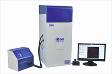 In vivo imaging of small animals for pre-clinical research using fluorescent markers is a novel approach to visualize cellular processes occurring at the micron level. There is growing interest in understanding what occurs at the level of tumor metastasis through direct visualization...
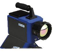 The FLIR SC7000 Series of Infrared Cameras from FLIR Advanced Thermal Solutions (ATS) provide the high performance and facilities for thermal imaging of high speed events. These high-speed, high-resolution research grade cameras provide Gigabit Ethernet, Camera Link and USB interfaces for maximum flexibility and performance. They also feature simultaneous digital and analogue outputs, and are available in multiple wavebands, detector resolutions, and lens configurations...
 JPK Instruments, a world-leading manufacturer of nanoanalytic instrumentation for research in life sciences and soft matter is pleased to announce the appointment of Thomas Neicke and Dr. Elmar Hartmann as new members of the sales team...
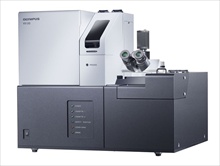 Olympus has released the latest version of its virtual slide (VS) product range, the VS120. The system can rapidly scan a slide in less than two minutes, creating a digital ‘virtual slide' that is an exact copy of the original. This is easily created and navigated using an intuitive graphical interface. The new unit provides semi-automatic and manual focusing modes for working with challenging samples, enhanced image clarity using the new Sample Detection Mask for automatic slide scanning, and increased optimisation of user workflow....
 Carl Zeiss has received a license from the University of California in San Francisco (UCSF) for the commercialization of "Multidirectional Selective Plane Illumination Microscopy" (mSPIM), an advanced illumination technique for light sheet fluorescence microscopy...
 China Medical Technologies, Inc. (CMED) (Nasdaq: CMED), a leading China-based advanced in-vitro diagnostic ("IVD") company, and Leica Biosystems, a division of Leica Microsystems, a world leader in microscopes and scientific instruments, today announced that they have established a sales, research and development collaboration to co-develop and market automated FISH kits to be used on the Leica BOND system. CMED will sell the Automated FISH Kits in China and Leica will have an option to sell the FISH kits in the rest of the world...
 SICM, a high resolution microscopy technique, has been used by a team of researchers to identify a drug that could one day be used to prevent abnormal heart rhythms, or arrhythmia. The study, published in Hepatology[1] this week, was enabled by SICM, which is the result of 7 years of research and development by ionscope Ltd. The global market for SICM equipment is supplied using patented technology from ionscope's offices in Cambridge, UK...
 Syngene, a world-leading manufacturer of image analysis solutions is pleased to introduce ChemiFast, a new chemiluminescence substrate specifically designed for use with all Syngene's chemiluminescence imaging systems. ChemiFast is highly sensitive and produces an enhanced signal that is stable for up to 24 hours, making it ideal for detecting even faint protein bands on Western blots...
 Market leaders in temperature controlled microscopy, Linkam Scientific Instruments report the use of their 1400°C high temperature stage to study silicate melt inclusions in the Fluids Research Group of the Geosciences Department at Virginia Tech. Researchers in the Fluids Research Group at Virginia Tech are concerned with the distribution, properties and role of fluids in and on the Earth, from its surface (shallow Earth's crust) to its deep interior (Earth's mantle)...
 Malvern Instruments has released a new version of software for the company's Morphologi G3 particle characterization system. This fully automated image analysis-based system delivers particle size and shape information for wet and dry samples, and for the analysis of foreign particulate matter collected on filters. Among the new applications in the version 7.40 software are facilities for user-defined light settings and automated dark-field measurements...
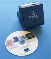 Warner Instruments is pleased to introduce its NEW line of ProgRes® Digital CMOS and CCD Microscope Cameras. These cameras are the result of more than two decades experience in the development and production of high-end solutions for digital imaging. The highly versatile and cost effective ProgRes® CMOS Camera range allows for quick and precise setting of both specimen and microscope...
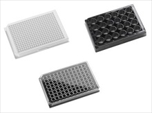 Precision manufactured to be extremely flat (+/- 15 microns across base) - KrystalTM glass bottom plates from Porvair Sciences provide unmatched performance for whole-plate CCD imaging and laser detection applications. Available in a choice of 24-, 96- and 384-well formats - Krystal glass bottom plates combine the advantageous optical properties of glass, low background and low birefringence, with the versatility of a microplate...