Channels
Special Offers & Promotions
The University of California, Los Angeles (UCLA), USA, Combines Raman Microscopy with Scanning Electron Microscopy (SEM) to Study Archaeological Textiles and Fibres
Renishaw, a world leader in metrology and spectroscopy, reports on the use of its SEM-SCA system to study textiles and fibres, non-destructively, at the University of California, Los Angeles, USA.
 The full paper describing the work has been published by the Journal of Raman Spectroscopy (John Wiley & Sons, Ltd.).
The Department of Materials Science and Engineering at UCLA delivers novel and highly innovative research that advances basic and applied knowledge in materials. This is achieved through interactions with the external community, and educational outreach and industrial collaborations. This is illustrated by a paper joint-authored by scientists from UCLA, the Los Angeles County Museum of Art, the Cotsen Institute of Archaeology and instrumentation manufacturer, Renishaw1. The paper “New Advancements in SERS Dye Detection Using Interfaced SEM and Raman Spectromicroscopy (µRS)” is published in the Journal of Raman Spectroscopy. It describes how a SEM may be interfaced with µRS to provide a means of non-destructively identifying organic compounds in complex samples, such as single fibres, by extractionless analysis. This enables the characterisation of dyes from reference collections and archaeological textiles.
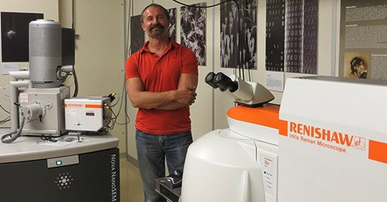
The lead author of the work is UCLA scientist, Dr Sergey V Prikhodko. He describes why Raman Spectromicroscopy has become a powerful analytical technique for the study of artistic, historical and archaeological materials: “As a non-destructive method that may also be interfaced with other techniques, it makes the sample reusable for subsequent analyses after performing µRS. In situ morphological characterization, elemental identification and structural analysis integrates µRS with SEM and energy dispersive X-ray spectroscopy (EDS) in the ‘hyphenated’ SEM-EDS-µRS system, at UCLA. This vital capability was brought together with the Renishaw SEM-SCA interface, where a Nova 230 SEM (FEI) is coupled to the Renishaw inVia confocal Raman microscope to provide structural and chemical analysis in situ.”
Dr Prikhodko's results on samples of various fibres clearly illustrate the potential of this non-destructive method. Interfacing SEM with µRS provides a very powerful tool to analyse unique and irreplaceable samples and artefacts quickly and in a cost-effective manner.
Media Partners


