Channels
Special Offers & Promotions
Renishaw's inVia Confocal Raman Microscope System is Being Used in Conservation Activities at the Rijksmuseum in Amsterdam, the Netherlands
Renishaw, a world leader in metrology and spectroscopy technologies, reports on how its Raman microscope assists in the conservation work of the Rijksmuseum in Amsterdam, the Netherlands.
 The Rijksmuseum is the iconic museum of the Netherlands and in 2013, following extensive renovation and restoration, it re-opened its doors to the public. At the museum, art and history takes on a new meaning for a broad-based, contemporary national and international audience. As a national institute, the Rijksmuseum offers a representative overview of Dutch art and history-from the Middle Ages to the 20th century-and of major aspects of European and Asian art. The Rijksmuseum has a long history and not only keeps artistic and historical objects, but also conserves, restores, researches, prepares and presents them, both on its own premises and elsewhere.
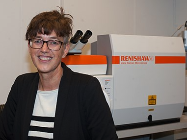
Jolanda van Iperen works as a research technician in the Conservation Department of the Rijksmuseum, where she carries out technical research on objects of art. She is involved in several research projects, including studies of red ground paint layers, chalk in frames and showcase materials. Jolanda works in collaboration with the Faculty of Humanities at the University of Amsterdam and with the research centre of the Dutch Cultural Heritage Agency. Her main interest is understanding material use in the context of history, and unravelling the underlying chemical processes.
They use several analytical techniques, including micro Raman spectroscopy, to solve research questions. Raman can solve a multitude of questions such as: Which pigments did the painter use? What is the corrosion product in the damaged part of the enamel? Does the treatment of this bronze leave residues on the surface? From where does the clay in the ceramic object originate?
The Conservation Department purchased a Renishaw inVia confocal Raman microscope equipped with a polarized light microscope and 785 nm and 532 nm lasers. The polarized light microscope is essential for this work as cross sections of paint layers, which typically consist of multiple coloured pigment grains, are impossible to visualize in reflected light with bright field illumination only. By using Raman spectroscopy in combination with other analytical techniques, they are able to find relationships between contemporary artists' use of paint materials. This includes, for example, discovering whether artists worked in the same workshop or shared their knowledge or materials.
Speaking about their use of the inVia, Mrs van Iperen says, .“We like the polarized light option and we are also impressed with the sensitivity of the system, the reproducibility of the results and the stability of the 532 nm laser. I am very much looking forward to starting to use our recently purchased steerable arm with which we can directly analyse the surfaces of paintings.”
Media Partners


