Syngene, a world-leading manufacturer of image analysis solutions, is delighted to announce its GeneSys software has been upgraded to include an icon of pre-set optimised “stain-free” protein gel imaging conditions.
This simple to use software, available to download free of charge to existing users of specified Syngene imaging systems, will save set-up time and ensure accurate stain-free protein gel images every time. For use in Syngene’s G:BOX Chemi and PXi imaging systems, the new GeneSys software now includes a “stain-free” imaging icon....
FLIR Systems has announced the X6570sc high-speed infrared camera, the latest in FLIR's X6000sc series of cameras for engineers, researchers, and scientists.
This longwave infrared (LWIR) high performance camera offers the accuracy and microsecond-precision of FLIR's mercury cadmium telluride (MCT) detector paired with an intuitive user experience, so researchers can set-up quickly and get working faster. FLIR engineered the X6570sc to provide researchers with measurement accuracy, advanced processing algorithms, and connectivity...
The Royal Microscopical Society have announced Allison Winton as their new Chief Executive.
Since 1839, the RMS has been championing microscopy, supporting scientists at all stages of their career and promoting the science to a wider audience. But behind the scenes, a dedicated team of office staff ensure the Society is serving its members and hosting high quality events to scientists all over the world....
Read MoreThermal Imaging for Bat Conservation & ResearchJan 26, 2017
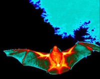
FLIR Systems reports on how its T1030sc thermal imaging camera is helping bat conservation and research efforts.
Bats play a major role in our ecosystem, but due to numerous threats bat populations have declined significantly over the years. FLIR thermal imaging technology is now playing a key role in helping experts understand, and conserve, bat populations in the UK. A video report from bat conservation advisors, Simon Holmes and Joe Nunez-Mino, from the Bat Conservation Trust in the UK, explains the important role that FLIR’s T1030sc thermal imaging cameras play in...
Powerful microscopes help researchers understand innovative material structures at the atomic scale
The Princeton Institute for the Science and Technology of Materials (PRISM) recently unveiled its new Micro/Nano Fabrication Laboratory (MNFL) and Imaging and Analysis Center (IAC) facilities, which are now home to a powerful suite of advanced analytical instrumentation from Thermo Fisher Scientific....
JPK Instruments announce the release of the world’s first combined system to provide optical tweezers and atomic force microscopy on a single inverted light microscope platform.
Based on years of leadership in the fields of atomic force microscopy and optical tweezers for applications in nanotechnology for life science, JPK have brought the technologies together on a single inverted light microscope platform. The OT-AFM Combi-System pairs the exceptional surface force measurement and imaging capabilities of AFM with the ability of optical tweezers to apply and measure smallest forces in 3D....
Olympus’ DP74 microscopy camera enhances return on investment by efficiently delivering sensitive fluorescence and brightfield imaging in one camera.
With a 60 frames per second live image and a 20.7 megapixel resolution it delivers a lifelike experience on-screen while also introducing intelligent features to make everyday operation faster and more convenient. Helping to save time and increase comfort at the microscope, Olympus’ new DP74 camera for brightfield and fluorescence delivers intelligent imaging....
BioTek’s BioSpa Live Cell Imaging System fully automates live cell imaging workflows for robust, real-time results without the need for manual intervention.
The BioSpa System has a compact footprint for use on benchtops and in biosafety cabinets, and consists of the BioSpa™ 8 Automated Incubator and Cytation™ 5 Cell Imaging Multi-Mode Reader. BioTek’s liquid handling instruments may also be linked with the BioSpa System for complete, walk away automation from sample preparation through image analysis....
Phenom-World announces a Eucentric Sample Holder with a 6-axes sub-stage for eucentric tilting and compucentric rotation.
The stage is designed for use in the Phenom XL desktop SEM and allows users to move samples around easily without losing sight of the sample detail. “A eucentric stage is typically an expensive option on large ‘floor model’ SEMs. This product changes all of that,” explains Emile Asselbergs, CEO of Phenom-World....
Sixth Annual Competition Recognizes Videos Capturing Hidden Details of Microscopic World
Nikon Instruments Inc. today unveiled the winners of the sixth annual Nikon Small World in Motion Photomicrography Competition, awarding First Place to William Gilpin of Stanford University for his video depicting an eight-week-old starfish larva churning the water around its body as it searches for food....
FLIR Systems X6540sc thermal imaging camera is being used by the Photonics Packaging Group at the Tyndall National Institute (Cork, Ireland) as part of a thermal microscope system to image the silicon photonic optical network unit (ONU) for next generation passive optical networks (NG-PONs) developed in the European FP7-Project "FABULOUS”.
Researchers at Tyndall are developing a next generation passive optical network (PON) demonstration module for high-speed fibre-to-home internet connectivity. At the heart of the PON is a silicon photonic integrated circuit (Si-PIC) that receives information on an incoming optical-signal (downloading), before reflecting the optical signal back, after encoding extra information (uploading)....
ZEISS will provide 1 million euros for innovative research projects at the EPFL
ZEISS will support innovative research projects at the Swiss Federal Institute of Technology in Lausanne (École polytechnique fédérale de Lausanne, abbr. EPFL). The company will make one million euros available for new research projects in key technology fields such as biomedical research, medical diagnostics and visualization as well as optical metrology and inspection...
Oxford Nanoimaging Limited manufacture and sell microscopes offering super-resolution and single-molecule performance to research users.
Today, the company reports on the work of early-adopters for their Nanoimager technology at the MRC Centre for Molecular Bacteriology and Infection located at Imperial College, London. The MRC Centre for Molecular Bacteriology and Infection is uniquely focused on disease-causing bacteria. Ramesh Wigneshweraraj is a Professor of Microbiology leading...
The University of Glasgow, in partnership with NHS Greater Glasgow & Clyde, has taken delivery of Scotland’s first ultra-powerful 17.5 tonne 7 Tesla (7T) MRI scanner at the new Imaging Centre of Excellence (ICE) on the site of the Queen Elizabeth University Hospital (QEUH).
The new ultra-high resolution scanner – one of the world’s most powerful magnetic resonance imaging (MRI) machines – is also the first scanner of its kind in the UK to be located in a clinical setting. It will be situated on the grounds of Glasgow’s new super hospital, the QEUH. The £10m 7T MRI scanner will be used to advance critical clinical research and will allow scientists and clinicians to study the human body...
Market leaders in temperature controlled microscopy, Linkam Scientific Instruments, announce the launch of their Optical DSC450 which enables simultaneous visualization of thermal processes for improved materials characterisation.
Linkam's engineers are continually working to develop constructive solutions to practical problems. One of the latest developments is the Optical DSC450, a Differential Scanning Calorimeter for measurements up to 450 °C. Differential scanning calorimetry, DSC, is a technique used to measure temperatures and heat flow associated with thermal transitions in a material....
Quorum Technologies, market and technology leaders in electron microscopy coating and cryogenic preparation products, report on the work of the Agricultural Research Service of the US Department of Agriculture where their PP2000 Cryo-SEM preparation system is in use to prepare soft bodied organisms including mites & ticks for study using cryo-SEM
Dr Gary Bauchan is the Director of the Electron and Confocal Microscopy Unit at the Agricultural Research Service (ARS), the principal in-house research agency of the United States Department of Agriculture. The Unit is a core facility with the responsibility of providing collaborative assistance to scientists from ARS, Northeast Area and Beltsville Agricultural Research Center (BARC) who have microscopy applications that require high resolution imaging...
JPK Instruments, a world-leading manufacturer of nanoanalytic instrumentation for research in life sciences and soft matter, reports on the use of their NanoTracker™ 2 Optical Tweezers system at the Centre for Cancer Research in Marseilles, a part of CNRS and INSERM located at the Aix-Marseille Université.
The research team lead by Dr Mauro Modesti at the Centre for Cancer Research, Marseilles (CRCM), focuses on the study and understanding of the molecular mechanisms that assure DNA repair and the maintain of genome integrity in human cells. Failure to detect and/or repair of DNA genotoxic lesions, such as double-strand breaks, can lead to the appearance of mutations, genomic instability...
Providing automatic panoramic images on any manual microscope and improving software and hardware integration, Olympus’ cellSens imaging software simply helps users do more
Committed to fast and reliable image acquisition in life science, Olympus has updated its cellSens imaging software (now version 1.16). The update provides cost-effective solutions by expanding the capabilities of manual microscopes: fast, detailed, real-time panoramic imaging is now possible without an expensive motorised or encoded stage thanks to the Instant Multiple Image Alignment (Instant MIA) function....
HeliScan MicroCT designed to allow materials scientists to nondestructively visualize and characterize internal structure down to the micrometer scale
Materials scientists can now gain valuable insight into the relationships between a material’s structure at the microscopic level and its bulk properties using a new multi-scale imaging solution that is designed to provide large-scale, high-fidelity three-dimensional (3D) images of the sample....
UK based MR Solutions’, revolutionary pre-clinical scanner MRS 7000, is an award winner at the US-based R&D 100 Awards.
The 54th annual awards, held in Washington in November 2016, recognise the 100 most innovative technologies and services of the past year. The compact MRS 7000 series is the world’s first 7.0T (T is for tesla, the power of the magnet), helium-free, preclinical MRI system, which can be combined with PET (photon emission computed tomography) and SPECT (single photon emission computed tomography) imaging modules....
Scientists at Tokyo Institute of Technology have developed a portable and wearable terahertz scanning device made using arrays of carbon nanotubes, for non-invasive inspection of three-dimensional objects without requiring bulky peripheral optical components.
The device is expected to have wide ranging applications including the noninvasive inspections of medical and drug delivery equipment such as syringes, as well as in medicine for imaging cancer cells, blood clots, sweat glands, and teeth. The findings are published in Nature Photonics, November 2016....
Synoptics, a world leader in the development and manufacture of innovative digital imaging systems for life science and clinical applications, is pleased to announce the appointment of Dr Brian Stammers as its new CEO.
Brian’s wealth of experience in the life science industry will help guide Synoptics’s strategic direction to deliver innovative and commercially successful products. As the CEO of Synoptics, which is part of the AIM quoted UK company Scientific Digital Imaging (SDI), Dr Stammers will oversee the commercial and technical direction of Synoptics’s three divisions, Syngene, Synbiosis and Synoptics Health....

 This longwave infrared (LWIR) high performance camera offers the accuracy and microsecond-precision of FLIR's mercury cadmium telluride (MCT) detector paired with an intuitive user experience, so researchers can set-up quickly and get working faster. FLIR engineered the X6570sc to provide researchers with measurement accuracy, advanced processing algorithms, and connectivity...
 Since 1839, the RMS has been championing microscopy, supporting scientists at all stages of their career and promoting the science to a wider audience. But behind the scenes, a dedicated team of office staff ensure the Society is serving its members and hosting high quality events to scientists all over the world....
 Bats play a major role in our ecosystem, but due to numerous threats bat populations have declined significantly over the years. FLIR thermal imaging technology is now playing a key role in helping experts understand, and conserve, bat populations in the UK. A video report from bat conservation advisors, Simon Holmes and Joe Nunez-Mino, from the Bat Conservation Trust in the UK, explains the important role that FLIR’s T1030sc thermal imaging cameras play in...
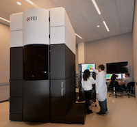 The Princeton Institute for the Science and Technology of Materials (PRISM) recently unveiled its new Micro/Nano Fabrication Laboratory (MNFL) and Imaging and Analysis Center (IAC) facilities, which are now home to a powerful suite of advanced analytical instrumentation from Thermo Fisher Scientific....
 Based on years of leadership in the fields of atomic force microscopy and optical tweezers for applications in nanotechnology for life science, JPK have brought the technologies together on a single inverted light microscope platform. The OT-AFM Combi-System pairs the exceptional surface force measurement and imaging capabilities of AFM with the ability of optical tweezers to apply and measure smallest forces in 3D....
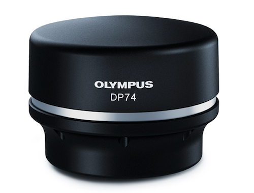 With a 60 frames per second live image and a 20.7 megapixel resolution it delivers a lifelike experience on-screen while also introducing intelligent features to make everyday operation faster and more convenient. Helping to save time and increase comfort at the microscope, Olympus’ new DP74 camera for brightfield and fluorescence delivers intelligent imaging....
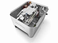 The stage is designed for use in the Phenom XL desktop SEM and allows users to move samples around easily without losing sight of the sample detail. “A eucentric stage is typically an expensive option on large ‘floor model’ SEMs. This product changes all of that,” explains Emile Asselbergs, CEO of Phenom-World....
 Nikon Instruments Inc. today unveiled the winners of the sixth annual Nikon Small World in Motion Photomicrography Competition, awarding First Place to William Gilpin of Stanford University for his video depicting an eight-week-old starfish larva churning the water around its body as it searches for food....
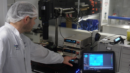 Researchers at Tyndall are developing a next generation passive optical network (PON) demonstration module for high-speed fibre-to-home internet connectivity. At the heart of the PON is a silicon photonic integrated circuit (Si-PIC) that receives information on an incoming optical-signal (downloading), before reflecting the optical signal back, after encoding extra information (uploading)....
 ZEISS will support innovative research projects at the Swiss Federal Institute of Technology in Lausanne (École polytechnique fédérale de Lausanne, abbr. EPFL). The company will make one million euros available for new research projects in key technology fields such as biomedical research, medical diagnostics and visualization as well as optical metrology and inspection...
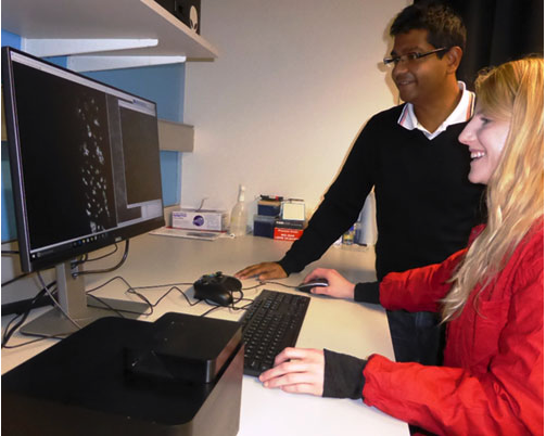 Today, the company reports on the work of early-adopters for their Nanoimager technology at the MRC Centre for Molecular Bacteriology and Infection located at Imperial College, London. The MRC Centre for Molecular Bacteriology and Infection is uniquely focused on disease-causing bacteria. Ramesh Wigneshweraraj is a Professor of Microbiology leading...
 The new ultra-high resolution scanner – one of the world’s most powerful magnetic resonance imaging (MRI) machines – is also the first scanner of its kind in the UK to be located in a clinical setting. It will be situated on the grounds of Glasgow’s new super hospital, the QEUH. The £10m 7T MRI scanner will be used to advance critical clinical research and will allow scientists and clinicians to study the human body...
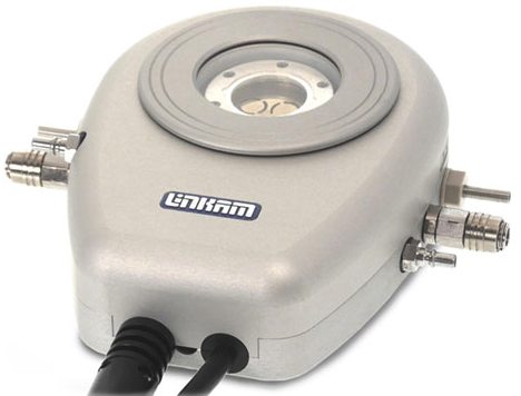 Linkam's engineers are continually working to develop constructive solutions to practical problems. One of the latest developments is the Optical DSC450, a Differential Scanning Calorimeter for measurements up to 450 °C. Differential scanning calorimetry, DSC, is a technique used to measure temperatures and heat flow associated with thermal transitions in a material....
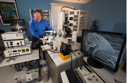 Dr Gary Bauchan is the Director of the Electron and Confocal Microscopy Unit at the Agricultural Research Service (ARS), the principal in-house research agency of the United States Department of Agriculture. The Unit is a core facility with the responsibility of providing collaborative assistance to scientists from ARS, Northeast Area and Beltsville Agricultural Research Center (BARC) who have microscopy applications that require high resolution imaging...
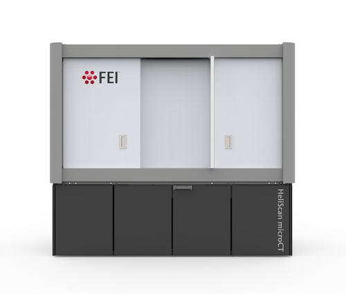 Materials scientists can now gain valuable insight into the relationships between a material’s structure at the microscopic level and its bulk properties using a new multi-scale imaging solution that is designed to provide large-scale, high-fidelity three-dimensional (3D) images of the sample....
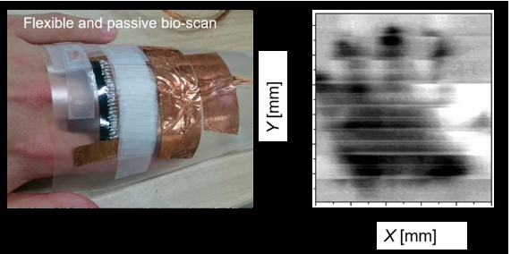 The device is expected to have wide ranging applications including the noninvasive inspections of medical and drug delivery equipment such as syringes, as well as in medicine for imaging cancer cells, blood clots, sweat glands, and teeth. The findings are published in Nature Photonics, November 2016....