Market leaders in temperature controlled microscopy and established manufacturers of advanced freeze drying microscopes, Linkam Scientific Instruments, announce the launch of the FDVS platform, a lyophilisation system to replicate large scale freeze drying processes.
Replicating large scale industrial freeze drying processes as smaller test runs is an effective way to save time, cut costs and perfect the technicalities of an operation run. With this in mind, Linkam engineers have created the new Freeze Drying Vial System - FDVS. Building on the experience gained from the FDCS Freeze Drying stage....
International medical imaging IT company Sectra (STO: SECT B) and digital pathology company Visiopharm A/S have reached agreement on an open exchange of pathology images between their respective digital pathology systems.
The companies agree that open solutions and the free exchange of information between the various healthcare IT systems without “internal company formats” are of benefit to both customers and providers. The integration of the two systems will enable customers to work in the platform they consider best for the respective application....
FLIR Systems announces a web page that brings together the opportunity to sign-up for forthcoming free live online tutorials and review recent popular thermal imaging webinars, seminars and events.
Due to air on December 20th 2016, the 'Tips for Capturing Great Thermal Images' tutorial will address how to capture quality thermal data with your infrared camera, covering the key basics of infrared imaging in an easy to understand presentation. The 'How to Create a Report with FLIR Tools' live tutorial, taking place....
Automated Microscopy Platform Offers High Flexibility and Throughput
ZEISS introduces a new system for automated microscopy in life sciences research. ZEISS Celldiscoverer 7 combines the user-friendly automation features of a boxed microscope with the image quality and flexibility of a classic inverted research microscope. Scientists acquire better data in shorter times with 2D or 3D cell cultures, tissue sections or small model organisms.
The NORLUX Neuro-Oncology Laboratory, an international research center which is part of the Luxembourg Institute of Health (LIH) has installed MR Solution’s - MRS 3017 - 3T cryogen-free MRI preclinical scanner to further cancer research with a particular focus on malignant brain tumours.
Not only does this 17 cm bore scanner provide state of the art images but its small footprint – the size of a desk - means it can easily be accommodated while the integrity of the specific pathogen free (SPF) environment remains intact as there is no need for an emergency liquid helium venting system. It is also the only 3T scanning system which has the ability to operate at various clinical field strengths allowing easy translation of the researchers’ applications to the clinic. Further...
Specialised Imaging Ltd reports as of 1st October 2016 the formation of a new division - Wegapixel focusing on the design and development of advanced CMOS imaging sensors.
Wegapixel has been set-up as a turn-key solutions provider for custom CMOS image sensors. In addition to conventional imager design, the company brings together unique expertise in many application areas including high speed imaging, electron microscopy, X-ray digital imaging as well as UV and IR imaging....
EM Resolutions, manufacturers and suppliers of tools and accessories for users of electron microscopes, launches the EM-Storr vacuum sample storage container.
EM Resolutions announces the EM-Storr vacuum sample storage container which has been specifically developed to protect EM samples under vacuum during storage and transportation. EM-Storr has a modern space saving design and superior construction using high vacuum compatible materials to reduce outgassing and to hold vacuum for extended periods....
Until now, the only way to diagnose disease accurately using a microscope has been to take samples and send them back to a laboratory for expensive and time-consuming tests, carried out by experienced pathologists.
With the arrival of the ioLight microscope however, vets can immediately look at the sample and be able to either reassure pet owners or farmers , or prioritise laboratory diagnosis and treatment, if they are concerned. Launched in 2016, the ioLight microscope is the first professional-quality pocket digital microscope....
Quorum Technologies report on the use of their Q150T ES combined sputter coating and carbon coating system selected as a general purpose workhorse facility for the Biomedical Imaging Unit at Southampton University.
The Biomedical Imaging Unit (BIU) is a core facility which provides a diagnostic and research service in high quality/high resolution microscopy. It is a joint facility run for the benefit of, and jointly funded by, the University of Southampton and Southampton University Hospitals NHS Trust with eight full time staff....
Siemens Healthineers has been collaborating with the University of Cambridge to install a new type of MRI scanner, which will allow researchers to see detail in the brain as tiny as a grain of sand.
The Siemens 7T Terra MRI has the potential to bring a new era of personalised treatments to major illnesses, including cancer, dementia and mental health conditions. The cutting-edge machinery gives researchers the opportunity to study how the brain encodes information, including things like individual memories. This means the scanner will now be able to now pick up on extremely small structures, including those associated with the early stages of Alzheimer...
The open and flexible inverted microscope platform for living and fixed specimens
ZEISS introduces a new inverted microscope platform for life science research. The ZEISS Axio Observer family consists of three stable and modular microscope stands for flexible and efficient imaging. Scientists benefit from reproducible results from their experiments and high quality image data from a whole range of samples in a variety of conditions....
FLIR Systems has posted a recorded version of a popular webinar that compares two common instruments used for measuring temperature: thermocouples and infrared (IR) cameras.
The webinar provides an informative introduction to temperature measurement with the technologies available today, and explains the basics of how thermocouples and IR cameras work. To provide a direct comparison of the two measurement solutions an experiment is conducted using thermocouples and an IR camera to measure temperature and the results of this experiment are reviewed....
EM Resolutions, manufacturers and suppliers of tools and accessories for users of electron microscopes, announce the availability of new cryo microgripper for cryo-FIB lift-out, a product of Kleindiek Nanotechnik.
As UK and Irish distributors for Kleindiek Nanotechnik, EM Resolutions is pleased to announce the launch of a new cryo microgripper specifically designed for cryo Focussed Ion Beam (cryo-FIB) lift out procedures. Used with a MM3A-EM micromanipulator the specially insulated gripper provides a reliable way to perform lift out of TEM lamella down to liquid nitrogen temperatures....
LaVison BioTec, developers of advanced microscopy solutions for the life sciences, report on the latest work of Nicolas Renier, a Post-Doctoral Fellow in the laboratory of Marc Tessier-Lavigne at the Rockefeller University in New York where he applies light sheet microscopy to measure activity in a mouse brain from a single snapshot.
Drs Nicolas Renier and Zhuhao Wu are post-doctoral fellows in the laboratory of Marc Tessier-Lavigne, Carson Family Professor and head of the Laboratory of Brain Development and Repair and President of Rockefeller University in New York. Over recent years, they have co-developed methodologies for new imaging techniques applying light sheet microscopy....
EM Resolutions, manufacturers and suppliers of tools and accessories for users of electron microscopes, report on the research of Peter Martin from the University of Bristol.
He is applying Kleindiek micromanipulators in the characterisation of materials resulting from the accident at the Japanese nuclear power station. PhD student, Peter Martin, is a member of the School of Physics at the University of Bristol. His research focuses on the March 2011 incident at the Fukushima Daiichi Nuclear Power Plant (FDNPP) in Japan and its effects at both the metre and micron scales. He has used an unmanned aerial vehicle to investigate the...
Quorum Technologies, market and technology leaders in electron microscopy coating and cryogenic preparation products, report on how their PP3010T Cryo-SEM preparation system is being used to assist polymer and self-assembled materials research at the Institut Charles Sadron, CNRS-University of Strasbourg.
The Institute Charles Sadron is a CNRS-Institute located at the University of Strasbourg performing fundamental and applied research on polymers and self-assembled systems. Dr Marc Schmutz works in the Electron Microscopy facility where he uses a Hitachi SU8010 Ultra High Resolution Field Emission Scanning Electron Microscope in conjunction with a PP3010T cryo-SEM preparation system from Quorum Technologies. It is used to study a broad variety of materials...
Proprietary, cost-effective process restores performance, while new optics deliver high reflectivity and precision
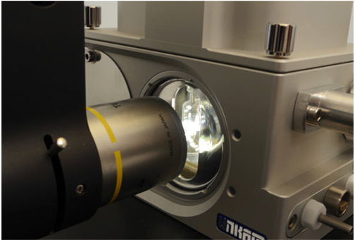
Rigaku Innovative Technologies (RIT), a global supplier of high performance multilayer optics, announces that it cleans and recoats old synchrotron optics to either retask the optics for new applications or to refurbish and repair the coatings to enhance performance. This proprietary process restores the optics’ performance with less lead time and at a far lower cost than purchasing new optics....
Visit Booth # 401 to see innovative imaging solutions for materials science applications
ZEISS announces they will be showcasing the latest microscopy innovations and advancements at Materials Science & Technology (MS&T) 2016. At Booth #401 will be ZEISS Axio Observer.Z1m inverted microscope, ZEISS Axio Lab A.1 MAT manual routine microscope for materials, ZEISS Smartzoom 5 digital microscope, and ZEISS Stemi 508 stereo microscope...
Linkam Scientific Instruments, report on the use of their world-leading THMS600 heating/cooling stage for the study of water anomalies by looking at the Brillouin spectroscopy of fluid inclusions proposed as a paleothermometer for subsurface rocks.
Frédéric Caupin is a Professor in the Liquids and Interfaces Group at the Institute of Light and Matter CNRS at Université Claude Bernard Lyon 1. The main motivation of his research is the study of water anomalies. Indeed, water is the most common liquid, but also the most anomalous. For instance, it expands upon cooling below 4 °C. Water anomalies are due to the complex hydrogen bonded network present. These anomalies get even more pronounced in the...
JPK Instruments reports on the use of their NanoWizard® AFM and CellHesion® systems in the Department of Physiology, Development & Neuroscience at the University of Cambridge.
Dr Kristian Franze is a lecturer in the Department of Physiology, Development and Neuroscience at the University of Cambridge. His major goal is to understand when, where and how mechanical signals, such as forces and local tissue stiffness, are involved in controlling cell development and function in the nervous system....
MR Solutions has received an order for a high-powered 7T, cryogen-free, preclinical PET-MRI multi-modality imaging system from the newly formed pre-clinical imaging resource at the Zilkha Neurogenetic Institute (ZNI) at the Keck School of Medicine of the University of Southern California (USC).
The simultaneous acquisition of PET and MR images marries the exquisite sensitivity of PET with the high spatial resolution and soft tissue contrast of MRI. MR Solutions is the only company in the world to be able to deliver this ground breaking cryogen free technology. Dr. Russell Jacobs, Professor of Research Physiology and Biophysics at the Zilkha Neurogenetic Institute commented...
Research in optical materials is rapidly moving towards stable, high-power laser sources emitting in the mid-infrared (mid-IR) region.
Despite their promising applications, there are currently not many commercially available lasers for this range of wavelengths (2 μm ? 8 μm). One of their main applications is in absorption spectroscopy for the detection of trace gases and atmospheric pollutants: many organic compounds have characteristic mid-IR bands, so the detection in this region is highly selective....

 Replicating large scale industrial freeze drying processes as smaller test runs is an effective way to save time, cut costs and perfect the technicalities of an operation run. With this in mind, Linkam engineers have created the new Freeze Drying Vial System - FDVS. Building on the experience gained from the FDCS Freeze Drying stage....
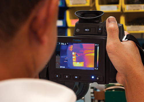 Due to air on December 20th 2016, the 'Tips for Capturing Great Thermal Images' tutorial will address how to capture quality thermal data with your infrared camera, covering the key basics of infrared imaging in an easy to understand presentation. The 'How to Create a Report with FLIR Tools' live tutorial, taking place....
 ZEISS introduces a new system for automated microscopy in life sciences research. ZEISS Celldiscoverer 7 combines the user-friendly automation features of a boxed microscope with the image quality and flexibility of a classic inverted research microscope. Scientists acquire better data in shorter times with 2D or 3D cell cultures, tissue sections or small model organisms.
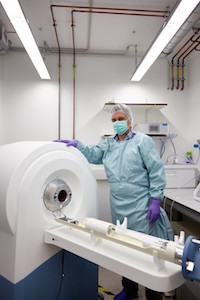 Not only does this 17 cm bore scanner provide state of the art images but its small footprint – the size of a desk - means it can easily be accommodated while the integrity of the specific pathogen free (SPF) environment remains intact as there is no need for an emergency liquid helium venting system. It is also the only 3T scanning system which has the ability to operate at various clinical field strengths allowing easy translation of the researchers’ applications to the clinic. Further...
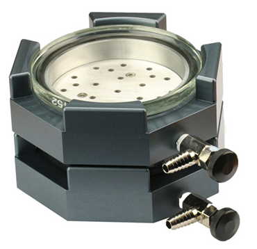 EM Resolutions announces the EM-Storr vacuum sample storage container which has been specifically developed to protect EM samples under vacuum during storage and transportation. EM-Storr has a modern space saving design and superior construction using high vacuum compatible materials to reduce outgassing and to hold vacuum for extended periods....
 With the arrival of the ioLight microscope however, vets can immediately look at the sample and be able to either reassure pet owners or farmers , or prioritise laboratory diagnosis and treatment, if they are concerned. Launched in 2016, the ioLight microscope is the first professional-quality pocket digital microscope....
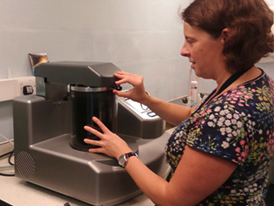 The Biomedical Imaging Unit (BIU) is a core facility which provides a diagnostic and research service in high quality/high resolution microscopy. It is a joint facility run for the benefit of, and jointly funded by, the University of Southampton and Southampton University Hospitals NHS Trust with eight full time staff....
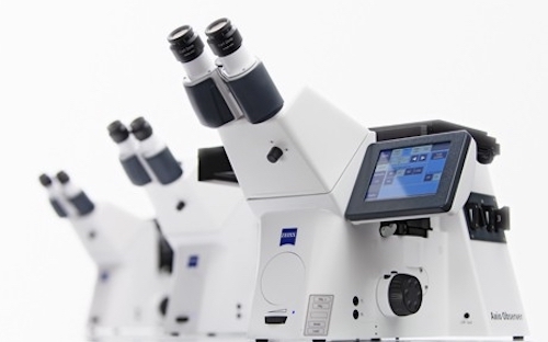 ZEISS introduces a new inverted microscope platform for life science research. The ZEISS Axio Observer family consists of three stable and modular microscope stands for flexible and efficient imaging. Scientists benefit from reproducible results from their experiments and high quality image data from a whole range of samples in a variety of conditions....
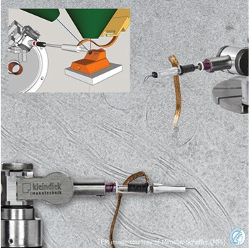 As UK and Irish distributors for Kleindiek Nanotechnik, EM Resolutions is pleased to announce the launch of a new cryo microgripper specifically designed for cryo Focussed Ion Beam (cryo-FIB) lift out procedures. Used with a MM3A-EM micromanipulator the specially insulated gripper provides a reliable way to perform lift out of TEM lamella down to liquid nitrogen temperatures....
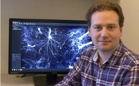 Drs Nicolas Renier and Zhuhao Wu are post-doctoral fellows in the laboratory of Marc Tessier-Lavigne, Carson Family Professor and head of the Laboratory of Brain Development and Repair and President of Rockefeller University in New York. Over recent years, they have co-developed methodologies for new imaging techniques applying light sheet microscopy....
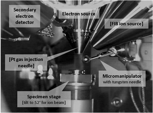 He is applying Kleindiek micromanipulators in the characterisation of materials resulting from the accident at the Japanese nuclear power station. PhD student, Peter Martin, is a member of the School of Physics at the University of Bristol. His research focuses on the March 2011 incident at the Fukushima Daiichi Nuclear Power Plant (FDNPP) in Japan and its effects at both the metre and micron scales. He has used an unmanned aerial vehicle to investigate the...
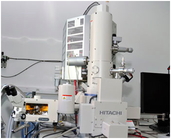 The Institute Charles Sadron is a CNRS-Institute located at the University of Strasbourg performing fundamental and applied research on polymers and self-assembled systems. Dr Marc Schmutz works in the Electron Microscopy facility where he uses a Hitachi SU8010 Ultra High Resolution Field Emission Scanning Electron Microscope in conjunction with a PP3010T cryo-SEM preparation system from Quorum Technologies. It is used to study a broad variety of materials...
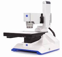 ZEISS announces they will be showcasing the latest microscopy innovations and advancements at Materials Science & Technology (MS&T) 2016. At Booth #401 will be ZEISS Axio Observer.Z1m inverted microscope, ZEISS Axio Lab A.1 MAT manual routine microscope for materials, ZEISS Smartzoom 5 digital microscope, and ZEISS Stemi 508 stereo microscope...
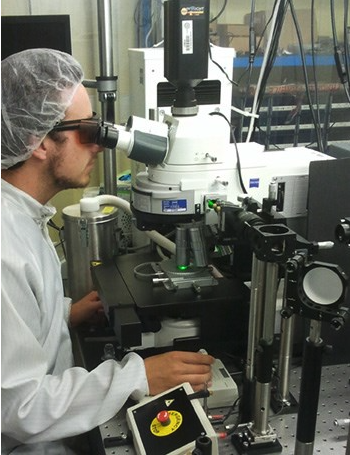 Frédéric Caupin is a Professor in the Liquids and Interfaces Group at the Institute of Light and Matter CNRS at Université Claude Bernard Lyon 1. The main motivation of his research is the study of water anomalies. Indeed, water is the most common liquid, but also the most anomalous. For instance, it expands upon cooling below 4 °C. Water anomalies are due to the complex hydrogen bonded network present. These anomalies get even more pronounced in the...
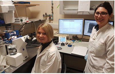 Dr Kristian Franze is a lecturer in the Department of Physiology, Development and Neuroscience at the University of Cambridge. His major goal is to understand when, where and how mechanical signals, such as forces and local tissue stiffness, are involved in controlling cell development and function in the nervous system....
 The simultaneous acquisition of PET and MR images marries the exquisite sensitivity of PET with the high spatial resolution and soft tissue contrast of MRI. MR Solutions is the only company in the world to be able to deliver this ground breaking cryogen free technology. Dr. Russell Jacobs, Professor of Research Physiology and Biophysics at the Zilkha Neurogenetic Institute commented...
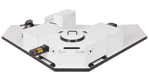 Despite their promising applications, there are currently not many commercially available lasers for this range of wavelengths (2 μm ? 8 μm). One of their main applications is in absorption spectroscopy for the detection of trace gases and atmospheric pollutants: many organic compounds have characteristic mid-IR bands, so the detection in this region is highly selective....