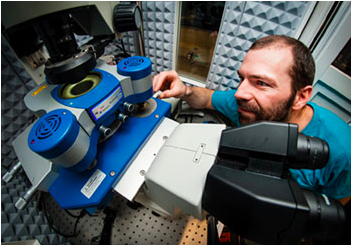
JPK Instruments works closely with users at the University of Sheffield where their NanoWizard® AFM systems are being used to further understand soft matter and biological systems at the molecular scale in the Hobbs SPM Group in the Department of Physics.
Dr Nic Mullin is a senior experimental officer in the SPM Group of Professor Jamie Hobbs in the Department of Physics & Astronomy at the University of Sheffield. His research is based around instrument development for scanning probe microscopy for the study of soft matter and biological systems at the molecular scale....
Highlighting Olympus’ workflow-oriented design quality across its range of life science microscopes, the FLUOVIEW FV3000 and the CX23 microscope have been recognised with the international iF award for exceptional product design.
Globally renowned as a symbol of design excellence, the iF design awards celebrate the best in user-focused, ergonomic and efficient design. With over 5,000 submissions from 70 countries, Olympus Scientific Solutions Division has received two of these prestigious awards for its CX23 upright microscope and FLUOVIEW FV3000 confocal laser scanning...
Tecan’s updated Spark® reader combines the benefits of a multimode microplate reader and a bright field imaging system in a compact package.
Unlike other multimode readers, Spark lets you actually see what’s happening to your cells, offering automated cell imaging, confluence measurements, cell counting and viability assessments to simplify cell biology protocols and enable long-term, walkaway experiments. Spark has been developed specifically to address the needs of cell-based workflows...
ioLight is delighted to announce that Bayer plc has purchased 10 microscopes for its Practice Support Advisors.
The ioLight microscope is the first professional quality pocket digital microscope. It is a laboratory grade microscope that fits in a jacket pocket, is simple to use and robust. It unfolds quickly to record and share 5MP still images and real time HD video on a tablet or phone....
Clariscan builds on GE Healthcare’s wide range of imaging products, services and support delivered to radiology professionals worldwide
Growing its range of magnetic resonance imaging (MRI) contrast media options available to patients and radiologists, GE Healthcare announced the launch of Clariscan™ (gadoteric acid) at the European Congress of Radiology (ECR) 2017 meeting. Clariscan is a gadolinium-based contrast agent (GBCA) designed to support effective visualisation of...
Thermo Fisher Scientific’s Cryo-TEM provides critical information for small molecule and biologic drug discovery
A new contract research laboratory operated by France-based NovAliX will provide pharmaceutical companies with access to high-resolution cryo-transmission electron microscopy (cryo-TEM) by Thermo Fisher Scientific for facilitating small molecule and biologic drug discovery. NovAliX’s new laboratory will use the cryo-TEM to provide two-dimensional...
The Lumen 200 Fluorescence Illumination System from Prior Scientific offers a powerful, cost effective alternative to traditional short arc Mercury vapour lamps and bulbs used in fluorescence microscopy.
Mercury vapour lamps and bulbs suffer from a number of limiting disadvantages including an operational lifetime rarely exceeding 200 hours and the need for time consuming alignment procedures during installation. In addition Mercury vapour bulbs are susceptible to flickering, limiting the use of these light sources in quantitative fluorescent microscopy....
Deben have supplied leading German instrument manufacturers, JPK Instruments, with a tensile stage to use in conjunction with the world-renowned NanoWizard® AFM platform.
Dr Torsten Mueller is a member of the development team with German nanoscience instrument makers, JPK Instruments. Dr Mueller and his colleagues are based in Berlin supporting a worldwide user network which is continuously testing JPK with requests for new capabilities for their life science and materials nanoscience systems....
JPK Instruments reports on how STM is being used to study surface plasmons in the Molecular Nanoscience Group at ISMO – Institut des Sciences Moléculaires d’Orsay – CNRS and the Université Paris-Sud.
One of the research goals of the Molecular Nanoscience Group at ISMO is to work towards circuits and devices in which surface plasmons (not electrons or photons) are used to transfer and manipulate information. However, why develop plasmonics when there are already solutions using electronics and photonics?....
Second Crowdcube campaign for growth and international expansion
Following on from an extremely successful fund raising campaign in 2015, and a product launch in July 2016, ioLight is delighted to announce that its second crowd funding campaign is now live on the Crowdcube platform. The ioLight microscope is the first professional quality pocket digital microscope....
In a move to serve customers with ready-to-use solutions for organ-on-a-chip model development and analysis, MIMETAS and Molecular Devices will combine marketing efforts on the OrganoPlate® organ-on-a-chip platform and ImageXpress® Micro Confocal high-content imaging systems.
This agreement was announced at the SLAS2017 conference in Washington. “OrganoPlates® are designed for ease-of-handling at any throughput, and provide unsurpassed physiological relevance,” said Jos Joore, Managing Director of MIMETAS, “As such, they are perfectly suited for the high-content imaging systems from Molecular Devices....
BioTek has been granted US patent 9,557,217 for imaging and microplate reading in one instrument.
The intellectual property protected by this patent is used in BioTek’s award winning line of Cytation™ Cell Imaging Multi-Mode readers. Other patents are pending. Cytation offers fluorescence, brightfield, color brightfield and phase contrast imaging up to 60x, and multi-mode detection optics that include fluorescence intensity, luminescence, absorbance and advanced measurements such as fluorescence polarization, time-resolved fluorescence and AlphaScreen...
Deben, a leading provider of in-situ testing stages together with innovative accessories and components for electron microscopy, reports on the use of a cathodoluminescence detector to understand structure of geological specimens collected in South East Asia by the research team of Professor Robert Hall of Royal Holloway University of London.
Professor Robert Hall leads field based research into the geology of South East Asia and the western Pacific. Samples of sedimentary rocks are brought back to the UK where they are analysed by heavy and light mineral analyses as well as uranium-lead (U-Pb) zircon geochronology for provenance studies and pathway reconstruction....
Oxford Nanoimaging Limited manufacture and sell custom microscopes offering super-resolution and single-molecule capabilities to research users.
The multidisciplinary bioimaging unit, Micron Oxford, are using the Nanoimager instrument to advance their cellular imaging techniques for both their facilities and research programs. Micron Oxford is a collaborative, multidisciplinary bioimaging unit working with biomedical researchers in the Oxford area and beyond, to apply advanced light microscopy imaging techniques to address key biological questions....
Quorum Technologies, market and technology leaders in electron microscopy coating and cryogenic preparation products, report on how their PP3010T Cryo-SEM preparation system is being used in the preparation of hydrated whole cells to be imaged using electron cryotomography in the Jensen Laboratory located at HHMI Caltech.
Alasdair McDowall is the EM Center Director in the Jensen Laboratory at the Howard Hughes Medical Institute located at Caltech. Headed by Professor Grant Jensen, the Lab uses Electron Cryotomography (ECT) to study the molecular architecture of microbial cells and HIV in their native state....
Market leaders in temperature controlled microscopy and established manufacturers of advanced freeze drying microscopes, Linkam Scientific Instruments, announce the launch of the CMS196M for enhanced cryo-correlative microscopy with greatly improved work flow.
Linkam has been developing cryo stages for correlative microscopy for many years and continues to be at the forefront of cryo-correlative microscopy with the latest update of their LINK software for the CMS196M, providing improved imaging capabilities and the new liquid nitrogen autofill system providing longer run times. Cryo-correlative microscopy has become an established technique in recent years....
Quorum Technologies, market and technology leaders in electron microscopy coating and cryogenic preparation products, hear from experienced electron microscopist, David McCarthy, about his work using Quorum vacuum coaters and Cryo-SEM preparation equipment in a career spanning five decades.
David McCarthy has been working in the world of electron microscopy since the early 70s. In this time, he has seen many changes in instrumentation, techniques and sample preparation methods. His career started back at the London Hospital Medical School for four and a half years before a move that would see him start and manage the Electron Microscopy Unit at the School of Pharmacy, University College London for thirty eight years....
Magritek, a leading provider of compact NMR and MRI instruments, reports on how a research group at the Johnson Matthey Technology Centre at Sonning Common is using their Spinsolve benchtop NMR spectrometer.
Dr Andrew York is a Senior Principal Scientist at the Johnson Matthey Technology Centre at Sonning Common, UK. His current role includes defining projects on catalysis and reaction engineering both internally and with external partners especially Cambridge University’s Chemical Engineering and Biotechnology Department. Dr York's group purchased the Magritek Spinsolve benchtop NMR spectrometer to characterize materials of importance to the businesses in the...
The Geological Institute of Romania, located in Bucharest, was founded in 1906.
It is famous for its museum which hosts a collection of more than 80,000 samples of rocks, fossils and minerals from all over Romania. Oana-Claudia Barbu and Daniel Bîrg?oanu are both PhD students and research assistants in the electron microscopy laboratory, known as MICROCOSMOS, where they study inorganic and mineral samples....
Resolve Optics announces a new brochure that provides an informative introduction to radiation resistant optics, their capabilities, areas of use and specifications of standard lenses available off-the-shelf.
Drawing upon approaching 30 years experience - Resolve Optics has built a strong reputation for specialist lens design and manufacture of smaller production quantities of radiation-resistant (non-browning) lenses and optical products on time to strict quality and target price guidelines. All optical elements within Resolve Optics radiation resistant lens...
Size really does matter when laboratories specify preclinical MRI imaging technology – in fact space is at such a premium that insufficient floor area often precludes purchase.
The high field Flexiscan and Powerscan cryogen-free systems from MR Solutions have a compact design and are simply wheeled in and connected to the mains and other services whilst providing state of the art imaging. And - to please the finance team - the capital and running costs of this system are substantially less than a larger, wet magnet system....
International medical imaging IT and cybersecurity company Sectra (STO: SECT B) announces a new distribution agreement with Opta-Tech.
Under the agreement, Opta-Tech will provide Sectra’s digital pathology solution to the Polish market. Digitizing pathology images increases reviewing efficiency due to fast image access and digital measurement tools. Since Sectra’s solutions for handling medical images are built on the same technical platform, medical images can easily be shared across disciplines. This enhanced ability of collaboration allows clinicians to benefit from integrated diagnostics...

 Dr Nic Mullin is a senior experimental officer in the SPM Group of Professor Jamie Hobbs in the Department of Physics & Astronomy at the University of Sheffield. His research is based around instrument development for scanning probe microscopy for the study of soft matter and biological systems at the molecular scale....
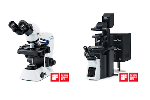 Globally renowned as a symbol of design excellence, the iF design awards celebrate the best in user-focused, ergonomic and efficient design. With over 5,000 submissions from 70 countries, Olympus Scientific Solutions Division has received two of these prestigious awards for its CX23 upright microscope and FLUOVIEW FV3000 confocal laser scanning...
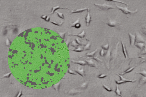 Unlike other multimode readers, Spark lets you actually see what’s happening to your cells, offering automated cell imaging, confluence measurements, cell counting and viability assessments to simplify cell biology protocols and enable long-term, walkaway experiments. Spark has been developed specifically to address the needs of cell-based workflows...
 The ioLight microscope is the first professional quality pocket digital microscope. It is a laboratory grade microscope that fits in a jacket pocket, is simple to use and robust. It unfolds quickly to record and share 5MP still images and real time HD video on a tablet or phone....
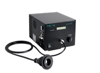 Mercury vapour lamps and bulbs suffer from a number of limiting disadvantages including an operational lifetime rarely exceeding 200 hours and the need for time consuming alignment procedures during installation. In addition Mercury vapour bulbs are susceptible to flickering, limiting the use of these light sources in quantitative fluorescent microscopy....
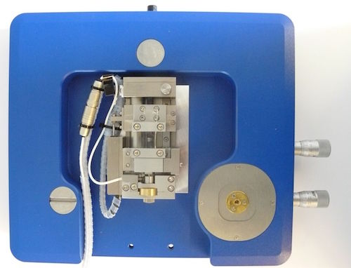 Dr Torsten Mueller is a member of the development team with German nanoscience instrument makers, JPK Instruments. Dr Mueller and his colleagues are based in Berlin supporting a worldwide user network which is continuously testing JPK with requests for new capabilities for their life science and materials nanoscience systems....
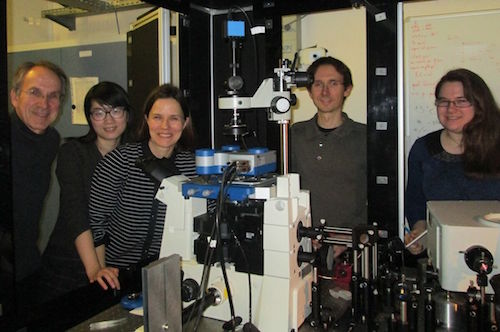 One of the research goals of the Molecular Nanoscience Group at ISMO is to work towards circuits and devices in which surface plasmons (not electrons or photons) are used to transfer and manipulate information. However, why develop plasmonics when there are already solutions using electronics and photonics?....
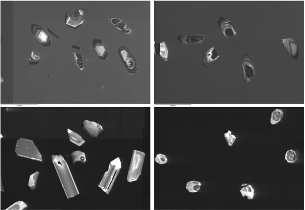 Professor Robert Hall leads field based research into the geology of South East Asia and the western Pacific. Samples of sedimentary rocks are brought back to the UK where they are analysed by heavy and light mineral analyses as well as uranium-lead (U-Pb) zircon geochronology for provenance studies and pathway reconstruction....
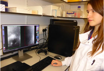 The multidisciplinary bioimaging unit, Micron Oxford, are using the Nanoimager instrument to advance their cellular imaging techniques for both their facilities and research programs. Micron Oxford is a collaborative, multidisciplinary bioimaging unit working with biomedical researchers in the Oxford area and beyond, to apply advanced light microscopy imaging techniques to address key biological questions....
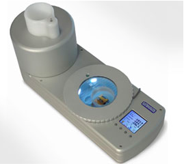 Linkam has been developing cryo stages for correlative microscopy for many years and continues to be at the forefront of cryo-correlative microscopy with the latest update of their LINK software for the CMS196M, providing improved imaging capabilities and the new liquid nitrogen autofill system providing longer run times. Cryo-correlative microscopy has become an established technique in recent years....
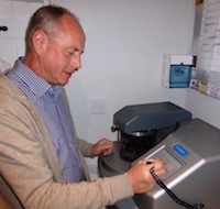 David McCarthy has been working in the world of electron microscopy since the early 70s. In this time, he has seen many changes in instrumentation, techniques and sample preparation methods. His career started back at the London Hospital Medical School for four and a half years before a move that would see him start and manage the Electron Microscopy Unit at the School of Pharmacy, University College London for thirty eight years....
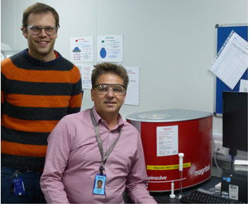 Dr Andrew York is a Senior Principal Scientist at the Johnson Matthey Technology Centre at Sonning Common, UK. His current role includes defining projects on catalysis and reaction engineering both internally and with external partners especially Cambridge University’s Chemical Engineering and Biotechnology Department. Dr York's group purchased the Magritek Spinsolve benchtop NMR spectrometer to characterize materials of importance to the businesses in the...
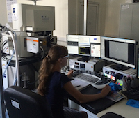 It is famous for its museum which hosts a collection of more than 80,000 samples of rocks, fossils and minerals from all over Romania. Oana-Claudia Barbu and Daniel Bîrg?oanu are both PhD students and research assistants in the electron microscopy laboratory, known as MICROCOSMOS, where they study inorganic and mineral samples....
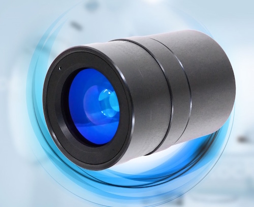 Drawing upon approaching 30 years experience - Resolve Optics has built a strong reputation for specialist lens design and manufacture of smaller production quantities of radiation-resistant (non-browning) lenses and optical products on time to strict quality and target price guidelines. All optical elements within Resolve Optics radiation resistant lens...
 The high field Flexiscan and Powerscan cryogen-free systems from MR Solutions have a compact design and are simply wheeled in and connected to the mains and other services whilst providing state of the art imaging. And - to please the finance team - the capital and running costs of this system are substantially less than a larger, wet magnet system....