Leica Microsystems, a leader in microscopy and scientific instrumentation, has released a new version of its AI-powered image analysis solution, Aivia. The new Aivia 11 features a new deep learning-based cell segmentation algorithm that offers advanced insight creation capabilities for all levels of users. Aivia 11 is based on the innovative Cellpose deep learning segmentation algorithm, which offers several pretrained models for the precise detection of cells from a wide range of image types....
JEOL, the global leader in the development of cutting-edge Electron Microscopes for materials characterization and analysis, introduces its latest Energy Dispersive Spectrometer (EDS), the Gather-X. This new windowless EDS answers the need for higher sensitivity and low-energy X-Ray detection in the Scanning Electron Microscope (SEM). It can collect the entire EDS range produced from the IT800 series Field Emission SEMs including low-energy X-rays down to Lithium.
Working with flow cells, shear stress, adhesion assays? The ibidi pump system can help. The ibidi pump system, combined with ibidi polymer channel slides, provides defined shear stress over long term cell culture periods. All available direct from Thistle Scientific. The Ibidi Pump System is ideal for simulation of various physiological conditions in blood vessels - a pump system for the cultivation of, e.g. endothelial, cells under flow. Compatible with ibidi heating and incubation systems as well as a range of channel slides...
TESCAN ORSAY HOLDING a.s. announces the installation of the AMBER X focused ion beam-scanning electron microscope (FIB-SEM) at the Institute for Factory Automation and Production Systems (FAPS) in Germany. The AMBER X offers a unique combination of plasma FIB with ultra-high resolution (UHR) field emission SEM for multiscale materials characterization. FAPS is using the FIB-SEM for research that will help to improve products used in a wide variety of industries...
Researchers at Swansea University have used a Linkam LTS420E-P stage to make temperature dependent measurements on organic photovoltaic (PV) cells, advancing our understanding of the PV cells that are the basis of solar power generation. Researchers at Swansea University, UK, have proven that it is possible to achieve near-unity charge generation quantum yields in organic solar cells...
Transmission electron microscopes (TEMs) have evolved throughout the years to offer ever greater resolution and magnification capabilities. However, for an instrument to work at its best, all external interferences that can affect the position of the electron beam – such as vibrations and stray magnetic fields – need to be removed. Researchers at Cataler Corporation have turned to Spicer Consulting to help them to optimise their equipment for investigations into non-homogeneous catalysts....
Ground-breaking technology unlocks label-free 3D and 4D live cell imaging on standard imaging plates for higher-throughput and automated screening applications. A novel optical microscope utilizing incoherent light to generate holographic images of unlabelled live cells is now available from Tomocube. Called HT-X1, the new microscope is ideally suited to higher-throughput and automated screening applications with its ability to image multi-well plate formats, large field-of-view, laser autofocus system, and very high performance 0.95NA objective...
CytoSMART Technologies, an Axion Bio company, announce the launch of a new Organoid Analysis Module for use with its flagship CytoSMART Omni live-cell imaging product line. The innovative software module, which incorporates next-generation machine learning capabilities, is designed to meet growing scientific demand from researchers and drug developers using complex three-dimensional (3D) in vitro models such as organoids, spheroids, and tumoroids to study disease mechanisms...
Prior Scientific, a manufacturer of microscopy solutions and precision optical and electromechanical equipment, announce the launch of the CS200 line of joysticks. These joysticks offer a simple, ergonomic, and cost-effective way to manually control your Prior XY stage and Z drive and replace the current PS3J100 joystick. Prior Scientific release the new CS200 series of joysticks and TS200 touchscreen display to control their ProScan and OptiScan...
Atik Cameras is thrilled to launch ChemiMOS 9.0. This 9-megapixel camera, with set point cooling of -20°C, has been optimised for long exposures. Hours of exposure time has previously only been available with CCD technology, but is now possible with CMOS technology thanks to the ChemiMOS zero-amp glow and low-noise design. The square format, 'K' grade sensor is guaranteed for?continuous?use, while the 3000 × 3000 resolution and 3.76 µm pixel size are perfect for multiple scientific applications...
To develop new drugs and vaccines, detailed knowledge about nature’s smallest biological building blocks – the biomolecules – is required. Researchers at Chalmers University of Technology, Sweden, are now presenting a groundbreaking microscopy technique that allows proteins, DNA and other tiny biological particles to be studied in their natural state in a completely new way...
SPT Labtech ("SPT" or "The Company") a global leader in the design and development of automated instrumentation and consumables for life science applications, announces its acquisition by EQT IX Fund ("EQT Private Equity"), a purpose-driven, global investment organization, from current owners Battery Ventures. The deal will underpin and accelerate SPT Labtech’s impressive trajectory of market growth and leadership in innovative product development...
Bruker Corporation displays its high-value scientific instrumentation, software and integrated solutions for applications in materials and energy research, biopharmaceuticals, applied markets, as well as life science and translational research at Analytica 2022. Frank H. Laukien, PhD, President and CEO of Bruker Corporation, commented: “Our differentiated high-value scientific instruments and solutions shown at Analytica 2022 demonstrate the diversity, flexibility and high performance that Bruker brings to the world of laboratory science....
Fast, large field-of-view MALDI HiPLEX-IHC imaging of targeted proteins, overlaid with unbiased small molecule MALDI Imaging on fresh frozen or FFPE tissues offers compelling innovation for spatial biology and cancer research. At the 70th ASMS Conference, Bruker Corporation announced key innovations for spatial multiomics of tissue and tumor microenvironments (TME). Following Bruker’s strategic partnership with AmberGen, key enhancements are introduced for MALDI HiPLEX-IHC mass spectrometry imaging...
Evident, a wholly owned subsidiary of Olympus Corporation, is proud to announce that its Olympus Provi™ CM20 incubation monitoring system is the 2022 recipient of the prestigious "Best of the Best" award in the Product Design category of the Red Dot Design Award. This Award is bestowed on a product that the Red Dot Jury deems as groundbreaking and is the highest honor given in this world-renowned design competition...
Leica Microsystems, a leading provider of microscopy and scientific instruments, has introduced a new generation of its multiplexed imaging solution, Cell DIVE, including software and hardware improvements. The more scalable and efficient multiplexing platform addresses spatial cell biology and function within the tissue microenvironment, offering researchers the freedom to select from...
Hyperpolarized water boosts signal intensities of proteins, DNA and membranes. A small group of researchers including Dennis Kurzbach from the Faculty of Chemistry of the University of Vienna just published in „Nature Protocols” an advanced NMR (Nuclear Magnetic Resonance) method to monitor fast and complicated biomolecular events such as protein folding...
Indica Labs, the leading provider of computational pathology software and services, is excited to announce a CE-IVD Mark for HALO Prostate AI, a deep learning-based screening tool designed to assist pathologists in identifying and grading prostate cancer in core needle biopsies. Prostate cancer is the most common cancer diagnosed in men. With over 1.4 million cases reported worldwide in 2020, the incidence rates continue to rise with wider availability of screening tests, such as PSA....
ZEISS releases the next generation of ZEISS Lattice Lightsheet 7. The microscope system was introduced to the market in December 2020 and opened up a new way for researchers to explore the dynamics of life at subcellular resolution. It was the first commercial, easy-to-use implementation of the lattice light-sheet technology known for enabling sample-preserving, long-term imaging of living cells...
Vision-Based Sports Analytics, Training, and Player Assessment for Europe’s Top Football Club. Football is not only the world’s favorite pastime, but also a multi-billion-dollar industry. Individual players can command millions of dollars in salaries and transfer fees. With so much at stake, coaches and scouts need to make the most informed personnel choices, then, keep their players on top condition...
Thermo Fisher Scientific Inc., the world leader in serving science, and TransMIT GmbH Center for Mass Spectrometric Developments have announced a co-marketing agreement to promote the use of a mass spectrometry imaging (MSI) platform for spatial multi-omics applications in pharma and clinical labs. As part of the relationship, TransMIT will combine its proprietary scanning microprobe matrix-assisted laser desorption/ionization (SMALDI) MSI and 3D-surface MSI technology...
At the ISMRM 2022 conference, Bruker announced the launch of innovative 7 Tesla and 9.4 Tesla conduction-cooled Maxwell magnets for its market-leading preclinical magnetic resonance imaging (MRI) systems portfolio. Following the success of its BioSpec® Maxwell 3 Tesla model, the new range of Maxwell 7 Tesla and 9.4 Tesla magnets eliminates the need for liquid Helium or Nitrogen refills, while offering high-field sensitivity and resolution for advanced preclinical MRI and PET/MR research...

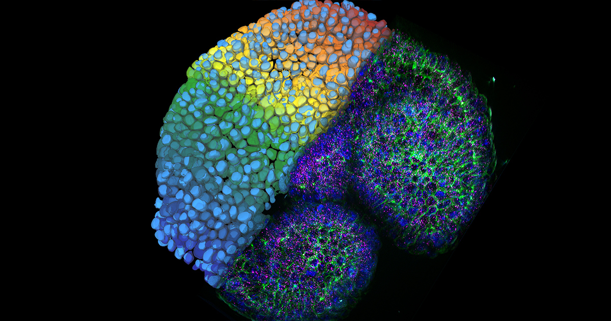 Leica Microsystems, a leader in microscopy and scientific instrumentation, has released a new version of its AI-powered image analysis solution, Aivia. The new Aivia 11 features a new deep learning-based cell segmentation algorithm that offers advanced insight creation capabilities for all levels of users. Aivia 11 is based on the innovative Cellpose deep learning segmentation algorithm, which offers several pretrained models for the precise detection of cells from a wide range of image types....
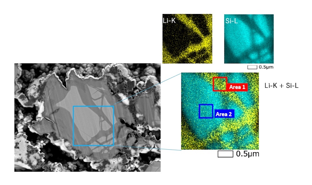 JEOL, the global leader in the development of cutting-edge Electron Microscopes for materials characterization and analysis, introduces its latest Energy Dispersive Spectrometer (EDS), the Gather-X. This new windowless EDS answers the need for higher sensitivity and low-energy X-Ray detection in the Scanning Electron Microscope (SEM). It can collect the entire EDS range produced from the IT800 series Field Emission SEMs including low-energy X-rays down to Lithium.
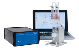 Working with flow cells, shear stress, adhesion assays? The ibidi pump system can help. The ibidi pump system, combined with ibidi polymer channel slides, provides defined shear stress over long term cell culture periods. All available direct from Thistle Scientific. The Ibidi Pump System is ideal for simulation of various physiological conditions in blood vessels - a pump system for the cultivation of, e.g. endothelial, cells under flow. Compatible with ibidi heating and incubation systems as well as a range of channel slides...
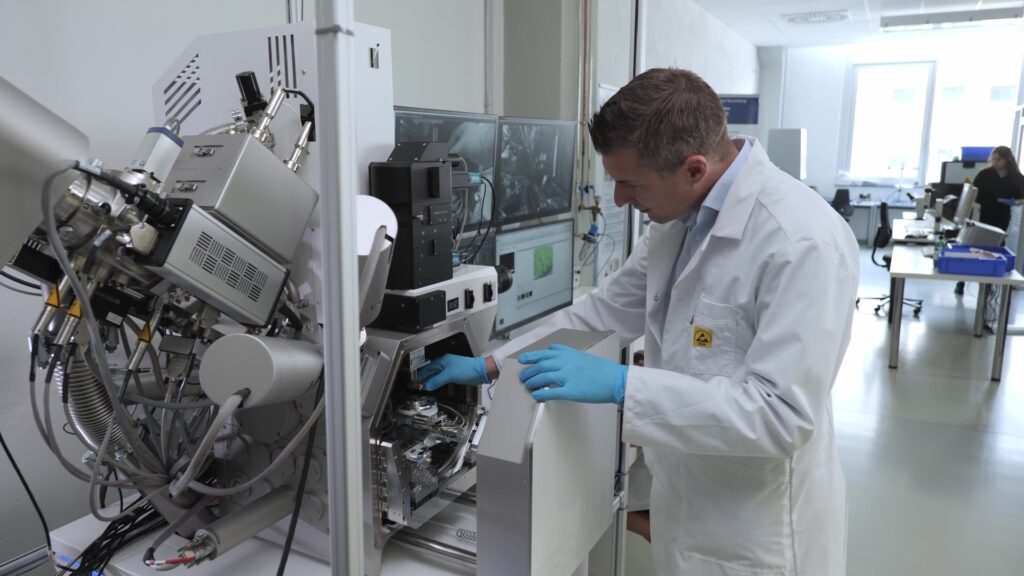 TESCAN ORSAY HOLDING a.s. announces the installation of the AMBER X focused ion beam-scanning electron microscope (FIB-SEM) at the Institute for Factory Automation and Production Systems (FAPS) in Germany. The AMBER X offers a unique combination of plasma FIB with ultra-high resolution (UHR) field emission SEM for multiscale materials characterization. FAPS is using the FIB-SEM for research that will help to improve products used in a wide variety of industries...
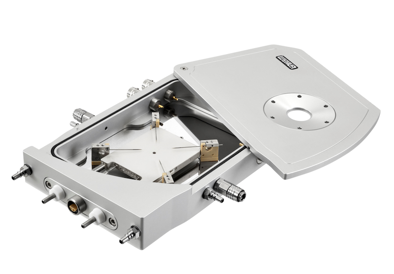 Researchers at Swansea University have used a Linkam LTS420E-P stage to make temperature dependent measurements on organic photovoltaic (PV) cells, advancing our understanding of the PV cells that are the basis of solar power generation. Researchers at Swansea University, UK, have proven that it is possible to achieve near-unity charge generation quantum yields in organic solar cells...
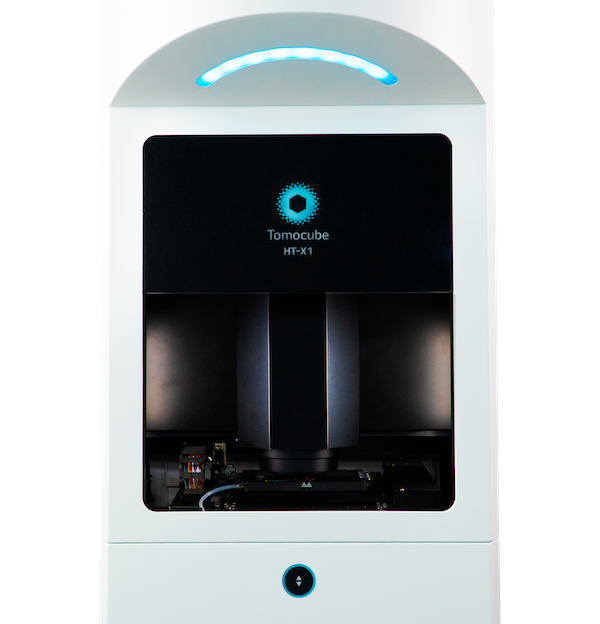 Ground-breaking technology unlocks label-free 3D and 4D live cell imaging on standard imaging plates for higher-throughput and automated screening applications. A novel optical microscope utilizing incoherent light to generate holographic images of unlabelled live cells is now available from Tomocube. Called HT-X1, the new microscope is ideally suited to higher-throughput and automated screening applications with its ability to image multi-well plate formats, large field-of-view, laser autofocus system, and very high performance 0.95NA objective...
 CytoSMART Technologies, an Axion Bio company, announce the launch of a new Organoid Analysis Module for use with its flagship CytoSMART Omni live-cell imaging product line. The innovative software module, which incorporates next-generation machine learning capabilities, is designed to meet growing scientific demand from researchers and drug developers using complex three-dimensional (3D) in vitro models such as organoids, spheroids, and tumoroids to study disease mechanisms...
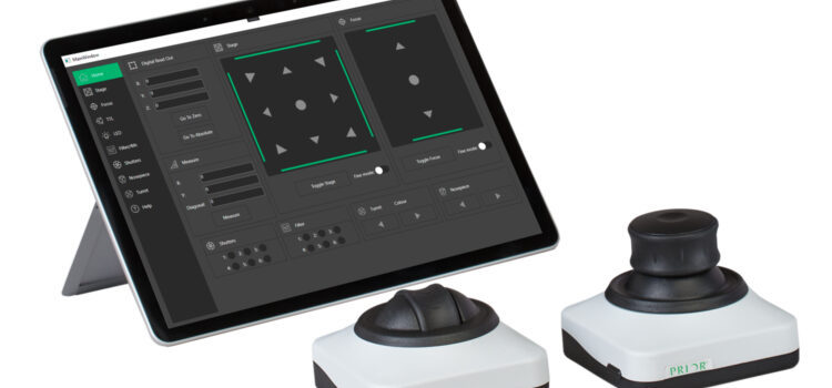 Prior Scientific, a manufacturer of microscopy solutions and precision optical and electromechanical equipment, announce the launch of the CS200 line of joysticks. These joysticks offer a simple, ergonomic, and cost-effective way to manually control your Prior XY stage and Z drive and replace the current PS3J100 joystick. Prior Scientific release the new CS200 series of joysticks and TS200 touchscreen display to control their ProScan and OptiScan...
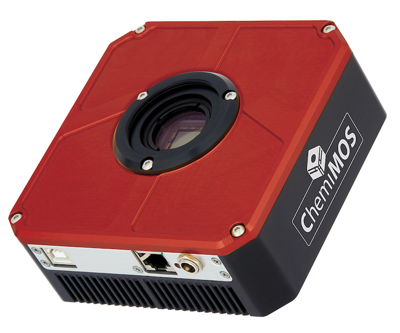 Atik Cameras is thrilled to launch ChemiMOS 9.0. This 9-megapixel camera, with set point cooling of -20°C, has been optimised for long exposures. Hours of exposure time has previously only been available with CCD technology, but is now possible with CMOS technology thanks to the ChemiMOS zero-amp glow and low-noise design. The square format, 'K' grade sensor is guaranteed for?continuous?use, while the 3000 × 3000 resolution and 3.76 µm pixel size are perfect for multiple scientific applications...
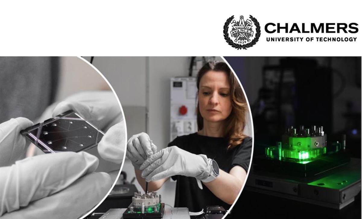 To develop new drugs and vaccines, detailed knowledge about nature’s smallest biological building blocks – the biomolecules – is required. Researchers at Chalmers University of Technology, Sweden, are now presenting a groundbreaking microscopy technique that allows proteins, DNA and other tiny biological particles to be studied in their natural state in a completely new way...
 Bruker Corporation displays its high-value scientific instrumentation, software and integrated solutions for applications in materials and energy research, biopharmaceuticals, applied markets, as well as life science and translational research at Analytica 2022. Frank H. Laukien, PhD, President and CEO of Bruker Corporation, commented: “Our differentiated high-value scientific instruments and solutions shown at Analytica 2022 demonstrate the diversity, flexibility and high performance that Bruker brings to the world of laboratory science....
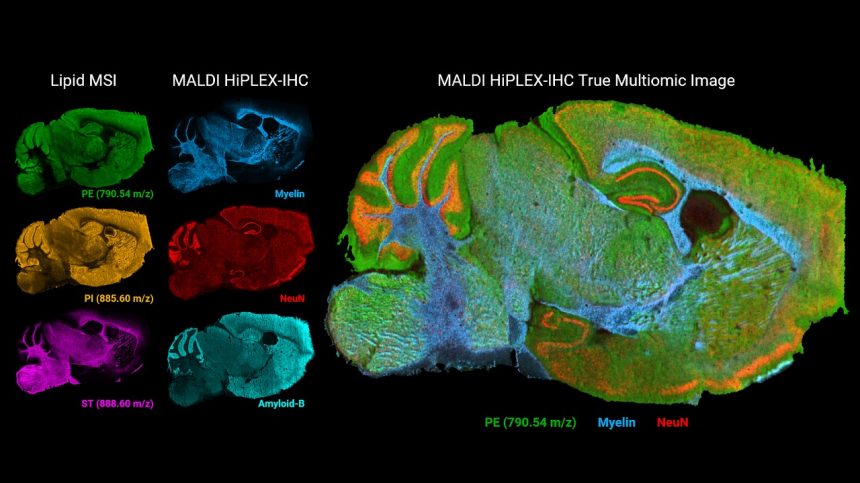 Fast, large field-of-view MALDI HiPLEX-IHC imaging of targeted proteins, overlaid with unbiased small molecule MALDI Imaging on fresh frozen or FFPE tissues offers compelling innovation for spatial biology and cancer research. At the 70th ASMS Conference, Bruker Corporation announced key innovations for spatial multiomics of tissue and tumor microenvironments (TME). Following Bruker’s strategic partnership with AmberGen, key enhancements are introduced for MALDI HiPLEX-IHC mass spectrometry imaging...
 Evident, a wholly owned subsidiary of Olympus Corporation, is proud to announce that its Olympus Provi™ CM20 incubation monitoring system is the 2022 recipient of the prestigious "Best of the Best" award in the Product Design category of the Red Dot Design Award. This Award is bestowed on a product that the Red Dot Jury deems as groundbreaking and is the highest honor given in this world-renowned design competition...
 Leica Microsystems, a leading provider of microscopy and scientific instruments, has introduced a new generation of its multiplexed imaging solution, Cell DIVE, including software and hardware improvements. The more scalable and efficient multiplexing platform addresses spatial cell biology and function within the tissue microenvironment, offering researchers the freedom to select from...
 Hyperpolarized water boosts signal intensities of proteins, DNA and membranes. A small group of researchers including Dennis Kurzbach from the Faculty of Chemistry of the University of Vienna just published in „Nature Protocols” an advanced NMR (Nuclear Magnetic Resonance) method to monitor fast and complicated biomolecular events such as protein folding...
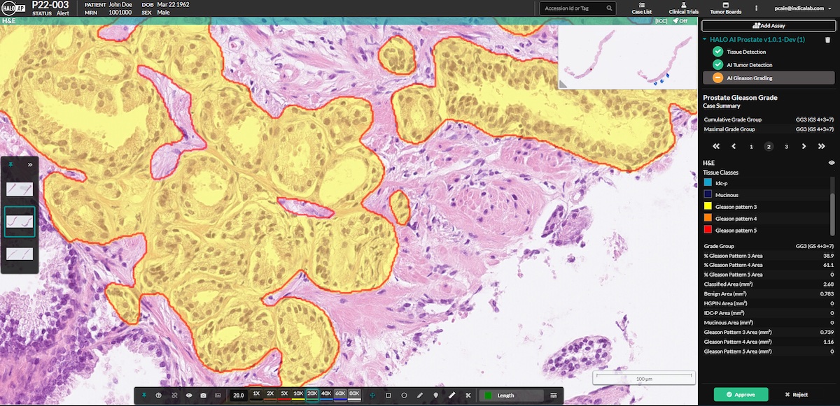 Indica Labs, the leading provider of computational pathology software and services, is excited to announce a CE-IVD Mark for HALO Prostate AI, a deep learning-based screening tool designed to assist pathologists in identifying and grading prostate cancer in core needle biopsies. Prostate cancer is the most common cancer diagnosed in men. With over 1.4 million cases reported worldwide in 2020, the incidence rates continue to rise with wider availability of screening tests, such as PSA....
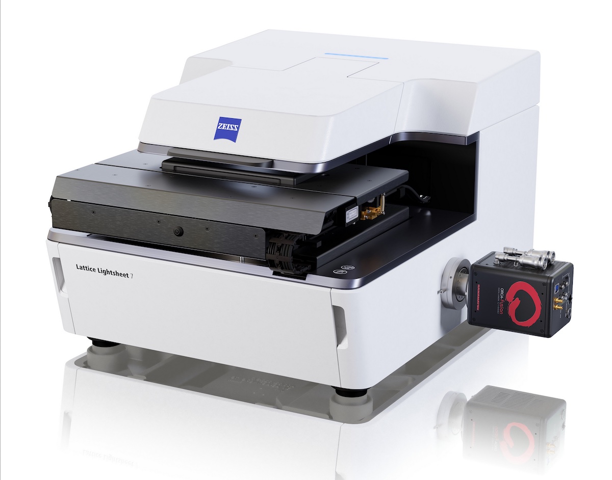 ZEISS releases the next generation of ZEISS Lattice Lightsheet 7. The microscope system was introduced to the market in December 2020 and opened up a new way for researchers to explore the dynamics of life at subcellular resolution. It was the first commercial, easy-to-use implementation of the lattice light-sheet technology known for enabling sample-preserving, long-term imaging of living cells...
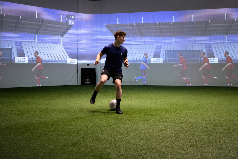 Vision-Based Sports Analytics, Training, and Player Assessment for Europe’s Top Football Club. Football is not only the world’s favorite pastime, but also a multi-billion-dollar industry. Individual players can command millions of dollars in salaries and transfer fees. With so much at stake, coaches and scouts need to make the most informed personnel choices, then, keep their players on top condition...
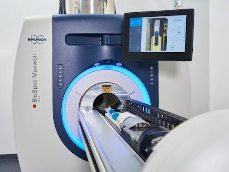 At the ISMRM 2022 conference, Bruker announced the launch of innovative 7 Tesla and 9.4 Tesla conduction-cooled Maxwell magnets for its market-leading preclinical magnetic resonance imaging (MRI) systems portfolio. Following the success of its BioSpec® Maxwell 3 Tesla model, the new range of Maxwell 7 Tesla and 9.4 Tesla magnets eliminates the need for liquid Helium or Nitrogen refills, while offering high-field sensitivity and resolution for advanced preclinical MRI and PET/MR research...