Hitachi announces the SU8200 – a new type of cold field emitter SEM
Hitachi High-Technologies announces the worldwide release of the new SU8200 series FE-SEMs – a range of high-performance instruments which provides the perfect fusion of high resolution imaging and analysis. Employing a new type of cold field emission (CFE) gun the SU8200 combines the inherent high resolution and brightness of conventional CFE with previously unseen beam stability and probe currents...
UK-based MR Solutions, a world leader in the development and manufacture of pre clinical MRI scanners, has developed the world’s first range of high-performance, 3T, scanners using super-conducting magnets which eliminate the need for liquid helium cooling. This advance has significant benefits for the scientific community as the global shortage of helium has been risking research projects across the world with experts predicting that the second most common element in the universe may be depleted by the middle of the century[1][1]. MR Solutions Chief Executive Dr David Taylor commented: “At MR Solutions we have developed a range of pre clinical MRI scanners which eliminate the need for helium...
NanoSight reports on how Nanoparticle Tracking Analysis, NTA, is being used in the research project "Engineering of Biomolecules" at the Lorraine University, based in Nancy.
Dr Jordane Jasniewski works at the Laboratory of Biomolecules Engineering (LIBio) at Lorraine University, Nancy, and teaches in Food Chemistry at ENSAIA, an engineering school. He is a member of the research team working on the engineering of biomolecules to understand their structure and function to help develop new molecular architectures, to be applied in the areas of foods & agrichemicals, nutrition, pharmacology and cosmetics...
New automated particle location and chemical characterization tool
HORIBA Scientific, a leading innovator in the manufacture of high performance scientific and analytical equipment, is announcing the launch of their new ParticleFinder module for the LabSpec 6 Spectroscopy Suite. In combination with the full range of HORIBA Raman microscopes ParticleFinderbrings a new level of automation and ease of use for particle analysts who require the unique chemical characterization tools that Raman can offer...
JPK Instruments, a world-leading manufacturer of nanoanalytic instrumentation for research in life sciences and soft matter, reports on the Yan Jie single molecule biophysics research group at the Mechanobiology Institute of NUS and their use of optical tweezers.
The Mechanobiology Institute (MBI) of the National University of Singapore (NUS) was created through joint funding by the National Research Foundation and the Ministry of Education with the goal of creating a new research center in mechanobiology to benefit both the discipline and Singapore. Its' primary focus is to identify, measure and describe how the forces for motility and morphogenesis are expressed at the molecular, cellular and tissue level...
Market leaders in temperature controlled microscopy, Linkam Scientific Instruments, report on the use of their popular THMSG600 and TS1500 heating stages which are being used for geological research at the Institute of Earth Sciences, Taiwan.
Founded in 1982, the Institute of Earth Sciences (IES) conducts research in two major disciplines: geophysics and geochemistry. The mineral and rock physics laboratory in the IES is one of the most prominent labs in mineral physics in Taiwan. It is one of nearly 30 institutes and centres that form the Academia Sinica (AS), the main governmental organization for basic research in all academic disciplines in Taiwan. Recent research has included mantle dynamics modelling, heat flow and space remote sensing...
Artemis CCD Limited, a leading manufacturer of cooled CCD cameras for low-light applications today launches the FS92 featuring 9.19 million effective pixels in a Type 1 3388 x 2712 pixel array. A combination of high resolution and high sensitivity increases significantly the application options for the scientific user and OEM customer. As with all members of the FS range of cameras a typical read noise of 5 electrons makes the FS92 a perfect choice for low light applications including fluorescence and chemi-luminescence imaging and with the asymmetric pixel binning function, a highly effective detector in optical spectroscopy...
Resolve Optics has supplied a high performance 8-channel optical module to Specialised Imaging (Tring, UK) to enable researchers to undertake UV imaging experiments at ultra high speed using their SIM ultra high-speed framing camera.
Using the new UV optical module and UV ICCD enables the SIM camera to optimally operate inthe 230-400nm region allowing scientists to study phenomena that emit ultraviolet radiation during the initial phase, such as corona from a pre-streamer in electrical discharge studies. Resolve Optics has a proven track record of developing state-of-the-art optical solutions that meet the highest standards and solve the most challenging problems for its customers...
AMSBIO has introduced a new range of 96-well format 3D Spheroid Cell Proliferation / Viability Assays, providing a new tool to allow cell-based assays to be carried out in 3D.
The new AMSBIO 3D Spheroid Proliferation/Viability Assay provides a useful tool for modeling tumor response in vitro. The kit utilises a 3D Culture Qualified 96 Well Spheroid Formation Plate alongside a specialised Spheroid Formation Extracellular Matrix to drive aggregation and/or spheroid formation of cells. Upon completion of spheroid formation, the spheroid may be treated with pharmacological agents to evaluate tumor viability after drug treatment...
ChemiDoc-ItTS2 Imager for Genomics and Proteomics Research now with Multiple Language Interface
UVP, LLC now offers the ChemiDoc-It®TS2 Imager software with multiple language options. English is the standard language format of the software and users can select from Chinese (simplified), Turkish, Japanese, Spanish, Korean and Russian for all screen text and buttons. "The language offerings will assist researchers in expediting and simplifying the image acquisition capabilities of the system. The integrated touch screen with software interaction in local languages makes the system highly intuitive and friendly," says Myrna Espiritu, International Sales Manager for UVP...
Worldwide the Laser Particle Sizers ANALYSETTE 22 ensures precise determination of particle size distributions, by Static Laser Scattering – in production and quality control as well as in research and development. Benefit from their decisive advantages: extremely simple operation and short analysis times for consistently reproducible and reliable results. AutoSampler is ideal for automation of measurement series: 26 positions for 40 ml containers are available for complete sample feeding, automatic dispersion, measurement and cleaning...
Market leaders in temperature controlled microscopy, Linkam Scientific Instruments report on the use of their dynamic MDSG600 stage for geological research at the Geological Survey of Finland.
Founded in 1886, the Geological Survey of Finland (GTK) was created to promote the systematic and sustainable use of the Earth's resources through the production and dissemination of geological information to society. The GTK is Finland's national geoscientific information centre and it participates actively in international research and project work, with the areas of focus falling into three main categories: mineral resources and exploration; energy supply and environment; and land use and construction...
NanoSight announces that Particle Technology Labs, PTL, has established cGMP compliance for Nanoparticle Tracking Analysis, NTA, and this is now available on a contract basis to the pharmaceutical industry.
PTL is the leading particle characterization research and advisory company in the United States, providing assistance to a wide range of industries. Nanoparticle Tracking Analysis, NTA, has been adopted for both research-based projects and as a quality control tool for regulated industries. PTL’s NanoSight LM10-HSB instrument is certified cGMP as of June 4, 2013. cGMP refers to the Current Good Manufacturing Practice regulations enforced by the US Food and Drug Administration (FDA)...
With the introduction of the next generation Phenom ProX desktop SEM Phenom-World confirms its positions in the top of the high-end table top for SEM imaging and analysis.
With a magnification range up to 100,000x and resolution down to 17nm the Phenom ProX delivers more detailed information than ever before. All Phenom-World scanning electron microscopes are intuitive to use, compact, fast to create results and built to high quality standards. The most extended solution in the range is the next generation Phenom ProX. The advanced system identifies different elements in a specimen by using the integrated Element Identification software with a specially designed EDS detector. With new techniques and software-developments, the magnification range has been extended from max. 45,000x to 100,000x magnification. Combined with a resolution of 17nm, the next generation Phenom ProX is a valuable instrument for a wide variety of applications....
Leica DMC2900 for Brightfield, Leica DFC3000 G for Fluorescence Applications
As of June, Leica Microsystems has launched two cameras that are ideal instruments for routine brightfield or fluorescence applications. Both cameras are equipped with sensors dedicated to the respective applications that allow live images of up to 30 frames per second and a USB 3.0 interface for fast data transfer. The Leica Application Suite (LAS) and the fluorescence counterpart LAS Advanced Fluorescence (LAS AF) software platform provide for image acquisition and support users in analysis and documentation...
Expanding from light and electron microscopy into X-ray microscopy solutions
ZEISS, the international leader in the fields of optics and optoelectronics today announced the planned acquisition of the US-based Xradia, Inc. Xradia is an medium-size company providing innovative 3D X-ray microscopes for industrial and academic research applications. The closing of the transaction is subject to the fulfillment of customary closing conditions including a required filing with the U.S. competition authorities. After closing, Xradia, Inc. will operate under the new name Carl Zeiss X-ray Microscopy, Inc...
Event Date: June 27, 2013 at 12:00 PM Eastern Daylight Time
Multi-mode microplate readers are a useful tool for cell-based assays. These instruments use PMT-based optics to provide a cell population-averaged assay signal relative to assay controls that can quantify biomolecule expression, activation, inhibition and post-translational modulation. Fluorescence microscopy allows the analysis of sub-populations of cells or even analysis of a single cell, depending on the magnification used...
Read MoreAny Assay, Any Wavelength, Any Bandwidth with the New CLARIOstar High Performance Microplate ReaderJun 24, 2013
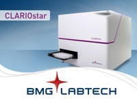
Any Assay, Any Wavelength, Any Bandwidth
Over the years, many different assays have been designed to work on many different microplate readers. As new techniques emerged, so did new microplate reader technology. The new CLARIOstar microplate reader from BMG LABTECH is the latest evolution in microplate reader instrumentation....
Olympus continues to expand the capabilities of its IX3 inverted microscope frames with the launch of cellVivo - a new, modular and flexible incubation system, including darkroom and laser-safety capabilities.
Fully adaptable for basic and high-end IX3 microscope systems alike, cellVivo offers precise control of environmental conditions, as well as user-friendly remote monitoring. Designed with the complete workflow in mind, Olympus has launched itscellVivo incubation system, for precise and ergonomic environmental control of advanced live cell imaging. Following the modular approach of the IX3 range of inverted microscope frames, cellVivo is based on a “one size fits all” concept, with adaptors for various IX73 and IX83 frame types, circumventing the need for multiple incubation systems for distinct microscope configurations...
FREE NanoSight Webinar on the use of Nanoparticle Tracking Analysis in Drug Delivery Research.
NanoSight, leading manufacturers of unique nanoparticle characterization technology, address the challenges of nanoparticle control and analysis in the latest of their popular webinar program. On Thursday 6th June, NanoSight hosted a webinar to look at how Nanoparticle Tracking Analysis (NTA) is used to as a characterization tool during the development of drug delivery systems. During the webinar NanoSight.....
FLIR Systems has announced the release of a new range of thermal imaging cameras optimised for industrial R&D applications including tyre / brake testing, on and through glass measurements, welding / soldering processes and fast moving process control applications.
FLIR A3500sc / A6500sc series thermal imaging cameras incorporate a cooled 3-5 micron MW-IR detector making R&D applications that need better image quality, more sensitivity and a higher frame rate routine. Achieving a high thermal sensitivity of <25 mK - FLIR A3500sc / A6500sc series cameras are able to capture the finest image details and temperature difference information. In addition, precise camera synchronization and triggering makes these cameras ideal for high-speed, high sensitivity applications...
The advanced Spectroline®Bi-O-Vision™ Series transilluminators feature two workstations, producing both 312nm ultraviolet and white light.
The TD-1000R model offers fixed-intensity while the TVD-1000R model offers variable-intensity control of either UV or white light. These units are continuously adjustable from 100% down to 50%. This enables life science researchers to select medium wavelength ultraviolet or white light illumination to view fluorescent gels or visible blots...

 Hitachi High-Technologies announces the worldwide release of the new SU8200 series FE-SEMs – a range of high-performance instruments which provides the perfect fusion of high resolution imaging and analysis. Employing a new type of cold field emission (CFE) gun the SU8200 combines the inherent high resolution and brightness of conventional CFE with previously unseen beam stability and probe currents...
 Dr Jordane Jasniewski works at the Laboratory of Biomolecules Engineering (LIBio) at Lorraine University, Nancy, and teaches in Food Chemistry at ENSAIA, an engineering school. He is a member of the research team working on the engineering of biomolecules to understand their structure and function to help develop new molecular architectures, to be applied in the areas of foods & agrichemicals, nutrition, pharmacology and cosmetics...
 HORIBA Scientific, a leading innovator in the manufacture of high performance scientific and analytical equipment, is announcing the launch of their new ParticleFinder module for the LabSpec 6 Spectroscopy Suite. In combination with the full range of HORIBA Raman microscopes ParticleFinder brings a new level of automation and ease of use for particle analysts who require the unique chemical characterization tools that Raman can offer...
 Founded in 1982, the Institute of Earth Sciences (IES) conducts research in two major disciplines: geophysics and geochemistry. The mineral and rock physics laboratory in the IES is one of the most prominent labs in mineral physics in Taiwan. It is one of nearly 30 institutes and centres that form the Academia Sinica (AS), the main governmental organization for basic research in all academic disciplines in Taiwan. Recent research has included mantle dynamics modelling, heat flow and space remote sensing...
 Artemis CCD Limited, a leading manufacturer of cooled CCD cameras for low-light applications today launches the FS92 featuring 9.19 million effective pixels in a Type 1 3388 x 2712 pixel array. A combination of high resolution and high sensitivity increases significantly the application options for the scientific user and OEM customer. As with all members of the FS range of cameras a typical read noise of 5 electrons makes the FS92 a perfect choice for low light applications including fluorescence and chemi-luminescence imaging and with the asymmetric pixel binning function, a highly effective detector in optical spectroscopy...
 Using the new UV optical module and UV ICCD enables the SIM camera to optimally operate inthe 230-400nm region allowing scientists to study phenomena that emit ultraviolet radiation during the initial phase, such as corona from a pre-streamer in electrical discharge studies. Resolve Optics has a proven track record of developing state-of-the-art optical solutions that meet the highest standards and solve the most challenging problems for its customers...
 The new AMSBIO 3D Spheroid Proliferation/Viability Assay provides a useful tool for modeling tumor response in vitro. The kit utilises a 3D Culture Qualified 96 Well Spheroid Formation Plate alongside a specialised Spheroid Formation Extracellular Matrix to drive aggregation and/or spheroid formation of cells. Upon completion of spheroid formation, the spheroid may be treated with pharmacological agents to evaluate tumor viability after drug treatment...
 UVP, LLC now offers the ChemiDoc-It®TS2 Imager software with multiple language options. English is the standard language format of the software and users can select from Chinese (simplified), Turkish, Japanese, Spanish, Korean and Russian for all screen text and buttons. "The language offerings will assist researchers in expediting and simplifying the image acquisition capabilities of the system. The integrated touch screen with software interaction in local languages makes the system highly intuitive and friendly," says Myrna Espiritu, International Sales Manager for UVP...
 Founded in 1886, the Geological Survey of Finland (GTK) was created to promote the systematic and sustainable use of the Earth's resources through the production and dissemination of geological information to society. The GTK is Finland's national geoscientific information centre and it participates actively in international research and project work, with the areas of focus falling into three main categories: mineral resources and exploration; energy supply and environment; and land use and construction...
 PTL is the leading particle characterization research and advisory company in the United States, providing assistance to a wide range of industries. Nanoparticle Tracking Analysis, NTA, has been adopted for both research-based projects and as a quality control tool for regulated industries. PTL’s NanoSight LM10-HSB instrument is certified cGMP as of June 4, 2013. cGMP refers to the Current Good Manufacturing Practice regulations enforced by the US Food and Drug Administration (FDA)...
 With a magnification range up to 100,000x and resolution down to 17nm the Phenom ProX delivers more detailed information than ever before. All Phenom-World scanning electron microscopes are intuitive to use, compact, fast to create results and built to high quality standards. The most extended solution in the range is the next generation Phenom ProX. The advanced system identifies different elements in a specimen by using the integrated Element Identification software with a specially designed EDS detector. With new techniques and software-developments, the magnification range has been extended from max. 45,000x to 100,000x magnification. Combined with a resolution of 17nm, the next generation Phenom ProX is a valuable instrument for a wide variety of applications....
 As of June, Leica Microsystems has launched two cameras that are ideal instruments for routine brightfield or fluorescence applications. Both cameras are equipped with sensors dedicated to the respective applications that allow live images of up to 30 frames per second and a USB 3.0 interface for fast data transfer. The Leica Application Suite (LAS) and the fluorescence counterpart LAS Advanced Fluorescence (LAS AF) software platform provide for image acquisition and support users in analysis and documentation...

 Over the years, many different assays have been designed to work on many different microplate readers. As new techniques emerged, so did new microplate reader technology. The new CLARIOstar microplate reader from BMG LABTECH is the latest evolution in microplate reader instrumentation....
 FLIR A3500sc / A6500sc series thermal imaging cameras incorporate a cooled 3-5 micron MW-IR detector making R&D applications that need better image quality, more sensitivity and a higher frame rate routine. Achieving a high thermal sensitivity of <25 mK - FLIR A3500sc / A6500sc series cameras are able to capture the finest image details and temperature difference information. In addition, precise camera synchronization and triggering makes these cameras ideal for high-speed, high sensitivity applications...