
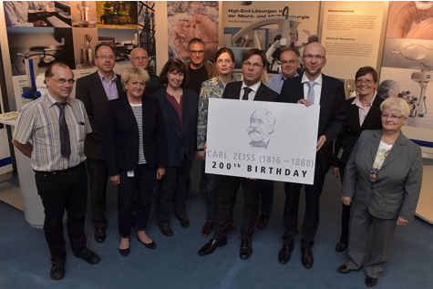
 Carl Friedrich Zeiss was born in the German city of Weimar on 11 September 1816. The company ZEISS is celebrating the 200th birthday of its founding father by organizing many different activities and events – in addition to the Carl Zeiss Day on 11 September 2016 in downtown Jena, there will also be a touring exhibition and a book. ZEISS will celebrate its prominent founder together with players from the city of Jena, the ZEISS...
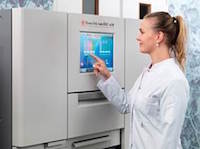 The AutoTEC a120 is a second-generation, fully automated tissue embedder that eliminates the labor intensive need to manually orient and embed tissue specimens and form tissue or cell paraffin blocks. The AutoTEC technology, combined with the Paraform® Sectionable Cassette System ensure that the orientation of specimens is locked from grossing to microtomy for all routine tissue types, thereby eliminating the risk of orientation mistakes...
 Bruker today introduces four important new preclinical imaging systems at the World Molecular Imaging Congress 2015 in Honolulu, Hawaii. The novel products launched at WMIC each deliver improved performance and convenience for routine imaging, and open new horizons for advanced translational research, while Bruker’s platform philosophy facilitates multimodal imaging projects. Researchers will gain a more complete picture and...
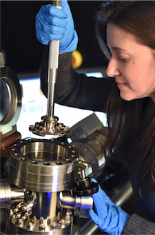 The US Naval Research Laboratory (NRL) has taken delivery of the US Department of Defense’s first Local Electrode Atom Probe (LEAP) microscope. The high- performance atom probe from CAMECA, a unit of AMETEK Materials Analysis, is used in advanced materials analysis to provide precise atom-by-atom identification, 3-D spatial positioning, and accurate atomic-scale reconstruction of a material’s microstructure. Since their development in the 1960s, atom probes have contributed to...
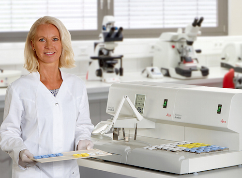 The independent Arcadia modules offer users the flexibility to organize the embedding workflow to their liking. HistoCore Arcadia is a combination of the paraffin dispensing module Arcadia H and the cold plate Arcadia C. Simple operation and precise control help improve the quality, reliability and speed of embedding work. The new station is designed with the user in mind and incorporates comfortable wrist-pads that increase comfort and stability...
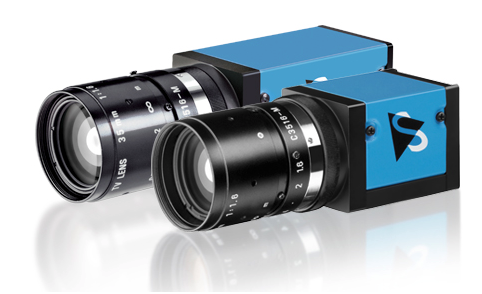 The Imaging Source, an international manufacturer of machine vision cameras, has just announced the immediate availability of new industrial cameras with the Sony Full HD WDR Sensor IMX236. The cameras feature compact, robust industrial housing with C/CS- or S-Mount and are available as GigE (PoE) and USB 3 versions in monochrome and color. With integrated WDR (Wide Dynamic Range) and a resolution from VGA to Full HD, they are...
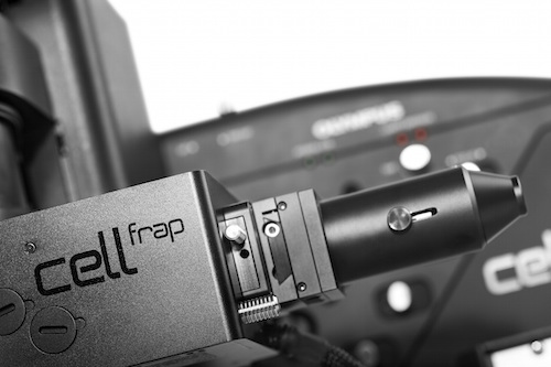 Enabling the easy addition of photomanipulation techniques to imaging platforms, Olympus has launched a new cellFRAP deck system for the popular IX3 ‘open source concept’ IX83 and IX73 microscopes. Olympus cellFRAP is designed with researchers in mind, providing highly accurate and flexible live-cell photomanipulation with various evaluation options for data presentation needs. Unlike conventional widefield-based FRAP systems, Olympus’ cellFRAP...
 Linkam's continued growth in sales worldwide has led to a further hire in the support team under sales and marketing manager, Duncan Stacey. He outlines the position: “With our sales and product development being driven by feedback from our users a Market leaders in temperature controlled microscopy, Linkam Scientific Instruments, are pleased to announce a new member of staff to join their growing sales and marketing support team. Linkam's continued...
 Nanolab Technologies Inc., a Silicon Valley-based analytical services lab, has purchased a new Local Electrode Atom Probe from CAMECA Instruments Inc. The high-performance atom probe from CAMECA, a unit of the AMETEK Materials Analysis Division, is used to provide advanced materials analysis, including precise atom-by-atom identification, 3-D spatial positioning, and accurate atomic-scale reconstruction of a material’s...
 This appointment became effective July 1, 2015. He succeeds Andries Peter Jan van den Broek who has left the company. Before joining Leica Microsystems, Markus Lusser was Vice President Global Sales and Customer Support with Sciex. Sciex produces analysis instruments and, like Leica Microsystems, belongs to the U.S. company, Danaher Corporation. Lusser holds an Electrical Engineering Degree in Telecommunications and Electronics from...
 Drs Christophe Demaille and Agnès Anne from CNRS work in the Laboratoire d'Electrochimie Moléculaire at the Université Paris Diderot as Group Leaders of a research team aiming to probe electron transport and communication in nanometric biostructures. Their studies are performed at the single nano-object scale using combined atomic force (AFM)-Electrochemical (SECM) microscopy. In this combined microscopy technique, the tip acts...
 XEI Scientific has appointed a new distributor for the UK and Irish markets. Founded in 2012, EM Resolutions offers a wide range of consumables and accessories for the electron microscopy market place. Steve Cham, Sales Director of EM Resolutions, is excited by the prospect of teaming up with XEI. “XEI is the technology leader with their Evactron De-Contaminator systems. The products are a natural fit to our current portfolio and we are confident...
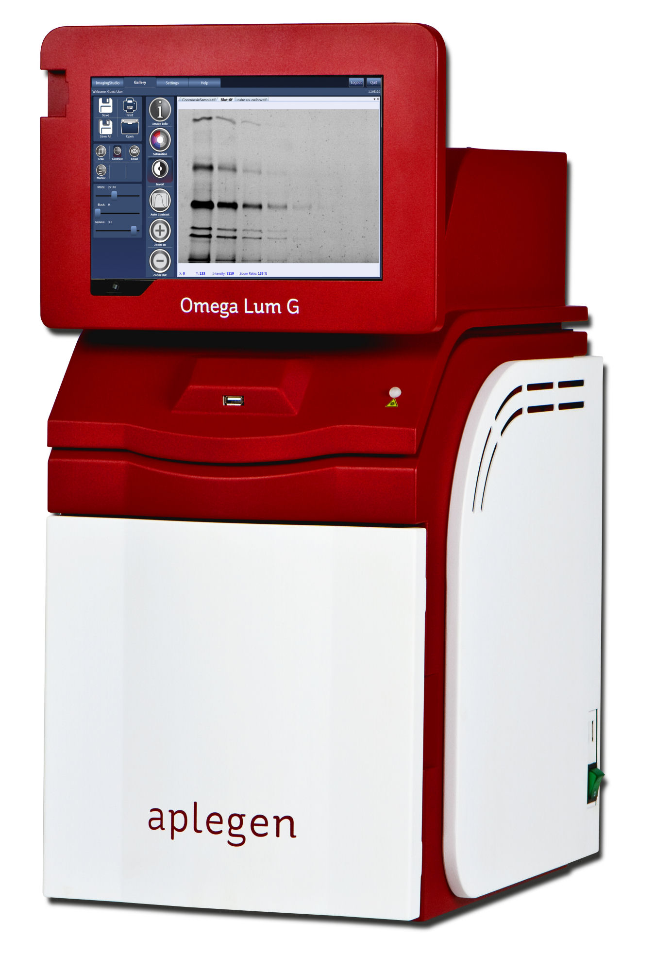 It comes complete with everything you need. Simply unpack it to get started capturing images of your gels and blots quickly. What will also surprise most people is the incredible low cost for such a high specification system. As with all Aplegen systems it is powered by the intelligent SmartCapture Technology which simplifies imaging to the point where the user just has to pick an application and the image is automatically captured with the optimum exposure.
 This powerful, new software successfully analyses chromogenic agars from major media supplier, E & O Laboratories and means microbiologists can use ProtoCOL 3 and Protos 3 systems to quickly and simply identify and count pathogens cultured on chromogenic plates from E & O Laboratories and CHROMagar. The new Chromogenic ID software module, ensures that the ProtoCOL 3 and Protos 3 systems can, in seconds, precisely identify, and at the same time count any bacteria...
 New microscopy technologies, such as light sheet fluorescence microscopy (LSFM) or clearing methods, allow the imaging of large samples at high resolution or high frame rates. Handling, processing, and analyzing these multi-terabyte data sets has become increasingly difficult for scientists, e.g. in developmental biology and neuroscience. New tools are needed to overcome these challenges. Therefore, ZEISS has teamed up with arivis AG to offer complete...
 The Leibniz Institute for Plasma Science and Technology eV (INP Greifswald) is the largest non-university research institute conducting basic research into and investigating the applications of low temperature plasmas. In addition to the application-oriented basic research, the INP promotes the development of plasma-supported processes and products. The Institute conducts research and development from idea to prototype. Using an optimised...
 The patent pending laser autofocus design puts all the required components into a single, user-installable cube that can be added at any time to a new or existing system. Laser autofocus offers speed, reproducibility and lowered risk of phototoxicity and photobleaching. With both laser autofocus and image-based autofocus available in Cytation, the system offers an important advantage in imaging – the ability to use the most appropriate autofocus...
 Made using a patent-pending process, CellMax™ Cell Pellet Slides have a highly consistent density and homogeneity across the entire cell pellet block that ensures you get accurate results time after time. Cells are harvested by a proprietary trypsin-free method preserving cell surface antigens and inflicting minimal physical damage that could lead to a loss of biomarkers. CellMax™ slides produce accurate and reproducible results...
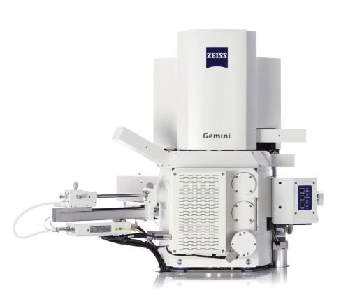 ZEISS celebrates the 50th anniversary of commercial scanning electron microscopy (SEM). In 1965, the first commercial SEM called Stereoscan was built by Cambridge Instrument Company, a UK based predecessor company of Carl Zeiss Microscopy Ltd. To mark the anniversary of this very first SEM sale 50 years ago, and celebrate the contributions to scientific research across so many fields and industries made possible through scanning electron microscopy...