Deben, a leading provider of in-situ testing stages together with innovative accessories and components for microscopy, reports on how amateur arachnologist Jeremy Poole uses the Deben Back-Scattered Electron Detector to image Britain’s spiders. There are approximately 700 species of spider in Britain, these can be divided into 34 families and categorised further into separate genera...
Syngene, a world-leading manufacturer of image analysis solutions, is pleased to announce it has entered a sponsorship agreement with Tufts Launchpad | BioLabs developed by Tufts University to be Boston’s premier fully equipped, co-working and shared lab space for biotech start-ups. Under the terms of the sponsorship, scientists working in biotech and life science companies located at Tufts Launchpad | BioLabs will have access to a G:BOX mini multi-application imaging system....
Development of the world’s first holotomographic microscope with 3D fluorescence recognised as photonics breakthrough by the 2019 Microscopy Today Innovation Award. Disruptive technology that combines the quantitative phase imaging (QPI) approach of label-free 3D refractive index (RI) tomography with fluorescence imaging to reveal the structure, volume, surface area, concentration, and dry matter mass of individual live cells in real-time....
ZEISS introduces four new high-quality CMOS cameras for digital imaging in light microscopy. These cameras complete the portfolio of proven ZEISS Axiocam models which stand for excellent performance in demanding microscopy applications. The microscope cameras ZEISS Axiocam 705 color and 712 color deliver the best possible image quality for histology, pathology or material research and analysis, thanks to excellent color rendition and greatly improved dynamic range....
Scientifica designs, manufactures and supplies microscopes to the research community worldwide. The company now offers its customers the option to use a Chromacity laser, particularly for multiphoton microscopy – a powerful technique used to image structures deep within thick samples, making it perfect for in vitro and in vivo imaging....
Digital Surf releases the Mountains® 8 software platform. This release builds extensively on the Mountains® 7 platform introduced in 2013 and now supplied by most major manufacturers with their instruments. It also sees the coming together of Mountains® (internationally renowned for profilometer and scanning electron microscope data analysis) and SPIP™ (widely recognized for scanning probe microscopy), thus setting a new standard for surface metrology and image analysis...
Research published in Nature by the Korea Advanced Institute of Science (KAIST) found that lymphatic vessels in the scull involved in the clearance of cerebrospinal fluid often become compromised with age. This finding was only possible through advances in imaging technology. Using MR Solutions’ 3T/17 preclinical liquid-helium free MRI system the KAIST researchers observed how the meningeal lymphatic vessels (mLVs), mainly in the basal part of the skull...
Thermo Fisher Scientific has introduced the Thermo Scientific Phenom ParticleX, a versatile and intuitive desktop scanning electron microscope (SEM) solution that is designed to provide automotive suppliers and additive manufacturing companies faster quality control analyses of materials used in development and production. The Phenom ParticleX includes a broad range of automated SEM analyses...
Prof. Mark Leake - University of York will present "Molecular precise optical microscopy for complex biological questions." Other leading researchers in the field will be giving presentations on a range of related topics, including novel, spectroscopy, nanoscale imaging, biomedical applications of novel microscopy techniques, image analysis and imaging frontiers. They will showcase the vibrant research and state-of-the-art in these areas...
Prior Scientific is proud to announce the release of the NanoScan OP400, a piezo based objective scanner. Prior Scientific acquired Queensgate in 2018 and the NanoScan OP400 combines Prior’s expertise in delivering microscopy solutions with Queensgate’s market leading nanopositioning technology. The NanoScan OP400 provides the fastest step and settle time of any objective positioner available....
Analytik Ltd. has launched a range of benchtop Time-Domain Nuclear Magnetic Resonance (TD-NMR) systems. Applications that yield valuable data using TD-NMR include solid fat content and oil seed analysis in the food industry, obesity research and MRI contrast agents in the medical / pharmaceutical industry and a growing number of measurements in the chemical and polymer sectors...
Deben, a leading provider of in-situ testing stages together with innovative accessories and components for microscopy, reports on how Bristol Composites Institute of University of Bristol use the Deben 200N in-situ tensile stage to study Advanced Composite Materials for Aerospace applications. The Bristol Composites Institute (ACCIS) is a department of the University of Bristol looking at materials...
HORIBA UK Ltd, Medical announces the publication of scientific studies which demonstrate the excellent performance of its new HELO high throughput fully automated haematology platform on body fluid and pathological samples. HORIBA’s Yumizen® H2500 and H1500 automated haematology analysers within the HELO platform deliver enhanced precision for complete blood counts and white blood cell....
Merck, a leading science and technology company, presented Professor David Alsteens (33), Catholic University of Louvain (UCLovain), Louvain-la-Neuve, Belgium with the 2019 Heinrich Emanuel Merck Award for Analytical Science. The award ceremony took place during the analytical conference Euroanalysis at Istanbul University in Turkey. “With his groundbreaking investigations revealing the molecular mechanisms established by viruses to hijack the cellular barrier and enter the cell....
Bruker Corporation has announced the world’s first 1.2 GHz high-resolution, protein nuclear magnetic resonance (NMR) data. Two 1.2 GHz superconducting magnets have now reached full field at Bruker’s Swiss magnet factory, setting the world record for stable, homogeneous NMR magnets for high-resolution and solid-state protein NMR applications in structural biology...
Bruker has announced the European launch of the Fourier 80 system, a next-generation, high-performance 80 MHz Fourier Transform nuclear magnetic resonance (FT-NMR) benchtop spectrometer. The Fourier 80 has been designed for organic or medicinal chemistry research, routine analysis, teaching or synthesis verification in any chemistry laboratory....
Cryo transfer can enable or improve the ability to collect atom probe data in difficult applications, like: Rapid oxidation studies (e.g. uranium, lithium, aluminum), Catalyst/reaction chamber studies of surface contamination, Characterization of hydrogen embrittlement of steel, Transport between various analysis modalities (e.g. FIB, TEM) under vacuum conditions and Analysis of "soft" (i.e. biological) materials...
The new lyophilized exosomes have various biology applications, such as assay calibration, control for exosome quantification, protein marker analysis, extraction and analysis of exosome nucleic acid, standardized positive controls for immunocapture performance evaluation, flow cytometry, and electron microscopy....
MR Solutions has received the prestigious Queen’s Award for Enterprise in recognition of the company’s innovative PET imaging technology for use in pre-clinical research. MR Solutions already holds Queens Awards for innovation in 2016 and for export achievement in 2017.
Agilent Technologies Inc.,has introduced a multimode real-time cell analyzer (RTCA)—the first of its kind—combining the best of non-invasive biosensor measurement with live cell imaging. "The xCELLigence RTCA eSight will revolutionize cell analysis in life science research," said Todd Christian, Agilent vice president, and general manager of the company's Cell Analysis Division...
Recently, a number of studies have been published demonstrating that label-free, 3-D imaging using holotomography (HT) microscopy enables researchers to observe morphological and chemical alterations of host cells due to the parasite infection without any transfection or dye staining. This powerful new tool allows parasites to be easily and quickly detected and monitored within the host cells and permits the intricacies of parasite infection mechanisms and the host cell/parasite life cycle to be studied....
Vision Engineering, a manufacturer of high quality visual inspection and measurement technologies, launches its latest, innovative flagship product Deep Reality Viewer (DRV-Z1) microscope. Designed at its Woking, UK HQ, the DRV-Z1 enables the user to view high definition 3D images under magnification without using a flat screen, or requiring operators to wear goggles or specialist glasses. Uniquely, by linking multiple DRV systems via wired or wireless technologies....

 Deben, a leading provider of in-situ testing stages together with innovative accessories and components for microscopy, reports on how amateur arachnologist Jeremy Poole uses the Deben Back-Scattered Electron Detector to image Britain’s spiders. There are approximately 700 species of spider in Britain, these can be divided into 34 families and categorised further into separate genera...
 Syngene, a world-leading manufacturer of image analysis solutions, is pleased to announce it has entered a sponsorship agreement with Tufts Launchpad | BioLabs developed by Tufts University to be Boston’s premier fully equipped, co-working and shared lab space for biotech start-ups. Under the terms of the sponsorship, scientists working in biotech and life science companies located at Tufts Launchpad | BioLabs will have access to a G:BOX mini multi-application imaging system....
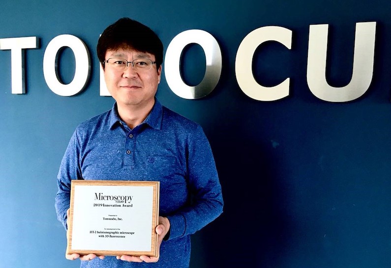 Development of the world’s first holotomographic microscope with 3D fluorescence recognised as photonics breakthrough by the 2019 Microscopy Today Innovation Award. Disruptive technology that combines the quantitative phase imaging (QPI) approach of label-free 3D refractive index (RI) tomography with fluorescence imaging to reveal the structure, volume, surface area, concentration, and dry matter mass of individual live cells in real-time....
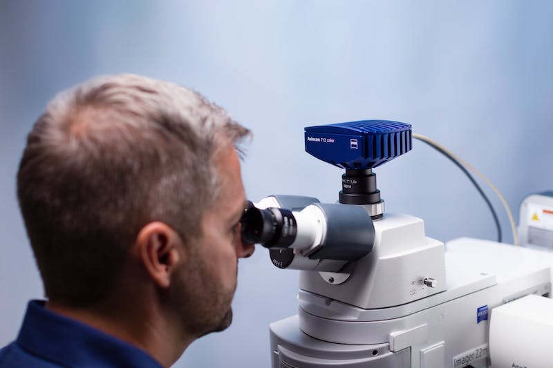 ZEISS introduces four new high-quality CMOS cameras for digital imaging in light microscopy. These cameras complete the portfolio of proven ZEISS Axiocam models which stand for excellent performance in demanding microscopy applications. The microscope cameras ZEISS Axiocam 705 color and 712 color deliver the best possible image quality for histology, pathology or material research and analysis, thanks to excellent color rendition and greatly improved dynamic range....
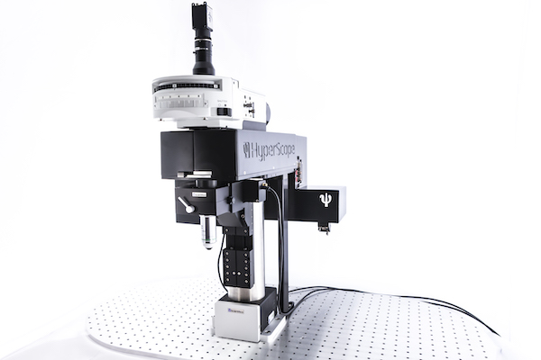 Scientifica designs, manufactures and supplies microscopes to the research community worldwide. The company now offers its customers the option to use a Chromacity laser, particularly for multiphoton microscopy – a powerful technique used to image structures deep within thick samples, making it perfect for in vitro and in vivo imaging....
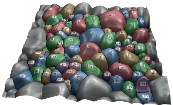 Digital Surf releases the Mountains® 8 software platform. This release builds extensively on the Mountains® 7 platform introduced in 2013 and now supplied by most major manufacturers with their instruments. It also sees the coming together of Mountains® (internationally renowned for profilometer and scanning electron microscope data analysis) and SPIP™ (widely recognized for scanning probe microscopy), thus setting a new standard for surface metrology and image analysis...
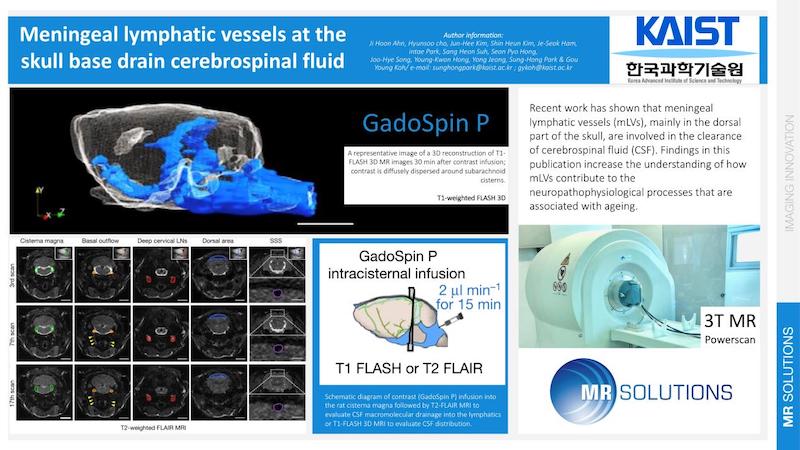 Research published in Nature by the Korea Advanced Institute of Science (KAIST) found that lymphatic vessels in the scull involved in the clearance of cerebrospinal fluid often become compromised with age. This finding was only possible through advances in imaging technology. Using MR Solutions’ 3T/17 preclinical liquid-helium free MRI system the KAIST researchers observed how the meningeal lymphatic vessels (mLVs), mainly in the basal part of the skull...
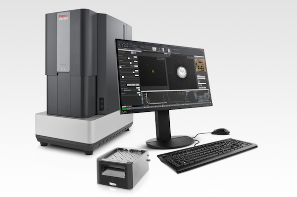 Thermo Fisher Scientific has introduced the Thermo Scientific Phenom ParticleX, a versatile and intuitive desktop scanning electron microscope (SEM) solution that is designed to provide automotive suppliers and additive manufacturing companies faster quality control analyses of materials used in development and production. The Phenom ParticleX includes a broad range of automated SEM analyses...
 Prior Scientific is proud to announce the release of the NanoScan OP400, a piezo based objective scanner. Prior Scientific acquired Queensgate in 2018 and the NanoScan OP400 combines Prior’s expertise in delivering microscopy solutions with Queensgate’s market leading nanopositioning technology. The NanoScan OP400 provides the fastest step and settle time of any objective positioner available....
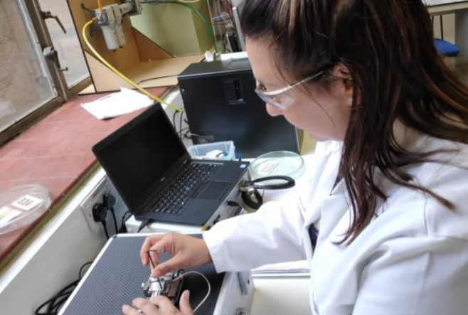 Deben, a leading provider of in-situ testing stages together with innovative accessories and components for microscopy, reports on how Bristol Composites Institute of University of Bristol use the Deben 200N in-situ tensile stage to study Advanced Composite Materials for Aerospace applications. The Bristol Composites Institute (ACCIS) is a department of the University of Bristol looking at materials...
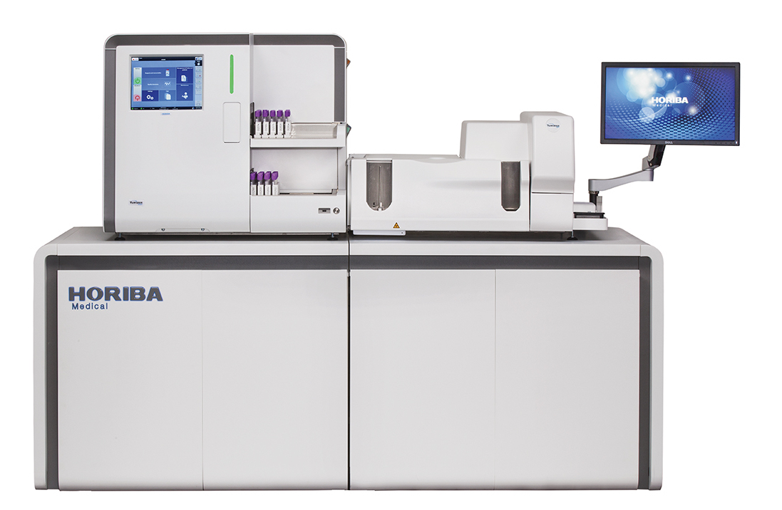 HORIBA UK Ltd, Medical announces the publication of scientific studies which demonstrate the excellent performance of its new HELO high throughput fully automated haematology platform on body fluid and pathological samples. HORIBA’s Yumizen® H2500 and H1500 automated haematology analysers within the HELO platform deliver enhanced precision for complete blood counts and white blood cell....
 Merck, a leading science and technology company, presented Professor David Alsteens (33), Catholic University of Louvain (UCLovain), Louvain-la-Neuve, Belgium with the 2019 Heinrich Emanuel Merck Award for Analytical Science. The award ceremony took place during the analytical conference Euroanalysis at Istanbul University in Turkey. “With his groundbreaking investigations revealing the molecular mechanisms established by viruses to hijack the cellular barrier and enter the cell....
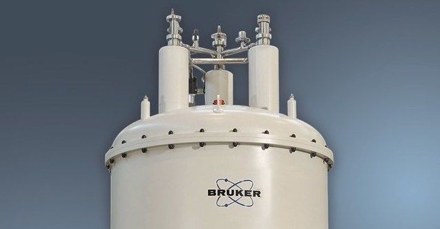 Bruker Corporation has announced the world’s first 1.2 GHz high-resolution, protein nuclear magnetic resonance (NMR) data. Two 1.2 GHz superconducting magnets have now reached full field at Bruker’s Swiss magnet factory, setting the world record for stable, homogeneous NMR magnets for high-resolution and solid-state protein NMR applications in structural biology...
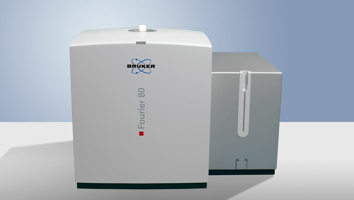 Bruker has announced the European launch of the Fourier 80 system, a next-generation, high-performance 80 MHz Fourier Transform nuclear magnetic resonance (FT-NMR) benchtop spectrometer. The Fourier 80 has been designed for organic or medicinal chemistry research, routine analysis, teaching or synthesis verification in any chemistry laboratory....
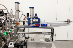 Cryo transfer can enable or improve the ability to collect atom probe data in difficult applications, like: Rapid oxidation studies (e.g. uranium, lithium, aluminum), Catalyst/reaction chamber studies of surface contamination, Characterization of hydrogen embrittlement of steel, Transport between various analysis modalities (e.g. FIB, TEM) under vacuum conditions and Analysis of "soft" (i.e. biological) materials...
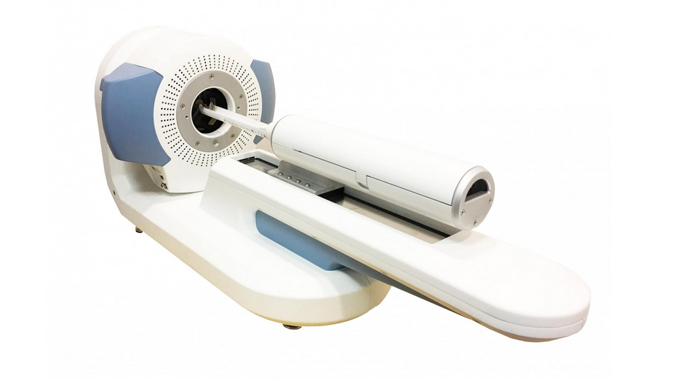 MR Solutions has received the prestigious Queen’s Award for Enterprise in recognition of the company’s innovative PET imaging technology for use in pre-clinical research. MR Solutions already holds Queens Awards for innovation in 2016 and for export achievement in 2017.
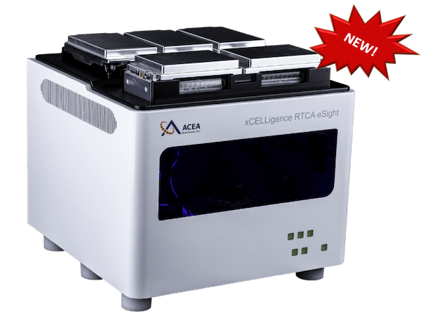 Agilent Technologies Inc.,has introduced a multimode real-time cell analyzer (RTCA)—the first of its kind—combining the best of non-invasive biosensor measurement with live cell imaging. "The xCELLigence RTCA eSight will revolutionize cell analysis in life science research," said Todd Christian, Agilent vice president, and general manager of the company's Cell Analysis Division...
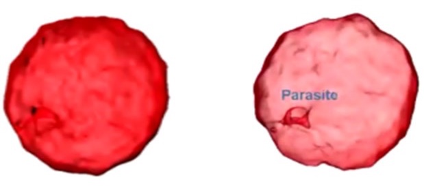 Recently, a number of studies have been published demonstrating that label-free, 3-D imaging using holotomography (HT) microscopy enables researchers to observe morphological and chemical alterations of host cells due to the parasite infection without any transfection or dye staining. This powerful new tool allows parasites to be easily and quickly detected and monitored within the host cells and permits the intricacies of parasite infection mechanisms and the host cell/parasite life cycle to be studied....
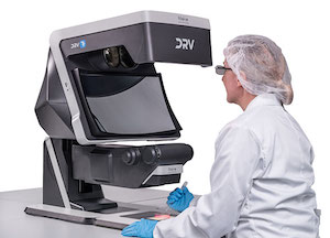 Vision Engineering, a manufacturer of high quality visual inspection and measurement technologies, launches its latest, innovative flagship product Deep Reality Viewer (DRV-Z1) microscope. Designed at its Woking, UK HQ, the DRV-Z1 enables the user to view high definition 3D images under magnification without using a flat screen, or requiring operators to wear goggles or specialist glasses. Uniquely, by linking multiple DRV systems via wired or wireless technologies....