Renishaw's latest version of its Virsa™ Raman analyser, with new WiRE™ 5.5 software, enables its users to analyse samples away from the confines of the laboratory microscope, using remote fibre-optic probes. Its features expand the use of Raman spectroscopy to new samples, applications, and environments. The new Virsa system has LiveTrack™ focus-tracking technology and the new Monitor™ software module....
Sensofar, a technology company specialising in the field of non-contact surface metrology, has developed a new technique for characterising the evolution of a sample’s surface topography with temperature using the S neox 3D optical profiler and Linnik interferometer coupled with Linkam’s LTS420 temperature-controlled chamber. The technique has been used to successfully map the changes in roughness and waviness of silicon wafers at temperatures up to 380°C...
The company will be celebrating its 175th anniversary through a variety of activities and events. Its close links to science are evident in projects such as the "ZEISS Beyond Talks" interview series. In these interviews, pioneers and eminent figures from across the globe, including climate researcher Professor Antje Boetius, speak about their work, their visions, their passion and the topics that are having a major impact on our world....
GenNext Technologies, Inc., a growth-stage company that provides instrumentation, software, and services to structural biology researchers within the biopharmaceutical industry, announce that nearly $7M (US) in NIH grant awards has led to a seminal US patent for its novel technology that vastly improves the utility of protein footprinting for the study of biopharmaceutical higher order structure....
Gemstones are pieces of mineral crystal cut and polished for use in the gem and jewellery industry. The term gemstone covers a wide variety of gems, roughly over 200 types of natural gemstones exist. Gemstones can be separated into two classifications: precious stones, such as sapphires, and semi-precious, such as garnet. The value of these stones depends on their colour, size, quality, and rarity...
Biosample Hub – a new ethical and transparent way of connecting Biobanks and Biotechs. Biosample Hub is a new worldwide platform providing a game-changing solution to enable researchers in the biotech and pharmaceutical industry to obtain the biosamples they need. Left-over clinical material like blood samples and excised tissue may be medical waste, but it can also be research treasure. This material is needed by researchers for the development of new drugs, diagnostics and vaccines....
CytoSMART Technologies has announced the release of its first fluorescence cell counter. The CytoSMART Exact FL is an automated, dual fluorescence cell counter with an expanded field of view and unmatched performance. Although the system’s footprint is very small, its specifications rival most, if not all alternative automated cell counters. The CytoSMART Exact FL sets new standards in terms of accuracy, speed, and performance thanks to a wide range of technological advancements....
ZEISS introduces SIM², a groundbreaking image reconstruction algorithm that increases the resolution and sectioning quality of structured illumination microscopy (SIM) data. With SIM² on the microscope system ZEISS Elyra 7, life science researchers can now double the conventional SIM resolution and discriminate the finest subcellular structures of living and fixed samples, even those no more than 60 nm apart....
Advanced MRI techniques may provide clues to how MS will progres. Research published in Brain1 has used new MRI techniques to show what is happening in the brains of people with multiple sclerosis (MS) in the early stages of their condition. Scientists say these previously unseen changes could have the potential to predict how disabled a person might become in the future....
Researchers have found that a natural molecule can effectively block the binding of a subset of human antibodies to SARS-CoV-2. The discovery may help explain why some COVID-19 patients can become severely ill despite having high levels of antibodies against the virus. In their research, published in Science Advances, teams from the Francis Crick Institute, in collaboration with researchers at Imperial College London, Kings College London and UCL (University College London), found that biliverdin and bilirubin...
NuNano Ltd. and TESCAN ORSAY HOLDING a.s. announce the successful installation of a TESCAN Field Emission Scanning Electron Microscope (FESEM) at NuNano’s Bristol facility, where it will be used for 100-percent automated quality control on atomic force microscope (AFM) probes with some of the tightest dimensional tolerances available today. NuNano, a developer of high-end AFM probes and cantilever-based sensor devices, has customized the TESCAN FESEM software for this highly automated imaging requirement...
In this application note, an Edinburgh Instruments RM5 Raman Microscope is used to highlight how Raman microscopy an essential tool for any material scientist researching graphene. First reported in Science in 2004, graphene is commonly termed the “wonder material” due to its impressive properties. Geim and Novoselov, the two scientists behind the first isolation of graphene, were awarded the 2010 Nobel Prize in Physics for their pioneering research into graphene....
Olympus unveiled the winners of their second Global Image of the Year Life Science Light Microscopy Award, an annual competition that recognizes the best in life science imaging. The winning images were selected from a record number of entries—nearly 700 submissions from 61 countries. Global Winner, Werner Zuschratter from Germany was selected as the global winner for his eye-catching image of a whole rat embryo captured with a confocal microscope....
With the release of ZEISS Axioscan 7, ZEISS presents its next-generation slide scanner for the automated digitization of microscopy samples. Following its successful predecessor ZEISS Axio Scan.Z1, ZEISS Axioscan 7 provides significant improvements in almost every aspect: a new acquisition engine for higher scan speeds, a broader range of imaging modes for more application flexibility...
New research, using cutting-edge cryo-electron microscopy (CryoEM), has revealed key insights into a vital DNA repair process, which is implicated in resistance to cancer treatments. Led by the University of Glasgow and published in Nature Structural Biology, the research is based on data and models collected from the Scottish Centre for Macromolecular Imaging (SCMI)...
EUROIMMUN, a PerkinElmer, Inc. company, announce the launch of the EUROPatternTM Microscope Live (EPML) compact immunofluorescence microscope, available with the fourth generation of the Company’s well-established EUROLabOfficeTM 4.0 (ELO 4.0) laboratory management software. The combined system of hardware and software allows for ultrafast automated immunofluorescence image acquisition, pattern recognition and titer estimation as well as modern diagnostics at the screen.
Nikon Corporation (Nikon) announce the release of the ECLIPSE Si biological microscope, which reduces the time spent on adjusting the light intensity when changing magnifications and which, by means of an ergonomic design, reduces physical strain during long hours of observations. The challenges in educational and clinical settings include complex operations during microscopic observations and accumulation of physical fatigue due to long hours of observations....
Particle analysis is essential: whether in production and quality control, research, development, laboratory or for controlling manufacturing processes. In all these cases fast, reliable and reproducible results are required. Find the perfect solution adapted to your application. Benefit from more than 35 years of experience in the field of particle technology! We help you to find the perfect measuring principle: static light scattering for analysis of particle size, or dynamic image analysis...
Olympus’ next virtual life science event, the Olympus Discovery Summit—Looking Forward: A New Era of Research will take place on April 27–29, 2021. The Olympus Life Science team has organized this opportunity for the microscopy community to virtually share its expertise and learn about important advances in technology. During this free three-day summit, attendees can enjoy a full schedule of customer presentations, tech talks, roundtable discussions and product demos....
Atik Cameras, a specialist designer and manufacturer of advanced scientific imaging solutions, has further expanded its collaboration with leading global Original Equipment Manufacturers (OEMs) of real time Polymerase Chain Reaction (PCR) DNA amplifiers, securing multiple new contracts to supply high-performing cameras for reliable COVID-19 testing...
With the release of ZEISS Correlative Cryo Workflow, ZEISS provides the life science research community with a new combined hardware and software solution for cryogenic microscopy. The workflow connects widefield, laser scanning, and FIB-SEM microscopes in a seamless and easy-to-use procedure....
Porvair Sciences new Krystal™ Glass Bottom microplate with high quality specialty glass from Schott provides unmatched performance for whole-plate CCD imaging and laser detection applications. Krystal™ Glass Bottom plates combine the advantageous optical properties of glass, low background and low birefringence, with the versatility of a microplate...

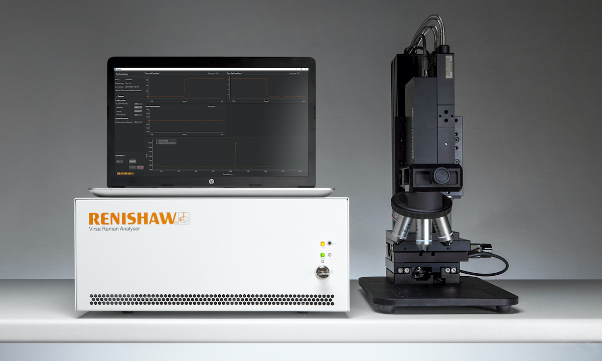 Renishaw's latest version of its Virsa™ Raman analyser, with new WiRE™ 5.5 software, enables its users to analyse samples away from the confines of the laboratory microscope, using remote fibre-optic probes. Its features expand the use of Raman spectroscopy to new samples, applications, and environments. The new Virsa system has LiveTrack™ focus-tracking technology and the new Monitor™ software module....
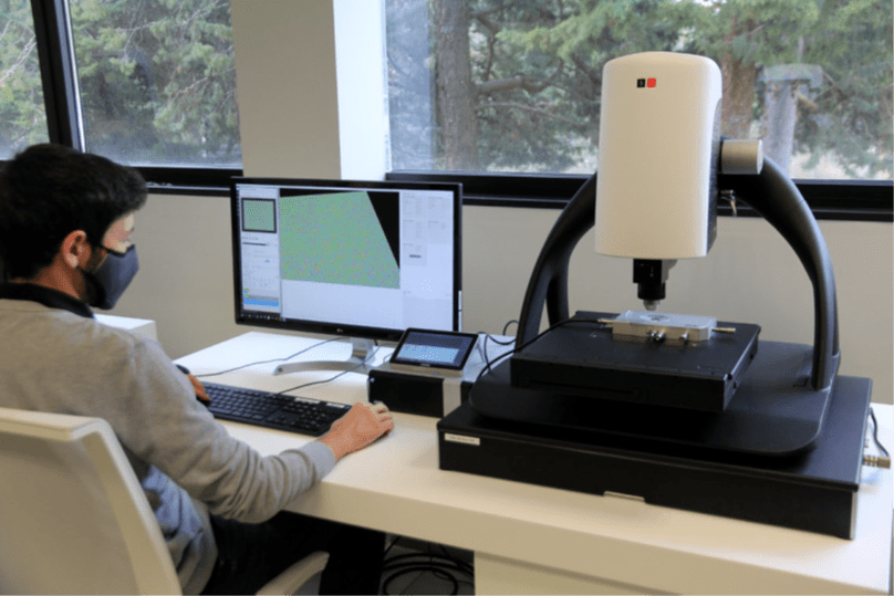 Sensofar, a technology company specialising in the field of non-contact surface metrology, has developed a new technique for characterising the evolution of a sample’s surface topography with temperature using the S neox 3D optical profiler and Linnik interferometer coupled with Linkam’s LTS420 temperature-controlled chamber. The technique has been used to successfully map the changes in roughness and waviness of silicon wafers at temperatures up to 380°C...
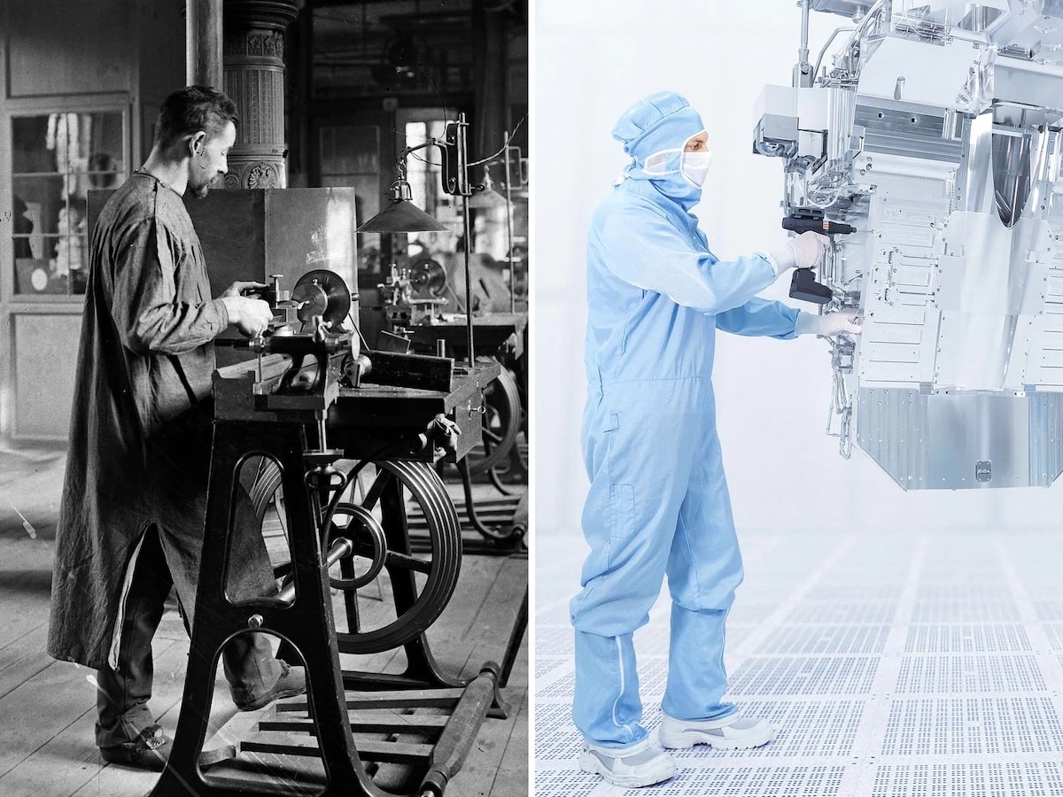 The company will be celebrating its 175th anniversary through a variety of activities and events. Its close links to science are evident in projects such as the "ZEISS Beyond Talks" interview series. In these interviews, pioneers and eminent figures from across the globe, including climate researcher Professor Antje Boetius, speak about their work, their visions, their passion and the topics that are having a major impact on our world....
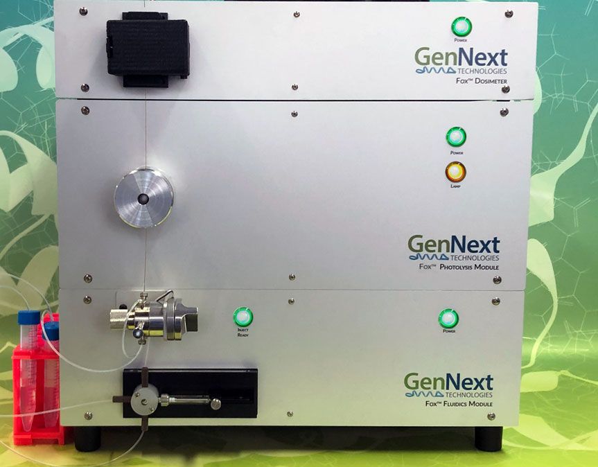 GenNext Technologies, Inc., a growth-stage company that provides instrumentation, software, and services to structural biology researchers within the biopharmaceutical industry, announce that nearly $7M (US) in NIH grant awards has led to a seminal US patent for its novel technology that vastly improves the utility of protein footprinting for the study of biopharmaceutical higher order structure....
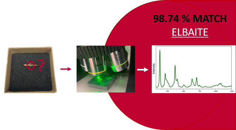 Gemstones are pieces of mineral crystal cut and polished for use in the gem and jewellery industry. The term gemstone covers a wide variety of gems, roughly over 200 types of natural gemstones exist. Gemstones can be separated into two classifications: precious stones, such as sapphires, and semi-precious, such as garnet. The value of these stones depends on their colour, size, quality, and rarity...
 Biosample Hub – a new ethical and transparent way of connecting Biobanks and Biotechs. Biosample Hub is a new worldwide platform providing a game-changing solution to enable researchers in the biotech and pharmaceutical industry to obtain the biosamples they need. Left-over clinical material like blood samples and excised tissue may be medical waste, but it can also be research treasure. This material is needed by researchers for the development of new drugs, diagnostics and vaccines....
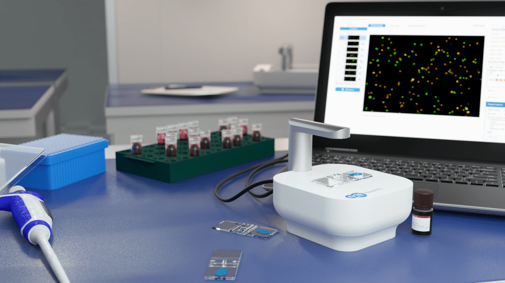 CytoSMART Technologies has announced the release of its first fluorescence cell counter. The CytoSMART Exact FL is an automated, dual fluorescence cell counter with an expanded field of view and unmatched performance. Although the system’s footprint is very small, its specifications rival most, if not all alternative automated cell counters. The CytoSMART Exact FL sets new standards in terms of accuracy, speed, and performance thanks to a wide range of technological advancements....
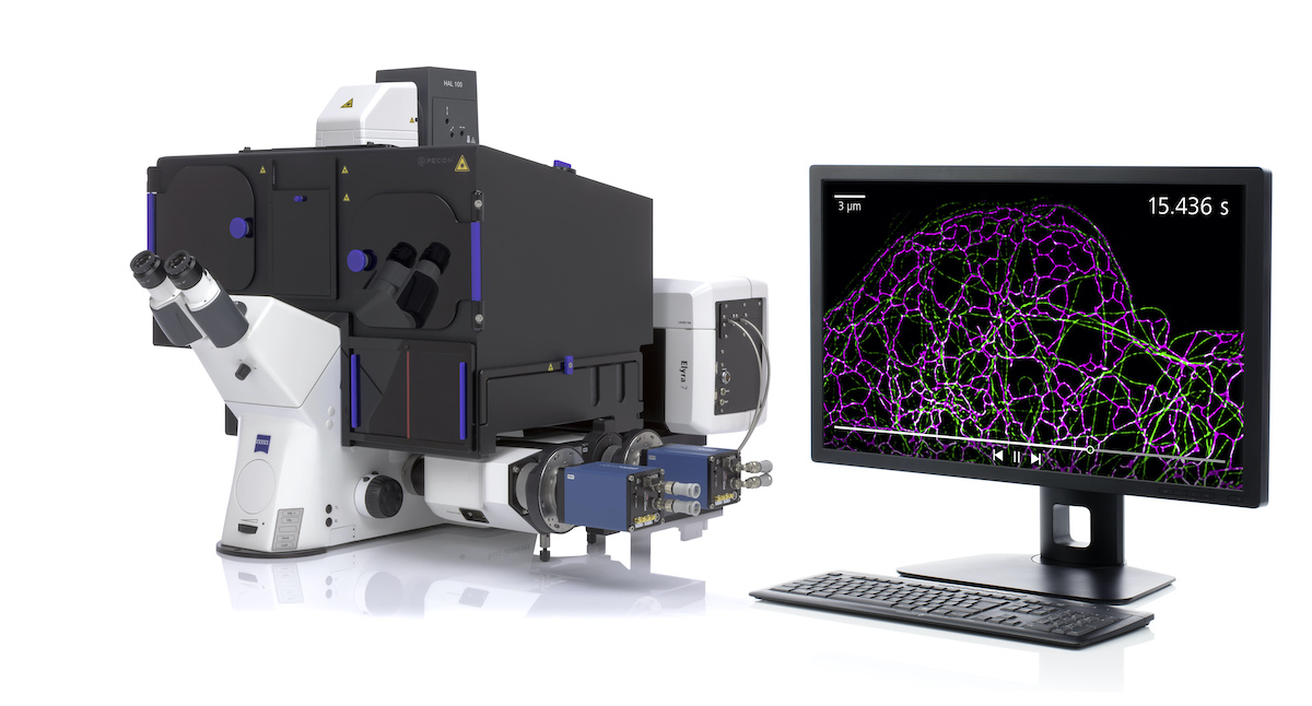 ZEISS introduces SIM², a groundbreaking image reconstruction algorithm that increases the resolution and sectioning quality of structured illumination microscopy (SIM) data. With SIM² on the microscope system ZEISS Elyra 7, life science researchers can now double the conventional SIM resolution and discriminate the finest subcellular structures of living and fixed samples, even those no more than 60 nm apart....
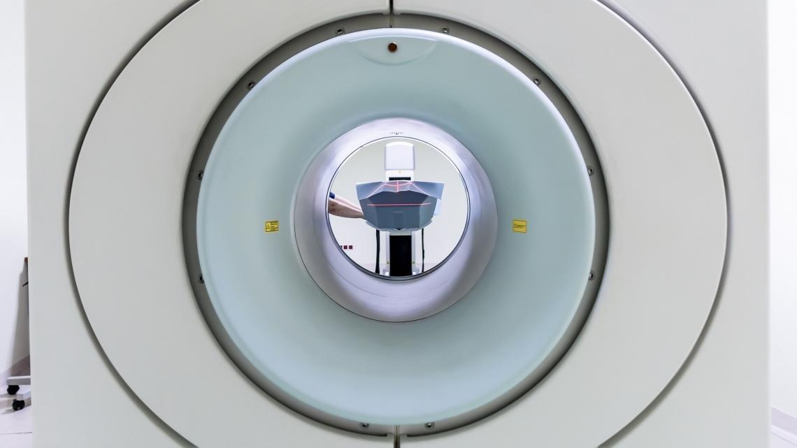 Advanced MRI techniques may provide clues to how MS will progres. Research published in Brain1 has used new MRI techniques to show what is happening in the brains of people with multiple sclerosis (MS) in the early stages of their condition. Scientists say these previously unseen changes could have the potential to predict how disabled a person might become in the future....
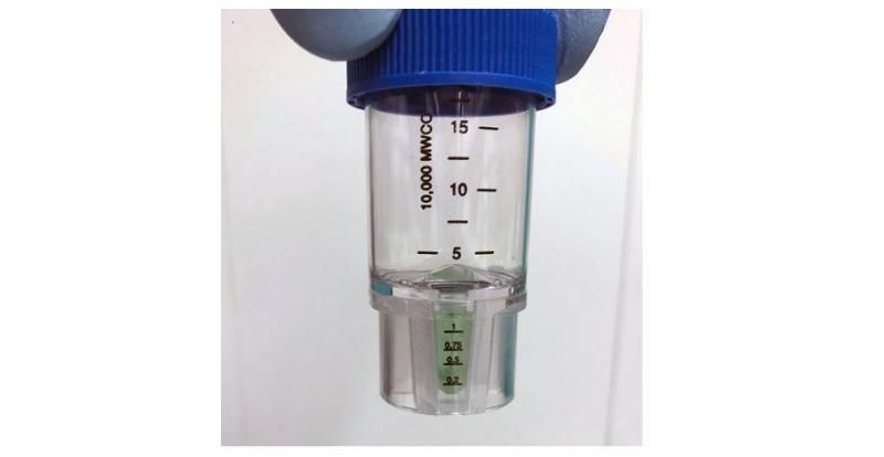 Researchers have found that a natural molecule can effectively block the binding of a subset of human antibodies to SARS-CoV-2. The discovery may help explain why some COVID-19 patients can become severely ill despite having high levels of antibodies against the virus. In their research, published in Science Advances, teams from the Francis Crick Institute, in collaboration with researchers at Imperial College London, Kings College London and UCL (University College London), found that biliverdin and bilirubin...
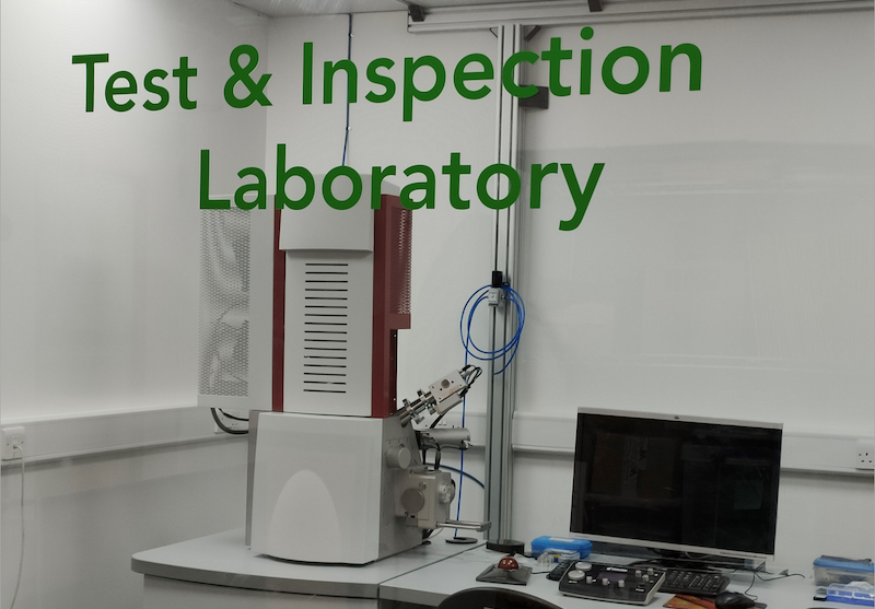 NuNano Ltd. and TESCAN ORSAY HOLDING a.s. announce the successful installation of a TESCAN Field Emission Scanning Electron Microscope (FESEM) at NuNano’s Bristol facility, where it will be used for 100-percent automated quality control on atomic force microscope (AFM) probes with some of the tightest dimensional tolerances available today. NuNano, a developer of high-end AFM probes and cantilever-based sensor devices, has customized the TESCAN FESEM software for this highly automated imaging requirement...
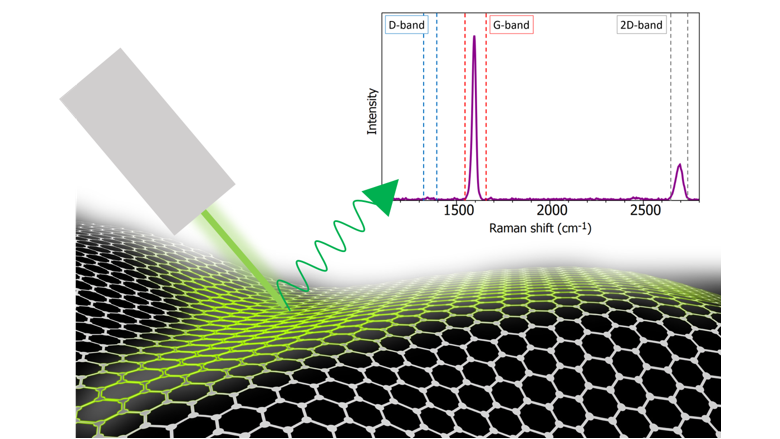 In this application note, an Edinburgh Instruments RM5 Raman Microscope is used to highlight how Raman microscopy an essential tool for any material scientist researching graphene. First reported in Science in 2004, graphene is commonly termed the “wonder material” due to its impressive properties. Geim and Novoselov, the two scientists behind the first isolation of graphene, were awarded the 2010 Nobel Prize in Physics for their pioneering research into graphene....
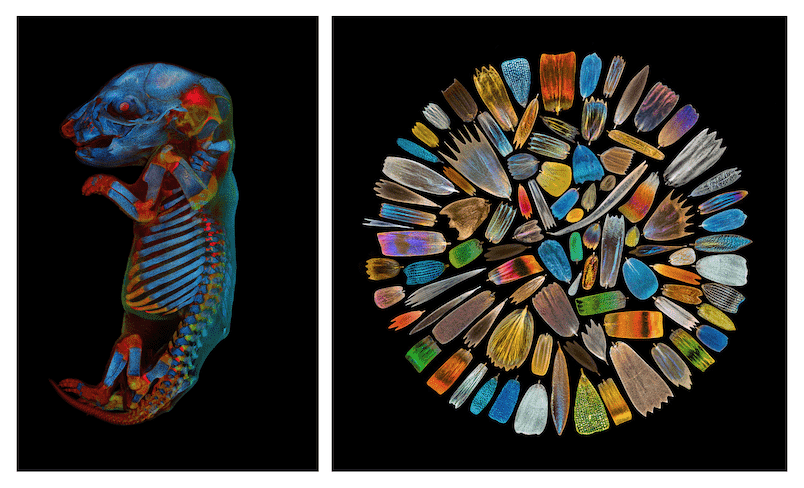 Olympus unveiled the winners of their second Global Image of the Year Life Science Light Microscopy Award, an annual competition that recognizes the best in life science imaging. The winning images were selected from a record number of entries—nearly 700 submissions from 61 countries. Global Winner, Werner Zuschratter from Germany was selected as the global winner for his eye-catching image of a whole rat embryo captured with a confocal microscope....
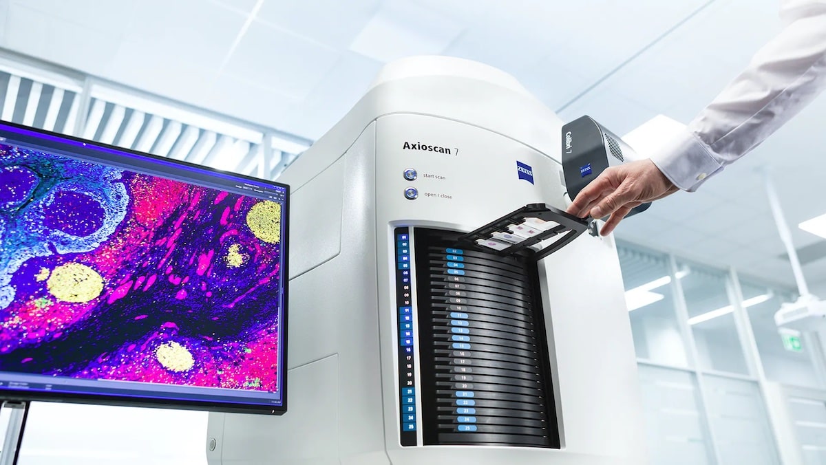 With the release of ZEISS Axioscan 7, ZEISS presents its next-generation slide scanner for the automated digitization of microscopy samples. Following its successful predecessor ZEISS Axio Scan.Z1, ZEISS Axioscan 7 provides significant improvements in almost every aspect: a new acquisition engine for higher scan speeds, a broader range of imaging modes for more application flexibility...
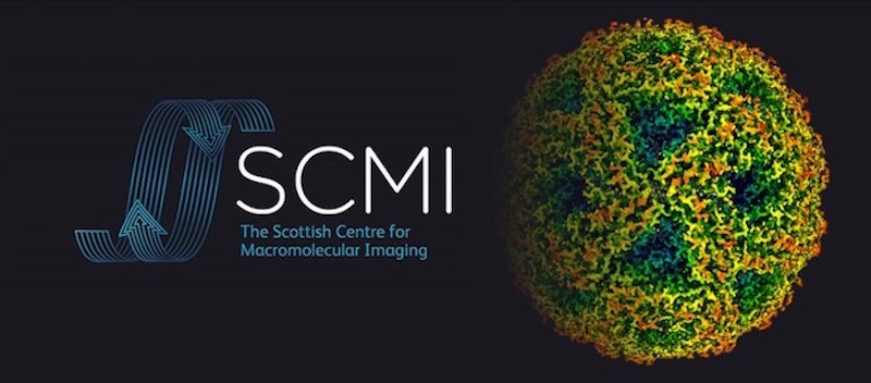 New research, using cutting-edge cryo-electron microscopy (CryoEM), has revealed key insights into a vital DNA repair process, which is implicated in resistance to cancer treatments. Led by the University of Glasgow and published in Nature Structural Biology, the research is based on data and models collected from the Scottish Centre for Macromolecular Imaging (SCMI)...
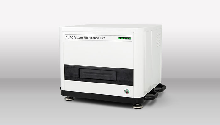 EUROIMMUN, a PerkinElmer, Inc. company, announce the launch of the EUROPatternTM Microscope Live (EPML) compact immunofluorescence microscope, available with the fourth generation of the Company’s well-established EUROLabOfficeTM 4.0 (ELO 4.0) laboratory management software. The combined system of hardware and software allows for ultrafast automated immunofluorescence image acquisition, pattern recognition and titer estimation as well as modern diagnostics at the screen.
 Nikon Corporation (Nikon) announce the release of the ECLIPSE Si biological microscope, which reduces the time spent on adjusting the light intensity when changing magnifications and which, by means of an ergonomic design, reduces physical strain during long hours of observations. The challenges in educational and clinical settings include complex operations during microscopic observations and accumulation of physical fatigue due to long hours of observations....
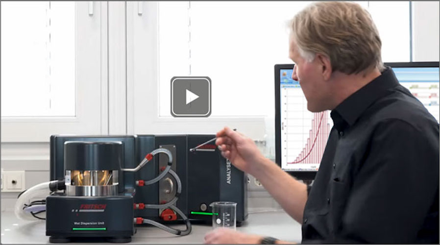 Particle analysis is essential: whether in production and quality control, research, development, laboratory or for controlling manufacturing processes. In all these cases fast, reliable and reproducible results are required. Find the perfect solution adapted to your application. Benefit from more than 35 years of experience in the field of particle technology! We help you to find the perfect measuring principle: static light scattering for analysis of particle size, or dynamic image analysis...
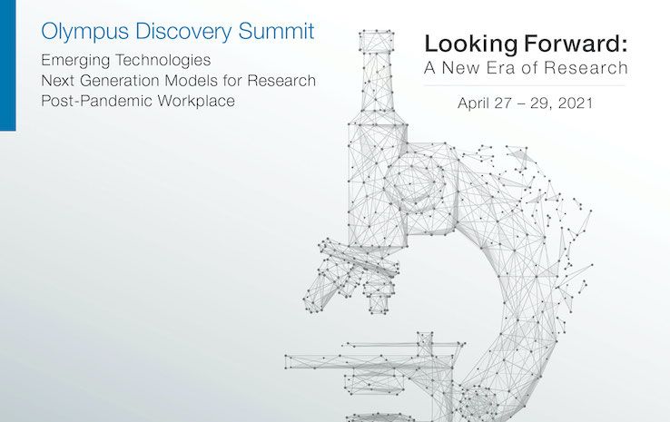 Olympus’ next virtual life science event, the Olympus Discovery Summit—Looking Forward: A New Era of Research will take place on April 27–29, 2021. The Olympus Life Science team has organized this opportunity for the microscopy community to virtually share its expertise and learn about important advances in technology. During this free three-day summit, attendees can enjoy a full schedule of customer presentations, tech talks, roundtable discussions and product demos....
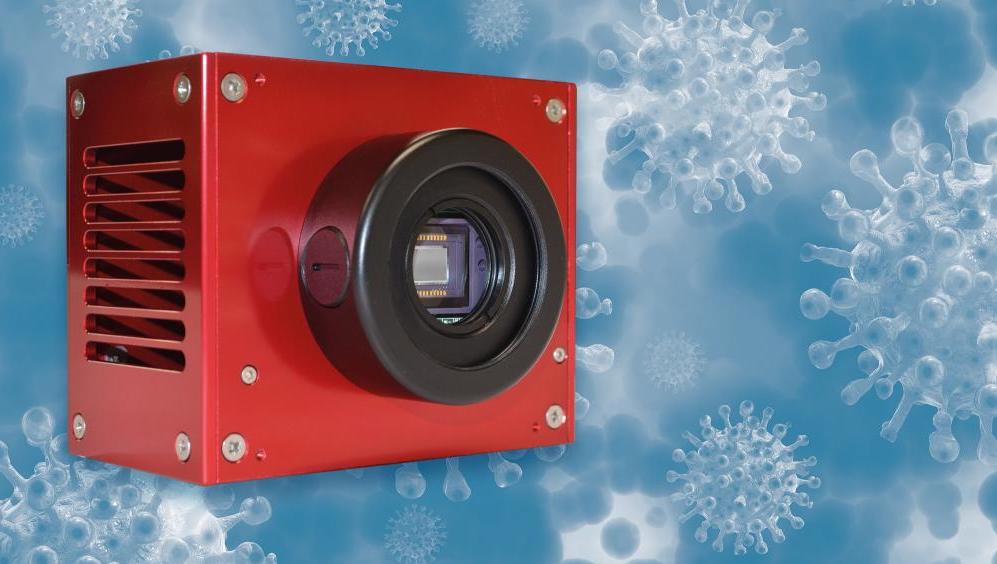 Atik Cameras, a specialist designer and manufacturer of advanced scientific imaging solutions, has further expanded its collaboration with leading global Original Equipment Manufacturers (OEMs) of real time Polymerase Chain Reaction (PCR) DNA amplifiers, securing multiple new contracts to supply high-performing cameras for reliable COVID-19 testing...
 With the release of ZEISS Correlative Cryo Workflow, ZEISS provides the life science research community with a new combined hardware and software solution for cryogenic microscopy. The workflow connects widefield, laser scanning, and FIB-SEM microscopes in a seamless and easy-to-use procedure....
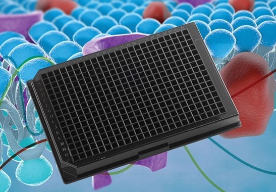 Porvair Sciences new Krystal™ Glass Bottom microplate with high quality specialty glass from Schott provides unmatched performance for whole-plate CCD imaging and laser detection applications. Krystal™ Glass Bottom plates combine the advantageous optical properties of glass, low background and low birefringence, with the versatility of a microplate...