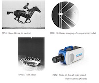
Specialised Imaging has produced a presentation that delivers an informative and unbiased review of the historical development of ultra high-speed imaging technology.
The first practical application of high-speed imaging was Eadweard Muybridge’s 1878 photographic investigation into whether a horses' feet were actually all off the ground at once during a gallop. The first photograph of a supersonic flying bullet was taken by the Austrian physicist Peter Salcher in 1886, a technique that was later used by Ernst Mach in his studies of supersonic motion. German weapons scientists later applied these high speed imaging...
Syngene, a world-leading manufacturer of image analysis solutions is delighted to announce the appointment of Matthew Dunne, as its new International Sales Manager.
The firm has appointed Matthew at this time to capitalise on the increasing demand for quality assured gel imaging systems, especially in the Middle East and Asia Pacific regions. Matthew comes to Syngene with a wealth of experience, having been active in sales management and sales for life science product suppliers, including a major international gel documentation company for over 16 years. His imaging expertise is supported by a masters...
Renishaw, a world leader in metrology and spectroscopy technologies, announce their participation at MMC 2015, which takes place from 29th June to 2nd July 2015 at the Manchester Central Convention Complex, Manchester, UK.
Renishaw Applications Scientist, Dr Katherine Lau, will make a presentation as part of the conference program and lead a workshop on Raman confocal microscopic imaging. MMC 2015, incorporating EMAG 2015, is hosted by the Royal Microscopical Society and takes place from 29th June to 2nd July 2015, at the Manchester Central Convention Complex, Manchester, UK. During this exhibition and conference, Applications Scientist, Dr Katherine Lau...
Microscopy tools optimized for use with emerging neurotechnologies provided for researchers
ZEISS announces that it is participating in a new public-private partnership with UC Berkeley as part of the Brain Microscopy Innovation Center (BrainMIC), a component of the Berkeley BRAIN Initiative. The Brain Research through Advancing Innovative Neurotechnologies (BRAIN) Initiative was launched by President Obama in 2013 and is focused on revolutionizing understanding of the human brain, with the goal of helping researchers uncover the...
Universities of Leicester and Nottingham to develop mini hybrid gamma ray camera to revolutionise identification and removal of tumours and lymph nodes
‘Hybrid technology’ mini camera combines optical and gamma imaging to improve diagnosis and lymph and tumour removal efficiency
Small size of camera allows for bedside diagnosis as well as for small organ imaging, surgical investigation and visualisation of drug delivery
Researchers are also investigating other clinical applications for the technology including thyroid, lymphatic drainage and ‘lacrimal drainage’
Prior Scientific has published a new 12-page catalogue that brings together its range of high performance LED and metal halide light sources proven to provide the best possible illumination for your microscopy work.
Regardless of microscopy technique, high quality imaging requires high performance illumination in order to obtain the best possible results. Prior Scientific has designed a range of precisely engineered, reliable and long lasting light sources tailored to the exacting requirements of scientists across a broad range of disciplines. The new catalogue details Prior’s expanding range of LED illumination sources that offer many advantages for brightfield, DIC, phase contrast and fluorescence microscopy...
Manchester University has chosen MR Solutions’ 3T MRI preclinical, cryogen free imaging solution for superior soft tissue contrast and molecular imaging research at the Wolfson Molecular Imaging Centre.
The scanner will be used in their Regenerative Medicine research programme which aims to develop a clearer understanding of the potential risks and hazards of therapies and to develop new methodologies to assess their risk. The aim of this research is to gain faster access to new medicines for patients. This new state of the art preclinical imaging system was developed, supplied and installed by MR Solutions of Guildford, Surrey and supported...
Linkam Scientific Instruments, have announced the launch of a new relative humidity controller for their range of temperature stages used for materials, pharmaceuticals and foods testing. The unit will make its debut at mmc2015, the largest European microscopy event of the year.
The new RH95 Relative Humidity Controller is the latest addition to provide precise environmental sample control to Linkam’s range of temperature stages. It has been developed based on the feedback of many Linkam users in the materials, foods and pharmaceutical industries where humidity plays an important role in product development, manufacture and even storage. It will be shown on stand number 411 at mmc2015, the biennial conference and exhibition organised by the Royal Microscopical Society.
FLIR Systems has announced a new version 4.2 of its ResearchIR thermal imaging software.
ResearchIR 4.2 provides researchers and scientists with a powerful tool for viewing, acquiring, analyzing, and sharing the thermal data captured with FLIR’s Scientific and R&D cameras. ResearchIR Max version 4.2 gives users direct access to their MATLAB scripts within ResearchIR for the first time. This will allow users to access their customized MatLab® scripts directly in ResearchIR for specially-tailored image analysis and processing tasks...
Specialised Imaging reports on how UK academic research groups have benefited from use of its SIM series ultra-high speed imaging camera.
The Engineering and Physical Sciences Research Council (EPSRC) has since 1985 offered UK academic institutions free loans of the latest scientific instruments to help further their research projects. In 2008 the EPSRC added a state-of-the-art SIM16 ultra-high speed imaging camera from Specialised Imaging Ltd. to its loan pool facility. Wai Chan, Managing Director of Specialised Imaging commented "Over the last 7 years the EPSRC Loan Pool SIM16...
The new product, launched with global distributor Applikon, offers an automated approach to gathering quantitative data
Ovizio Imaging System, an innovative quantitative microscopy company specializing in life science solutions, announces today the launch of its new product, the iLine F, an in-line suspension cell-monitoring microscope. In collaboration with global distributor, Applikon, the iLine F is being showcased at booth #74 and #77 at the European Society for Animal Cell Technologycongress (ESACT), held in Barcelona from May 31 to June 3...
A clinical MRI scanner can be transformed into an effective preclinical system using a simple to fit conversion kit from MR Solutions of Guildford, Surrey.
The transformation, which can be undertaken in around ten minutes, involves sliding in a carrier system linked to a separate state of the art EVO spectrometer providing superior soft tissue contrast and molecular imaging capabilities. This is a major breakthrough for clinical facilities which also conduct their own research as it enables existing technology to be utilised without the need for extensive investment in a separate system...
Market leaders in temperature controlled microscopy, Linkam Scientific Instruments, are working in partnership with the School of Pharmacy at the University of East Anglia to evaluate a new analytical technique known as TASC - Thermal Analysis by Structural Characterisation.
Dr Sheng Qi is a senior lecturer in pharmaceutics in the School of Pharmacy at the University of East Anglia. Sheng's current research interests focus on gaining a fundamental understanding of the behaviour of drug-polymer/lipid dispersions in solid (phase behaviour) and liquid (in biological fluids) states. She is also developing new approaches for stabilising supersaturated drug dispersions. By working with industrial partners as well as...
Market leaders in temperature controlled microscopy, Linkam Scientific Instruments, launch the WS37 Warm Stage at the 2015 Annual Meeting of the Association of Biomedical Andrologists (ABA).
At the 2015 and 10th anniversary annual meeting of the Association of Biomedical Andrologists, Linkam have introduced a new solution for embryologists seeking a better solution for the evaluation and quantification of sperm. Live cell research and, specifically, the testing of sperm in clinics and hospitals, has not been well served in terms of reliable instrumentation and testing protocols. Mr Stephen Harbottle is Chairman of the Association...
DiATOME have partnered with EM Resolutions, a Saffron Walden-based consumables and accessories supplier for electron microscopy, to supply their range of high quality diamond knives in the UK.
Whilst diamond knives are best known for producing high quality sections on an ultramicrotome, their use for surface preparation is less accepted. DiATOME diamond knives are used for the preparation of high quality surfaces for AFM and other imaging techniques requiring smooth surfaces. DiATOME ultra AFM knives are made from higher quality diamonds to produce extremely smooth sample surfaces and ensure the best possible...
LaVison BioTec, developers of advanced microscopy solutions for the life sciences, are presenting workshops on their 2-photon and light sheet microscope systems at the 15th Annual meeting of the European Light Microscopy Initiative in Sitges near Barcelona in Spain.
The 15th annual meeting of the European Light Microscopy Initiative continues the goals of the first meeting in 2001, namely to promote the quickly developing field of light microscopy as a fundamental research tool for the life sciences and to strengthen the channels of communication between researchers and industry. The combination of top level scientific presentations with practical hands-on workshops brings participants from all over...
Kleindiek Nanotechnik have partnered with EM Resolutions, a Saffron Walden-based consumables and accessories supplier for electron microscopy, to supply and install their range of MM3A nanomanipulators and force measurement systems to the UK research community.
For example, FMS-EM force measurement systems are enabling researchers at Imperial College London to investigate the root cause of failures in electrochemical devices such as fuel cells and batteries. Imperial College is also using them for testing during the development of nano robots and other nanostructures. The FMS-EM is a compact force readout tool that allows tensile measurements to be made inside an SEM/FIB. Whilst existing...
The imaging systems on display will suit all budgets and make producing amazing gel and blot imaging results incredibly simple.
For researchers wanting a dedicated chemiluminescence Western blot imager, Syngene’s experts demonstrated the new GeneGnomeXRQ. On the booth, they explained how this complete system with its sensitive, cooled camera and GeneSys software, allows scientists rapid walk-away imaging of even the faintest bands on Western blots....
Pie Medical Imaging announces its new release of CAAS MRV, for analysis of the heart to support diagnosis of cardiovascular conditions.
This new version contains intuitive and guided analysis workflows, optimized to clinical practice. The workflows will guide you through the required steps for Functional left and right ventricle, Viability and First Pass Perfusion analysis. The new viability workflow includes, besides analysis of Delayed Enhanced MR images, also analysis of edema on T2-weighted MR images. The combination of segmented infarcts and edema areas can identify...
LaVison BioTec, developers of advanced microscopy solutions for the life sciences, announces the dates and venue of the first international users' meeting on their UltraMicroscope light sheet microscopy products.
The first LaVision BioTec UltraMicroscope User Meeting will take place at the Max Planck Institute for Molecular Biomedicine in Münster, 21-22 September. Participants are cordially invited to join with all active and prospective UltraMicroscope users in a forum on the latest developments in light sheet microscopy. With this scientific gathering, LaVision BioTec is fostering the exchange of thoughts, experiences and views on this emerging and very active...
Deben, leading providers of in-situ testing stages together with innovative accessories and components for electron microscopy, report on the work of Microscopy & Histology Core Facility at the University of Aberdeen.
The Microscopy and Histology Facility provide access to assorted technologies to researchers at the University of Aberdeen. One core technique is Transmission Electron Microscopy (TEM) which was recently upgraded with the installation of a new JEOL JEM-1400 plus TEM with AMT camera system from Deben. The current research done using this equipment is varied and includes samples such as bone cells, yeast cells and macrophages. Some...
LaVison BioTec, developers of advanced microscopy solutions for the life sciences, report on the research of Dr Matteo Iannacone of the San Raffaele Scientific Institute in Milan where intravital microscopy is being applied to the study of host-viruses and associated immune responses.
Matteo Iannacone, MD, PhD is the Group Leader in the Division of Immunology at the San Raffaele Scientific Institute, Milan, Italy. His research program seeks to dissect the complex dynamics of host-virus interactions with a particular focus on the development and function of adaptive immune responses. Since it is still beyond the reach of even the most sophisticated in vitro methodology to simulate the complex interplay of physical...

 The first practical application of high-speed imaging was Eadweard Muybridge’s 1878 photographic investigation into whether a horses' feet were actually all off the ground at once during a gallop. The first photograph of a supersonic flying bullet was taken by the Austrian physicist Peter Salcher in 1886, a technique that was later used by Ernst Mach in his studies of supersonic motion. German weapons scientists later applied these high speed imaging...
 The firm has appointed Matthew at this time to capitalise on the increasing demand for quality assured gel imaging systems, especially in the Middle East and Asia Pacific regions. Matthew comes to Syngene with a wealth of experience, having been active in sales management and sales for life science product suppliers, including a major international gel documentation company for over 16 years. His imaging expertise is supported by a masters...
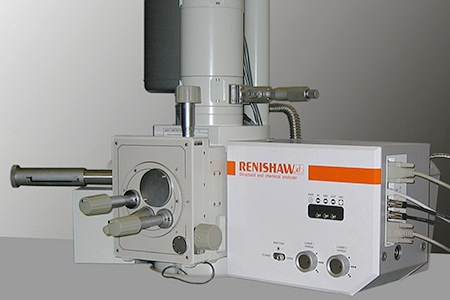 Renishaw Applications Scientist, Dr Katherine Lau, will make a presentation as part of the conference program and lead a workshop on Raman confocal microscopic imaging. MMC 2015, incorporating EMAG 2015, is hosted by the Royal Microscopical Society and takes place from 29th June to 2nd July 2015, at the Manchester Central Convention Complex, Manchester, UK. During this exhibition and conference, Applications Scientist, Dr Katherine Lau...
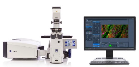 ZEISS announces that it is participating in a new public-private partnership with UC Berkeley as part of the Brain Microscopy Innovation Center (BrainMIC), a component of the Berkeley BRAIN Initiative. The Brain Research through Advancing Innovative Neurotechnologies (BRAIN) Initiative was launched by President Obama in 2013 and is focused on revolutionizing understanding of the human brain, with the goal of helping researchers uncover the...
 ‘Hybrid technology’ mini camera combines optical and gamma imaging to improve diagnosis and lymph and tumour removal efficiency
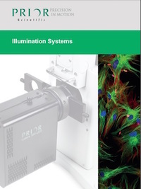 Regardless of microscopy technique, high quality imaging requires high performance illumination in order to obtain the best possible results. Prior Scientific has designed a range of precisely engineered, reliable and long lasting light sources tailored to the exacting requirements of scientists across a broad range of disciplines. The new catalogue details Prior’s expanding range of LED illumination sources that offer many advantages for brightfield, DIC, phase contrast and fluorescence microscopy...
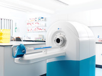 The scanner will be used in their Regenerative Medicine research programme which aims to develop a clearer understanding of the potential risks and hazards of therapies and to develop new methodologies to assess their risk. The aim of this research is to gain faster access to new medicines for patients. This new state of the art preclinical imaging system was developed, supplied and installed by MR Solutions of Guildford, Surrey and supported...
 The new RH95 Relative Humidity Controller is the latest addition to provide precise environmental sample control to Linkam’s range of temperature stages. It has been developed based on the feedback of many Linkam users in the materials, foods and pharmaceutical industries where humidity plays an important role in product development, manufacture and even storage. It will be shown on stand number 411 at mmc2015, the biennial conference and exhibition organised by the Royal Microscopical Society.
 ResearchIR 4.2 provides researchers and scientists with a powerful tool for viewing, acquiring, analyzing, and sharing the thermal data captured with FLIR’s Scientific and R&D cameras. ResearchIR Max version 4.2 gives users direct access to their MATLAB scripts within ResearchIR for the first time. This will allow users to access their customized MatLab® scripts directly in ResearchIR for specially-tailored image analysis and processing tasks...
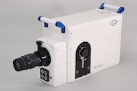 The Engineering and Physical Sciences Research Council (EPSRC) has since 1985 offered UK academic institutions free loans of the latest scientific instruments to help further their research projects. In 2008 the EPSRC added a state-of-the-art SIM16 ultra-high speed imaging camera from Specialised Imaging Ltd. to its loan pool facility. Wai Chan, Managing Director of Specialised Imaging commented "Over the last 7 years the EPSRC Loan Pool SIM16...
 Ovizio Imaging System, an innovative quantitative microscopy company specializing in life science solutions, announces today the launch of its new product,
 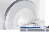 The transformation, which can be undertaken in around ten minutes, involves sliding in a carrier system linked to a separate state of the art EVO spectrometer providing superior soft tissue contrast and molecular imaging capabilities. This is a major breakthrough for clinical facilities which also conduct their own research as it enables existing technology to be utilised without the need for extensive investment in a separate system...
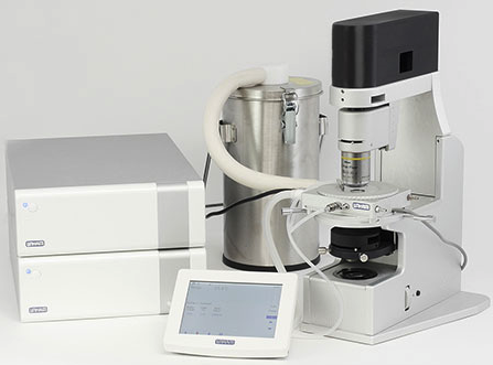 Dr Sheng Qi is a senior lecturer in pharmaceutics in the School of Pharmacy at the University of East Anglia. Sheng's current research interests focus on gaining a fundamental understanding of the behaviour of drug-polymer/lipid dispersions in solid (phase behaviour) and liquid (in biological fluids) states. She is also developing new approaches for stabilising supersaturated drug dispersions. By working with industrial partners as well as...
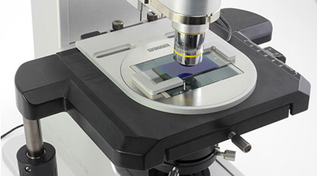 At the 2015 and 10th anniversary annual meeting of the Association of Biomedical Andrologists, Linkam have introduced a new solution for embryologists seeking a better solution for the evaluation and quantification of sperm. Live cell research and, specifically, the testing of sperm in clinics and hospitals, has not been well served in terms of reliable instrumentation and testing protocols. Mr Stephen Harbottle is Chairman of the Association...
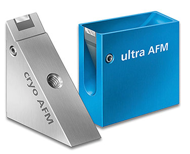 Whilst diamond knives are best known for producing high quality sections on an ultramicrotome, their use for surface preparation is less accepted. DiATOME diamond knives are used for the preparation of high quality surfaces for AFM and other imaging techniques requiring smooth surfaces. DiATOME ultra AFM knives are made from higher quality diamonds to produce extremely smooth sample surfaces and ensure the best possible...
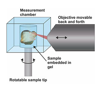 The 15th annual meeting of the European Light Microscopy Initiative continues the goals of the first meeting in 2001, namely to promote the quickly developing field of light microscopy as a fundamental research tool for the life sciences and to strengthen the channels of communication between researchers and industry. The combination of top level scientific presentations with practical hands-on workshops brings participants from all over...
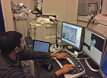 For example, FMS-EM force measurement systems are enabling researchers at Imperial College London to investigate the root cause of failures in electrochemical devices such as fuel cells and batteries. Imperial College is also using them for testing during the development of nano robots and other nanostructures. The FMS-EM is a compact force readout tool that allows tensile measurements to be made inside an SEM/FIB. Whilst existing...
 This new version contains intuitive and guided analysis workflows, optimized to clinical practice. The workflows will guide you through the required steps for Functional left and right ventricle, Viability and First Pass Perfusion analysis. The new viability workflow includes, besides analysis of Delayed Enhanced MR images, also analysis of edema on T2-weighted MR images. The combination of segmented infarcts and edema areas can identify...
 The first LaVision BioTec UltraMicroscope User Meeting will take place at the Max Planck Institute for Molecular Biomedicine in Münster, 21-22 September. Participants are cordially invited to join with all active and prospective UltraMicroscope users in a forum on the latest developments in light sheet microscopy. With this scientific gathering, LaVision BioTec is fostering the exchange of thoughts, experiences and views on this emerging and very active...
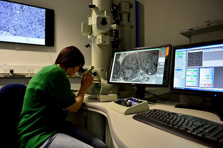 The Microscopy and Histology Facility provide access to assorted technologies to researchers at the University of Aberdeen. One core technique is Transmission Electron Microscopy (TEM) which was recently upgraded with the installation of a new JEOL JEM-1400 plus TEM with AMT camera system from Deben. The current research done using this equipment is varied and includes samples such as bone cells, yeast cells and macrophages. Some...
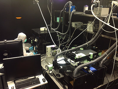 Matteo Iannacone, MD, PhD is the Group Leader in the Division of Immunology at the San Raffaele Scientific Institute, Milan, Italy. His research program seeks to dissect the complex dynamics of host-virus interactions with a particular focus on the development and function of adaptive immune responses. Since it is still beyond the reach of even the most sophisticated in vitro methodology to simulate the complex interplay of physical...