Members Login

Channels
Special Offers & Promotions
LaVision BioTec Reports on the Use of Light Sheet Microscopy in the Research as Part of the Miami Project to Cure Paralysis
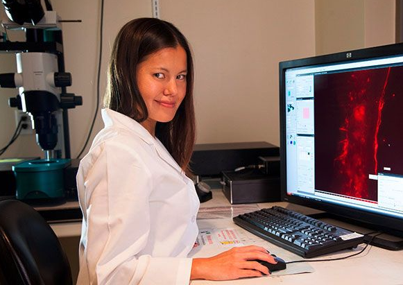
LaVison BioTec, developers of advanced microscopy solutions for the life sciences, report on users of their Ultramicroscope Light Sheet Fluorescence Microscope system to aid the research of the Miami Project to Cure Paralysis under the supervision of Professor Vance Lemmon, the Walter G. Ross Distinguished Chair in Developmental Neuroscience & Professor of Neurological Surgery at the University of Miami.
 In 2003, Professor Vance Lemmon accepted a position at The Miami Project to Cure Paralysis at the University of Miami. This centre has taken the philosophy that by promoting interactions between basic and clinical scientists, it will be possible to speed the finding of a cure for a devastating clinical problem. This research has focused on answering questions that help define human spinal cord injury and reveal strategies for the repair of damaged spinal tissue.
Professor Lemmon's lab studies nerve regeneration in the brain and spinal cord. Describing his work, he says "We hope to make nerve cells regenerate much better than they normally do. My colleagues study scar formation in the cord, neural cell translation and blood vessel formation after injury while another colleague studies optic nerve regeneration."
The biggest challenge to Professor Lemmon and his team is the ability to test literally thousands of samples each week in vitro and rapidly translating interesting "hits" into in vivo tests of efficacy. Imaging and analysis many in vivo samples is vital to this work and this led to the introduction of Light Sheet Fluorescence Microscopy (LSFM) to the team. As he says, "We always want to go faster! We needed to dramatically speed up the pace at which we can image large 3D volumes of the brain and spinal cord. Traditional methods are a bottle neck that limit discovery. While epifluorescence and confocal microscopies were useful, using LSFM enabled us to image way faster and much larger volumes can now be visualised."
The work is generating several high profile papers in the field of neuroscience. Professor Lemmon outilnes two. "In our group, we have demonstrated the applicability of LSFM for comprehensive assessment of optic nerve regeneration, providing in-depth analysis of the axonal trajectory and pathfinding. Our study indicates significant axon misguidance in the optic nerve and brain, and underscores the need for investigation of axon guidance mechanisms during optic nerve regeneration in adults.1 Other colleagues have applied genetic lineage tracing, light sheet fluorescent microscopy and antigenic profiling to identify collagen1α1 cells as perivascular fibroblasts that are distinct from pericytes. The results identify collagen1α1 cells as a novel source of the fibrotic scar after spinal cord injury and shift the focus from the meninges to the vasculature during scar formation.2"
Media Partners


