Channels
Special Offers & Promotions
LaVision BioTec Reports on the Award-Winning Scientist Peter Friedl and his Work Imaging Cancer Growth
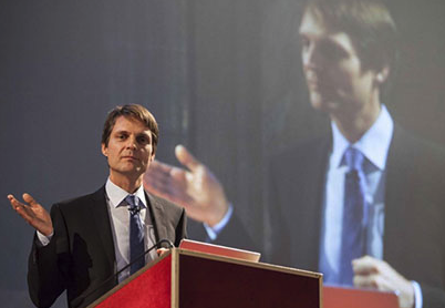
LaVison BioTec, developers of advanced microscopy solutions for the life sciences, report on the research of Dr Peter Friedl and his imaging of cancer growth using intravital microscopy.
 Peter Friedl holds the chair for Microscopical Imaging of the Cell at the St. Radboud University Nijmegen Medical Centre, University of Nijmegen, The Netherlands. He also holds a 20% joint-appointment as head of the imaging section at the David H. Koch Center, Department of Genitourinary Oncology, MD Anderson Cancer Center, Houston, TX, USA. His research work focuses on imaging cancer growth, metastasis and therapy response in the natural tumour environment - which is the living tissue in an intact body. As it is not possible to perform microscopy of tumours in patients, he uses experimental tumour models in live mice. Here, they implant fluorescent tumours; then follow individual tumour cells over time; and draft conclusions about their aggressiveness. The goal is to try to find therapeutic strategies to inhibit cancer progression.
His work has recently been recognised with the 13th City of Florence Prize for the Molecular Sciences. This recognises Dr Friedl and his team for the discovery/invention of a new technology to observe the dynamics of metastasis and to understand how cells divide and multiply. Thanks to his experience in the field of microscopy, Dr Friedl has developed a exciting new methodology with which to obtain 3D images of living tissue through low energy fluorescence irradiation. According to the team, special treatments using radiotherapy and immunotherapy may soon be visualized to a level required to better attack cancer and achieve a cure.
To generate these images, Dr Friedl selected the TriM Scope 2-photon microscope from LaVision BioTec. He describes his reasons for this decision: "The Trim Scope was the first user-friendly system on the market combining commonly used two-photon excitation with the new approach of infrared excitation. In addition, the science-driven approach of the engineers at LaVision BioTec allowed me as biomedical researcher to develop ideas and jointly find solutions to challenges in intravital microscopy that were, and are, beyond the state-of-the-art. This instigated more technical innovations which greatly enhance multi-parameter deep tissue imaging in cancer research, in particular, the implementation of dual- or triple-beam laser in-coupling and the combined use of fluorescence, second and third harmonic generation in live tissues. This strength and the versatility of the setup enabled my applications. None of the other systems I considered reached our needs for efficacy and flexibility."
The Trim Scope has many features vital for Dr Friedl's research. He lists a few: "First, the flexible placement of up to 8 detectors including specifically high-sensitivity detectors; second, the multi-laser beam capability; and third, the adaptability of the setup to different additional detectors such as a fluorescent life-time detector. The versatility of the table makes it straightforward to accommodate live animals. Lastly, I found the interaction with the LaVision instrument developers extremely positive. They listened and enabled the needs I identified."
Media Partners


