Members Login

Channels
Special Offers & Promotions
JPK reports on the characterization of nanostructures and nanosized systems using the NanoWizard AFM in the group of Giovanni Dietler at EPFL, Lausanne
JPK Instrument reports on how the NanoWizard AFM is being applied in the study of nanostructures and nano-sized systems
 JPK Instruments, a world-leading manufacturer of nanoanalytic instrumentation for research in life sciences and soft matter, reports on how the NanoWizard AFM is being applied in the study of nanostructures and nano-sized systems at the École Polytechnique Federale de Lausanne.
The Laboratory of the Physics of Living Matter (LPMV) at the École Polytechnique Federale de Lausanne, better known as EPFL, uses various techniques for the study of the nanosciences, an intrinsically interdisciplinary field providing many scientific challenges from different research fields including biosensors, life, materials and space sciences. These tools include Scanning Probe Microscopy, coupled with optical and fluorescence microscopy, spectroscopy (UV-Vis-IR, FTIR) and electron microscopy.
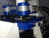
Dr Giovanni Longo is a Research Scientist in the LPMV under the supervision of Professor Giovanni Dietler. He works on a number of projects which require the accuracy and reproducible performance at high resolution of an atomic force microscope, AFM, and has chosen the JPK NanoWizard® 3 for his work. For example, he uses the AFM cantilevers for the rapid detection of bacterial resistance to antibiotics. The diagnostic tool has been conceived to study the dynamical movements of specimens (such as conformational changes in proteins, or small movements in bacteria or cells) in order to investigate their features and their response to external stimuli. Living, moving specimens are attached to an AFM cantilever and their movements are transduced by the lever to the AFM electronics and recorded. Dr Longo showed that these movements are related to the metabolism of the bacteria or cells attached to the lever and that this system can be used to define, in minutes, the resistance of bacteria to antibiotics. [Longo et al. Nature Nanotechnology 2013]
Describing this work, Dr Longo said "We recently moved this activity to the JPK NanoWizard microscope since it demonstrated an excellent sensitivity and since the optical microscope is invaluable to couple the macroscopic movements of the biological systems with the microscopic movements we detect using our nanomotion sensor." [Aghayee et al. Journal of Molecular Recognition, 2013]
Dr Longo describes a second project. "We also study osteoblasts for bone-integration and to characterize their response to microgravity conditions. This research activity is aimed to the study of the growth of osteoblasts with a focus on their interactions with differently prepared substrates and to different gravity conditions. In this field of research we have coupled the fluorescence microscopy capabilities of the Axiovert inverted microscope with the high-resolution AFM images to obtain stunning images of cells."
There are three main features of the NanoWizard which makes it the ideal tool for AFM studies. First, it operates with remarkable stability and the images it delivers are of high-quality and with a very high rate of repeatability. This is particularly impressive since the X/Y/Z ranges are not small and yet the images are of excellent quality both at very large and at very small ranges. Moreover, the system is capable of stable imaging for several hours with very little drift and a precise control of the imaging position.
The JPK Quantitative Imaging mode (QI™) is excellent allowing fast imaging of the specimens while, at the same time, collecting their mechanical properties. This is invaluable especially when characterizing many samples or when a large number of cells must be probed. For example, Dr Longo used it to determine the stiffness of hundreds of bacteria in different environmental conditions in just a few weeks, much more rapidly than could be achieved on older systems.
Lastly, when the microscope is coupled with an excellent optical inverted microscope, it is capable of fluorescence imaging. This allows producing wonderful correlative images and allows monitoring the evolution of living systems with the combination of two different microscopy techniques.
Media Partners


