Channels
Special Offers & Promotions
3D Imagery to Improve Surgical Visualisation of Liver Cancer
publication date: Dec 16, 2011
|
author/source: Acumen
 Holoxica, an Edinburgh-based 3D
holographic imaging company, has furthered the boundaries of biomedical imaging
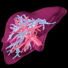
by creating the world's first 3D, full colour hologram of a human liver, paving
the way for a breakthrough in the way surgeons plan liver operations to remove
tumours.
Holoxica, an Edinburgh-based 3D
holographic imaging company, has furthered the boundaries of biomedical imaging
by creating the world's first 3D, full colour hologram of a human liver, paving
the way for a breakthrough in the way surgeons plan liver operations to remove
tumours.The human liver hologram will enable surgeons and oncologists to ‘look around' the ‘virtual' organ and marks a breakthrough for medical science which until now, has had to rely on two-dimensional screens to view three-dimensional information from CT, MRI and ultrasound scanning techniques. 3D models based on actual patient data can be used for training and simulation by surgeons, enabling the surgeon to visualise the intricacies of navigation within the organ.
It means that specialists can now find new ways of visualising the complete structure of a human liver in greater detail and to better understand tumour behaviour within the liver than would be the case from 2D images they currently use.
This helps to accurately pinpoint diseased liver tissue so surgeons can plan extraction operations armed with a fuller awareness of the surrounding tissues and organs. It is hoped that this will lead to improved chances of survival.
Javid Khan, managing director of Holoxica comments;
"This truly is a leap forward in ascertaining tumour characteristics within the liver or other organs. Holographic technology can now be used to great effect in the field of biomedical science. One scenario could be cancer treatment planning in radiation therapy. It's important that the radiation beam is concentrated directly on the tumour and not on surrounding tissues. A "true 3D" imaging hologram can help create a radiation plan that does just that!
It will also help with operation planning. Surgeons have always viewed the liver two dimensionally, yet can visualise a 3D model of the "scene". Therefore, a holographic image of the patient's target will give the surgeon a true perspective on where a tumour might be located and the best way to access it. Additionally, a holographic "print out" could be useful for archiving patient data for reference."
Holoxica has collaborated with the renowned Germany-based Fraunhofer Institute, Europe's largest application oriented research organisation, which has an annual research budget of over €1.65 billion, to develop this application. Liver imaging has been commonly undertaken in patients with cancer history because it is one of the most frequently involved organs vulnerable to the spread of tumours to other organs. To assist this, Holoxica plans to map a human liver in three conditions, so surgeons can acquaint themselves with a liver in healthy, diseased and post surgical states.
The goal of liver imaging includes liver tumour detection and characterisation and with new screening techniques continuing to evolve, the use of 3D modelling will give surgeons a far greater understanding of how a liver reacts to tumour and other chronic diseases.
Javid Khan, added;
"The life science community is benefitting from our technology in allowing them to access information in a manner which brings the subject - a human organ or any organism - to life in a more realistic manner they can gain a better understanding from it and better inform their subsequent course of action."
Holoxica has just won a Nexxus Scotland Collaboration Award sponsored by the Edinburgh Science Triangle. The award acknowledges collaboration to develop a technology, product or service involving life sciences and a company or organisation based in the East of Scotland/Edinburgh City Region.
For more information visit www.holoxica.com/scientific
Media Partners


