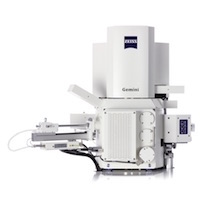
ZEISS industry-leading GeminiSEM 500 imaging system to enhance the MACES open access microscopy imaging facility
ZEISS announces that it is providing its GeminiSEM 500, customized for nanomaterial characterization, to the University of California Merced’s Merced Nanomaterials Center for Energy and Sensing (MACES). The University’s recent purchase will support the university’s strong nanomaterial-based research programs and aid in the Center’s research in functional nanomaterials for energy and sensing for NASA missions...
If you are still using film, you should consider some benefits of digital imaging for your gels and blots
The choice between digital imaging and using film for gel and blot documentation is generally based on what the lab has done historically and the availability of equipment and reagents within the lab. Depending on the scenario, the use of film for protein and DNA detection is still popular as it has been the gold standard for decades. However, if you are still using film, you should consider some benefits of digital imaging.
Magritek, a leading provider of compact NMR and MRI instruments, announce new 60 MHz capability for the family of benchtop NMR spectrometers.
Magritek launch their new Spinsolve 60 proton, fluorine and carbon systems at the ACS National Meeting in San Diego, 13th March, 2016. With an innovative high quality 60 MHz Halbach magnet squeezed into the compact Spinsolve® spectrometer form factor, Magritek's new Spinsolve 60 NMR spectrometers set the bar for 60MHz benchtop NMR instruments.
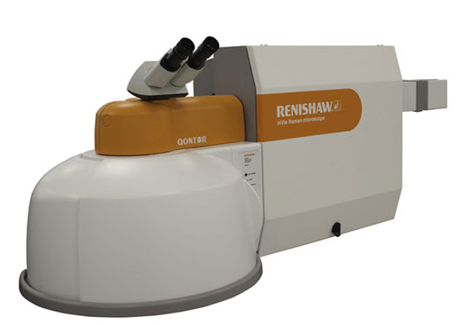
Read MoreRenishaw Launches the New inVia Qontor Confocal Raman McroscopeApr 11, 2016
The new inVia™ Qontor™ is Renishaw’s most advanced Raman microscope.
Building on the market-leading inVia Reflex, the inVia Qontor adds a new dimension to the performance and ease of use for which inVia is renowned. The inVia Qontor sees the addition of Renishaw's latest innovation, LiveTrack™ focus tracking technology, which enables users to analyse samples with uneven, curved or rough surfaces. Optimum focus is maintained in real time during data collection and white...
Thermo Scientific DXR2 line of Raman microscopes ideal for scientists working in advanced materials research, pharmaceuticals, polymers and the developing life science research market
New microscopes bring the power of Raman microscopy and high-resolution chemical imaging to more analytical laboratories and research groups who can benefit from having relevant sample information in the shortest time possible. The Thermo Scientific DXR2 Raman microscopes, which made their debut at Pittcon 2016 (booth 2239) at the Georgia World Congress Center, Atlanta, provide...
Newport Corporation, a global leader in high precision motion and photonics solutions, is introducing the XPS-RL, a cost effective, universal motion controller for up to four axes.
The new controller is based on Newport’s well-proven XPS platform. The XPS-RL focusses on user friendly operation with a newly designed graphical user interface, single-click stage configuration and TCP/IP auto-detection. Newport’s proprietary ESP plug-and-play compatibility instantly recognizes when an ESP-compatible stage is connected to the controller. All operating parameters are configured automatically without...
Specialised Imaging reports on a technical article, written by researchers from the King Abdullah University of Science and Technology, Saudi Arabia that describes the use of a Kirana ultra high-speed video system to undertake time-resolved imaging of a compressible air disk that forms under a drop impacting on a solid surface.
The capture of a bubble under an impacting drop can have detrimental effects on the uniformity of spray coatings and interfere with precision inkjet-based manufacturing such as are used in the printing of organic displays. When a drop impacts on a solid surface, its rapid deceleration is cushioned by a thin layer of air, which leads to the entrapment of a bubble under its centre. For large impact velocities the lubrication pressure in...
The Kircher laboratory at Memorial Sloan Kettering is developing novel nanoprobes for molecular imaging, image-guided therapy and theranostics.
Its ultimate goal is to develop a universal technology that allows precise determination of the actual spread of a tumour in vivo. Currently surgeons cannot see the microscopic extent of the tumour during a procedure, which is essential information for tumor removal and avoiding excess tissue excision. Physician-scientist Dr. Moritz Kircher is working on a new generation of nanometer-sized imaging beacons. These allow...
Dolomite has developed the Meros High Speed Digital Microscope specifically for microfluidic applications.
This high resolution imaging system allows users to conveniently monitor high speed microfluidic events, such as droplet formation, using a built-in stage. The Meros High Speed Digital Microscope features high magnification optics and a zoom lens to ensure that millimetre to micrometre scale features can be visualised clearly, with the added benefit of an extra-long working distance. Coupled with a high speed USB 3.0 camera...
We bring you more exciting news about Photonex London Roadshow.
The programme has now been finalised with an excellent calibre of speakers set to give presentations. Attend this event to broaden your knowledge in the world of photonics by attending a key meeting for photonics technologies in the life sciences. The technical committee have commented that this international quality 'invited' programme includes presentations by eminent and leading European research scientists...
FLIR Systems has introduced the new FLIR X6900sc as the world's fastest 640 x 512 pixel resolution thermal camera for high-speed science applications.
The new thermal imager is designed to record 1000 fps at full resolution onto the camera's RAM for up to 26 seconds. Whether measuring temperatures on fast moving objects or characterizing the thermal transient of objects as they heat up, this new camera offers the rapid frame speed, high resolution, and thermal sensitivity required to virtually stop motion enabling accurate temperature readings, and recording of gradients across...
MR Solutions’ new 3D in vivo confocal microscope for use in preclinical research provides a magnification range of up to 1000 times, allowing researchers to examine cellular details within a live small animal eliminating the need for a surgical biopsy - saving time and substantially reducing costs.
MR Solutions, the world leader in preclinical scanning, has partnered with Optiscan1 to introduce CellLiveTM, their second generation endomicroscopy platform to the preclinical market. The handheld Optiscan probe which is less than 3.6 mm in diameter delivers micron – smaller than a millionth of a metre - resolution. The Optiscan uses a fluorescence2, confocal3 imaging system to achieve this...
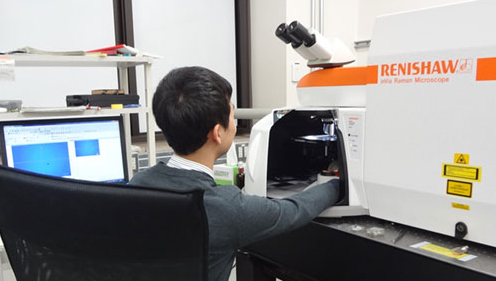
The University of Tokyo's Department of Mechanical Engineering was established in 1879, providing education based on four disciplines; mechanics, materials, hydrodynamics and thermodynamics.
Within the Department, the Maruyama-Chiashi Laboratory focuses its research on the synthesis and analysis of carbon nanotubes (CNT), graphene and other nano-materials. They study applications related to the development of energy related devices, such as solar cells. The laboratory uses scanning Raman spectroscopy as an important tool for the investigation of the synthesized materials and their structure...
JPK Instruments, a world-leading manufacturer of nanoanalytic instrumentation for research in life sciences and soft matter, reports on the use of their NanoTracker™ 2 optical tweezers system which is being used to study the physical and chemical properties of micro-bubbles in the Department of Mechanical Engineering at the Shibaura Institute of Technology under the leadership of Associate Professor, Dr Yoko Yamanishi.
Dr Yoko Yamanishi is an Associate Professor in the Department of Mechanical Engineering at Shibaura Institute of Technology, Tokyo, Japan. She leads the Yamanishi Laboratory - the Micro-nano Functional Fluid Laboratory. The Laboratory's goals aim to clarify unknown function of cells by using micro-nano technology based on mechanical, electrical and bio-medical engineering. It targets a contribution to the development of cellular scale...
Prior Scientific has announced the FB201 co-axial coarse and fine focusing block, designed for OEM microscopy applications where precise fine focus adjustment is required.
Rack and pinion mounting allows the FB201 to produce smooth, precise focusing over 29 mm of travel. A large coarse focus mechanism incorporates a slip clutch and tension adjustment. Fine focus adjustment control is graduated in two micron divisions throughout the coarse focus range. The focus block can be easily adapted to various mounting configurations and can support up to 5 kg. Motorised variants able to...
At last, an affordable solution for the scanning of histology and Cytology microscope slides.
The new Slide Scanner available from Eikonix now brings high quality slide scanning to a much wider market. This compact device utilises a host of the latest automated imaging technologies with a proven scanning engine to create an effective and reliable tool for the rapid acquisition of whole slide images from standard glass microscope slides. The easy to use system can be operated by anyone and requires no specialist training. The intuitive software effortlessly guides the user through each step to produce images of exceptional quality.
Fluorescence analysis workstations are one of many products manufactured by Spectronics Corporation to aid laboratory technicians.
Built to exacting standards, they’re used for viewing and analyzing fluorescent samples with both epi-illumination and trans-illumination light sources. The Spectroline® CM-10MP mini viewing cabinet is perfect for any type of laboratory application requiring high-contrast fluorescence analysis. It’s also excellent for viewing TLC plates and for quality control inspection of PC boards. The CM-10MP is an “all-in-one tool” for the laboratory technician...
Combines Accuracy with Affordability to Deliver Rapid Count Results
Synbiosis, a long-established, expert manufacturer of automated microbiological systems, is delighted to introduce the aCOLyte 3 HD its next generation, automated colony counter for microbiologists that demand a sensitive, budget system to quickly and accurately count colonies of all sizes and colours. The new aCOLyte 3 HD colony counter, features a high resolution megapixel CCD camera for accurate detection of different coloured...
Union Biometrica develops tools for model organism research.
The NEW VAST BioImager platform™ (Vertebrate Automated Screening Technology) is designed for zebrafish researchers who need to image large numbers of 2–7 day old sedated zebrafish larvae. VAST automation avoids the time consuming and tedious steps of manual manipulation of each larva during screens. VAST loads each larva into a capillary and then rotates it 360 degrees to determine the orientation of the fish...
ZEISS introduced two new digital microscope cameras during the annual meeting of the American Society for Cell Biology (ASCB 2015) in San Diego.
ZEISS Axiocam 702 mono and ZEISS Axiocam 512 color complement the current portfolio of high-speed USB 3.0 microscope cameras. With ZEISS Axiocam 702 mono ZEISS for the first time introduces a microscope camera with a scientific CMOS sensor. Users benefit from low read noise, excellent low light sensitivity and high speed for live cell imaging and acquisition of fast processes. ZEISS Axiocam 702 mono features a 1/1.2" (13.3 mm diagonal)...
LED fluorescence illumination devices that provide a simple solution for microscope users wishing to upgrade their microscope for fluorescence applications.
New from Eikonix, the imaging specialists, is a range of LED fluorescence illumination devices that provide a simple solution for microscope users wishing to upgrade their microscope for fluorescence applications. The MF-LED module which has built-in coloured LED’s can easily upgrade a traditional infinity upright microscope to give fluorescence functionality. It is optically compatible with many popular microscope brands such as Olympus, Nikon, Leica, Zeiss and others....
FLIR Systems announces a new addition to its RS-Series of long-range infrared camera systems designed for range tracking, target signature, research, and scientific applications.
The new FLIR RS8300 couples a proprietary high-speed HD MWIR detector with a 10x continuous zoom lens in a sealed, ruggedized enclosure. Combining reliability with leading edge performance, the FLIR RS8300 delivers stunning megapixel infrared imagery at up to 200 megapixels per second. To capture the most fleeting of events the camera is capable of lightning fast frame rates from full-frame resolution 14-bit...

 ZEISS announces that it is providing its GeminiSEM 500, customized for nanomaterial characterization, to the University of California Merced’s Merced Nanomaterials Center for Energy and Sensing (MACES). The University’s recent purchase will support the university’s strong nanomaterial-based research programs and aid in the Center’s research in functional nanomaterials for energy and sensing for NASA missions...
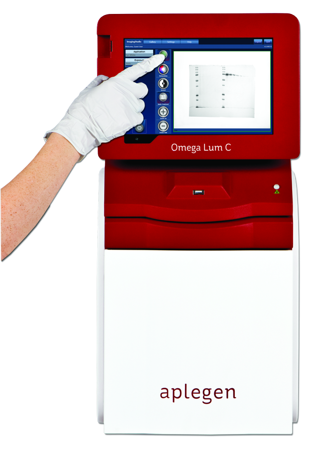 The choice between digital imaging and using film for gel and blot documentation is generally based on what the lab has done historically and the availability of equipment and reagents within the lab. Depending on the scenario, the use of film for protein and DNA detection is still popular as it has been the gold standard for decades. However, if you are still using film, you should consider some benefits of digital imaging.
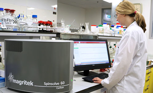 Magritek launch their new Spinsolve 60 proton, fluorine and carbon systems at the ACS National Meeting in San Diego, 13th March, 2016. With an innovative high quality 60 MHz Halbach magnet squeezed into the compact Spinsolve® spectrometer form factor, Magritek's new Spinsolve 60 NMR spectrometers set the bar for 60MHz benchtop NMR instruments.
 Building on the market-leading inVia Reflex, the inVia Qontor adds a new dimension to the performance and ease of use for which inVia is renowned. The inVia Qontor sees the addition of Renishaw's latest innovation, LiveTrack™ focus tracking technology, which enables users to analyse samples with uneven, curved or rough surfaces. Optimum focus is maintained in real time during data collection and white...
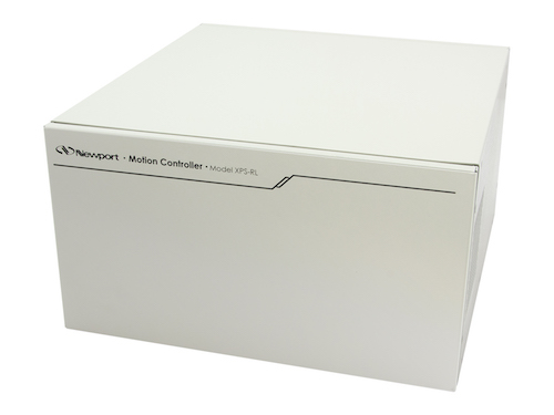 The new controller is based on Newport’s well-proven XPS platform. The XPS-RL focusses on user friendly operation with a newly designed graphical user interface, single-click stage configuration and TCP/IP auto-detection. Newport’s proprietary ESP plug-and-play compatibility instantly recognizes when an ESP-compatible stage is connected to the controller. All operating parameters are configured automatically without...
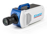 The capture of a bubble under an impacting drop can have detrimental effects on the uniformity of spray coatings and interfere with precision inkjet-based manufacturing such as are used in the printing of organic displays. When a drop impacts on a solid surface, its rapid deceleration is cushioned by a thin layer of air, which leads to the entrapment of a bubble under its centre. For large impact velocities the lubrication pressure in...
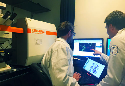 Its ultimate goal is to develop a universal technology that allows precise determination of the actual spread of a tumour in vivo. Currently surgeons cannot see the microscopic extent of the tumour during a procedure, which is essential information for tumor removal and avoiding excess tissue excision. Physician-scientist Dr. Moritz Kircher is working on a new generation of nanometer-sized imaging beacons. These allow...
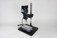 This high resolution imaging system allows users to conveniently monitor high speed microfluidic events, such as droplet formation, using a built-in stage. The Meros High Speed Digital Microscope features high magnification optics and a zoom lens to ensure that millimetre to micrometre scale features can be visualised clearly, with the added benefit of an extra-long working distance. Coupled with a high speed USB 3.0 camera...
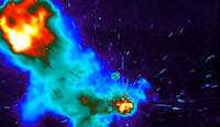 The new thermal imager is designed to record 1000 fps at full resolution onto the camera's RAM for up to 26 seconds. Whether measuring temperatures on fast moving objects or characterizing the thermal transient of objects as they heat up, this new camera offers the rapid frame speed, high resolution, and thermal sensitivity required to virtually stop motion enabling accurate temperature readings, and recording of gradients across...
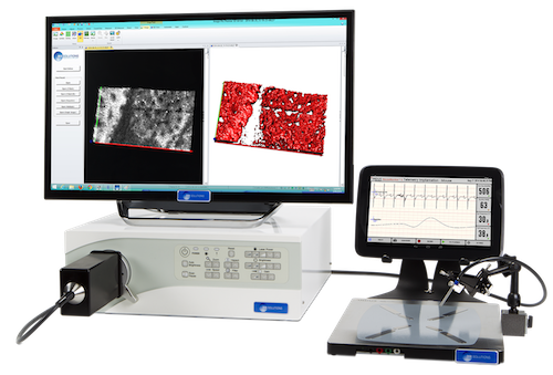
 Within the Department, the Maruyama-Chiashi Laboratory focuses its research on the synthesis and analysis of carbon nanotubes (CNT), graphene and other nano-materials. They study applications related to the development of energy related devices, such as solar cells. The laboratory uses scanning Raman spectroscopy as an important tool for the investigation of the synthesized materials and their structure...
 Dr Yoko Yamanishi is an Associate Professor in the Department of Mechanical Engineering at Shibaura Institute of Technology, Tokyo, Japan. She leads the Yamanishi Laboratory - the Micro-nano Functional Fluid Laboratory. The Laboratory's goals aim to clarify unknown function of cells by using micro-nano technology based on mechanical, electrical and bio-medical engineering. It targets a contribution to the development of cellular scale...
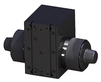 Rack and pinion mounting allows the FB201 to produce smooth, precise focusing over 29 mm of travel. A large coarse focus mechanism incorporates a slip clutch and tension adjustment. Fine focus adjustment control is graduated in two micron divisions throughout the coarse focus range. The focus block can be easily adapted to various mounting configurations and can support up to 5 kg. Motorised variants able to...
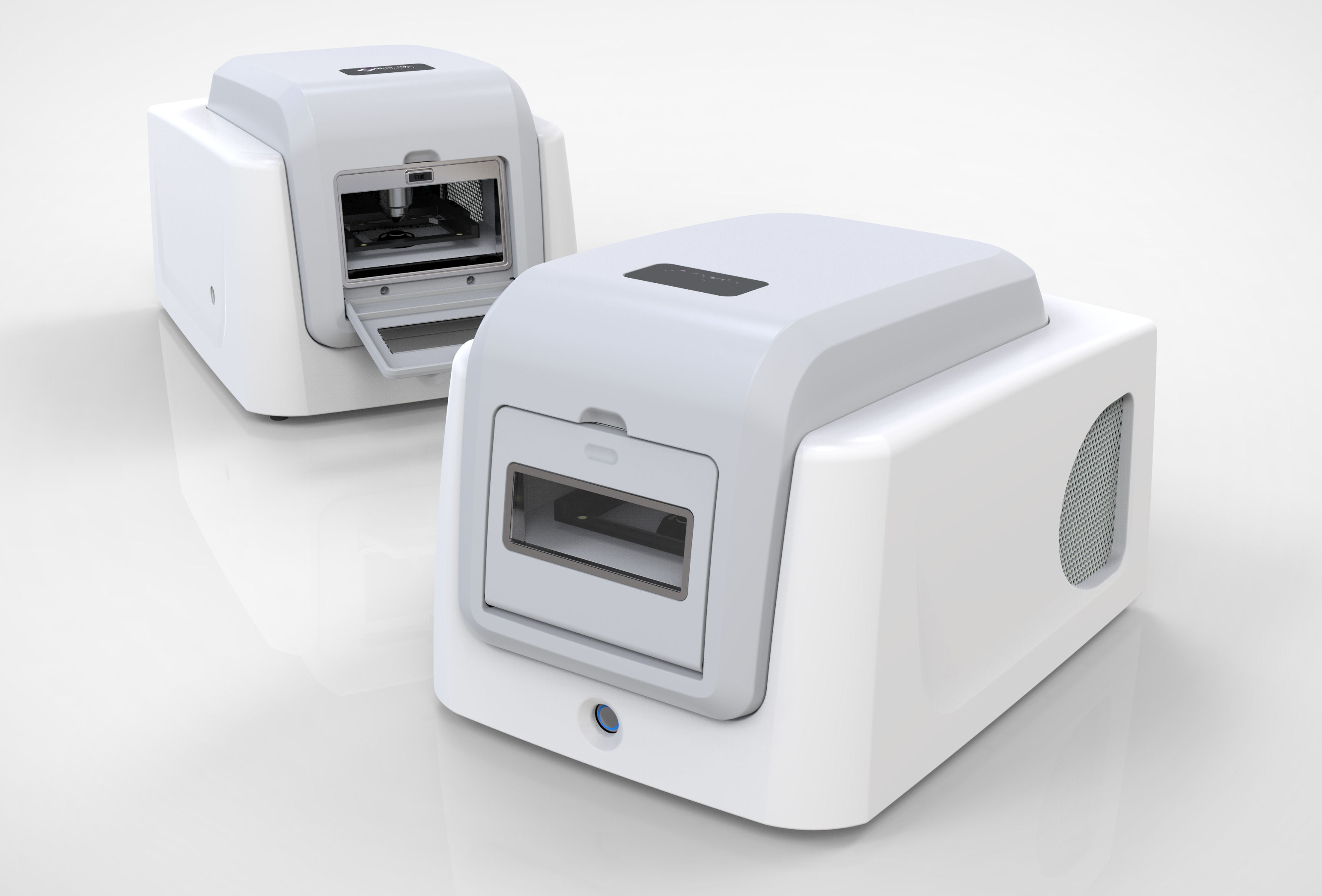 The new Slide Scanner available from Eikonix now brings high quality slide scanning to a much wider market. This compact device utilises a host of the latest automated imaging technologies with a proven scanning engine to create an effective and reliable tool for the rapid acquisition of whole slide images from standard glass microscope slides. The easy to use system can be operated by anyone and requires no specialist training. The intuitive software effortlessly guides the user through each step to produce images of exceptional quality.
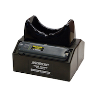 Built to exacting standards, they’re used for viewing and analyzing fluorescent samples with both epi-illumination and trans-illumination light sources. The Spectroline® CM-10MP mini viewing cabinet is perfect for any type of laboratory application requiring high-contrast fluorescence analysis. It’s also excellent for viewing TLC plates and for quality control inspection of PC boards. The CM-10MP is an “all-in-one tool” for the laboratory technician...
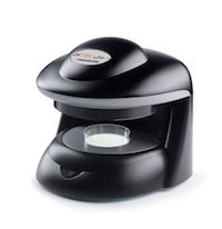 Synbiosis, a long-established, expert manufacturer of automated microbiological systems, is delighted to introduce the aCOLyte 3 HD its next generation, automated colony counter for microbiologists that demand a sensitive, budget system to quickly and accurately count colonies of all sizes and colours. The new aCOLyte 3 HD colony counter, features a high resolution megapixel CCD camera for accurate detection of different coloured...
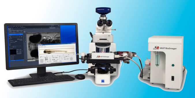 The NEW VAST BioImager platform™ (Vertebrate Automated Screening Technology) is designed for zebrafish researchers who need to image large numbers of 2–7 day old sedated zebrafish larvae. VAST automation avoids the time consuming and tedious steps of manual manipulation of each larva during screens. VAST loads each larva into a capillary and then rotates it 360 degrees to determine the orientation of the fish...
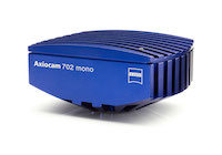 ZEISS Axiocam 702 mono and ZEISS Axiocam 512 color complement the current portfolio of high-speed USB 3.0 microscope cameras. With ZEISS Axiocam 702 mono ZEISS for the first time introduces a microscope camera with a scientific CMOS sensor. Users benefit from low read noise, excellent low light sensitivity and high speed for live cell imaging and acquisition of fast processes. ZEISS Axiocam 702 mono features a 1/1.2" (13.3 mm diagonal)...
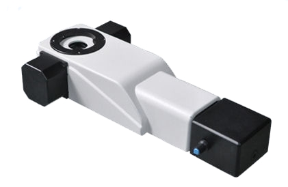 New from Eikonix, the imaging specialists, is a range of LED fluorescence illumination devices that provide a simple solution for microscope users wishing to upgrade their microscope for fluorescence applications. The MF-LED module which has built-in coloured LED’s can easily upgrade a traditional infinity upright microscope to give fluorescence functionality. It is optically compatible with many popular microscope brands such as Olympus, Nikon, Leica, Zeiss and others....
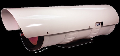 The new FLIR RS8300 couples a proprietary high-speed HD MWIR detector with a 10x continuous zoom lens in a sealed, ruggedized enclosure. Combining reliability with leading edge performance, the FLIR RS8300 delivers stunning megapixel infrared imagery at up to 200 megapixels per second. To capture the most fleeting of events the camera is capable of lightning fast frame rates from full-frame resolution 14-bit...