SI Sensors design novel CMOS image sensors for ultra-fast cameras that increase the velocity of the signal electrons allowing them to be swept out of the photodiode and be detected much faster. Developing ultra-high-speed CMOS image sensors is difficult because at very high frame rates, the integration time is extremely short and there is never enough light...
Indica Labs, the leading provider of AI-powered digital pathology solutions, proudly announces the launch of Lung Macrodissect AI, a groundbreaking tool that revolutionizes slide macrodissection and molecular pathology workflows. Lung Macrodissect AI represents a significant leap forward for the integration of artificial intelligence with digital pathology by offering pathologists a streamlined macrodissection workflow...
Flexibility in image analysis is key to addressing your diverse research questions. The ZEISS arivis software suite is designed to accelerate your research, offering advanced tools that ensure you reach reliable and reproducible results faster. No matter the size, format, or source of your image, ZEISS arivis can help you analyze it. And all without the need to code. Version 4.2 of arivis Pro is available now...
Oxford Instruments is delighted that its new Unity detector has been recognised as one of the ten best microscopy innovations of the year in the 2024 Microscopy Today Innovation Awards. Each year, Microscopy Today awards ten organizations or individuals for innovations they have launched or published in microscopy or microanalysis...
CrestOptics S.p.A., a manufacturer of high-end microscopy solutions and advanced systems for fluorescence microscopy and diagnostic applications, announce that its CICERO spinning disk confocal system has been named a ‘Top 10 Best Microscopy Innovation’ by Microscopy Today, an industry-leading microscopy publication from the Microscopy Society of America...
Indica Labs announces the expansion of their reseller network in the Asia Pacific (APAC) region. New distributorship agreements have been inked with MRL Cybertec Corporation, Pathology Ware International, Minh Khang Technical Service & Trading Co. Ltd., and Histocenter, for distribution in the Philippines, Thailand, Vietnam, and Malaysia, respectively. The new partnerships build upon Indica’s previous commercial success in China and Japan where sales have grown substantially in recent years...
Specialised Imaging has introduced a new version of its market-leading SIM family of ultra high-speed framing camera that makes hyperspectral imaging simpler with the option of user interchangeable filters. Though hyperspectral imaging was possible on previous models, the new SIM design was driven by requests from the field of energetic materials research which required a camera with simple and fast exchange of filters in all its operating channels...
Oxford Instruments Andor has announced the release of Imaris 10.2, the latest version of its market-leading AI-powered image analysis software for microscopic images. In the last decade, Imaris has constantly evolved to improve the visualisation and analysis of large images. Past releases have introduced AI (machine learning) to improve object identification and classification, spatial measurements, correlative data visualisation, and other tools for more precise segmentation...
Linkam Scientific Instruments is showcasing its cryo-correlative portfolio at this year’s M&M conference, and will be hosting tutorials and workshops at the event. Linkam will also be presenting two posters during the conference. Linkam’s CryoGenium plunger system, one of the company’s latest developments, will be shown on the booth. The CryoGenium is a fully automated system which offers a range of features for users of cryo-EM, cryo-tomography, single particle tomography (SPT) and cryo-CLEM methods....
TESCAN, a leading provider of scientific instrumentation, announces the launch of the TESCAN AMBER 2, the fourth generation of the TESCAN gallium FIB-SEM platform and the successor to the current AMBER generation, at the Microscopy & Microanalysis (M&M) Expo 2024. Alongside AMBER 2, TESCAN is also unveiling two new integrated innovative tools...
Evident’s popular PRECiV™ image analysis software now powers our DSX1000 digital microscope, bringing new levels of ease of use and usability. With the launch of PRECiV v. 2.1.1, a single, unified software program can now be used to control all our manual, motorized, and digital industrial microscopes. PRECiV DSX makes using the DSX1000 digital microscope even simpler, providing seamless control over all motorized functions for greater speed and accuracy...
Bio-Rad Laboratories, Inc., a global leader in life science research and clinical diagnostics products, announce the launch of the ChemiDoc™ Go Imaging System, the latest addition to its portfolio of ChemiDoc Imaging Systems. The system offers rapid, reliable, and highly sensitive gel and western blot imaging on a benchtop scale. The ChemiDoc Go Imaging System uses complementary metal oxide semiconductor (CMOS) digital imaging to capture gel and western blot images with the same high sensitivity as larger instruments...
Leica Biosystems and Navinci Partner to Accelerate Cancer Research Through First-Ever Fully Automated In Situ Proximity Ligation Assays. Leica Biosystems, a global leader in anatomic pathology, and Navinci, a leader in developing innovative solutions for studying protein interactions, announce a strategic collaboration to drive innovation in cancer patient care. This partnership enables automated in situ proximity ligation assays on the BOND RX Fully Automated Research Stainer...
Shimadzu Corporation has concluded a business partnership agreement with TESCAN GROUP, a.s. (TESCAN), a leading manufacturer of scanning electron microscopes (SEM) in the Czech Republic. TESCAN's SEM will be added to our core analytical measurement product lineup, and the product will be launched in Japan this fall, creating synergies with existing analytical and measurement instruments...
To support next-generation semiconductor and materials innovation, Thermo Fisher Scientific has announced the opening of its first electron microscopy demo center in Taiwan. This state-of-the-art facility, called a NanoPort, is strategically located in the region’s semiconductor manufacturing hub and designed to meet the area’s growing need for advanced analytical instruments and expertise. The new center is now one of six Thermo Fisher NanoPort facilities worldwide and will partner with semiconductor customers in the region...
Leica Microsystems, a leading provider of microscopy and scientific instrumentation, has released version 14 of Aivia, its flagship image analysis solution. This update introduces a suite of new features and enhancements for accurate deep-learning based cell segmentation, automated phenotyping and spatial data analysis in 3D multiplexed images..
Nikon Instruments Inc. (Nikon) is pleased to announce the opening of its Nikon BioImaging Lab (NBIL) in Lexington, Massachusetts. The NBIL is a fully equipped research facility specifically designed to provide contract imaging services to the biotech, pharmaceutical, and scientific research communities. This facility joins its sister location in Cambridge as part of a larger network of Nikon imaging centers and Nikon BioImaging Lab across the globe...
ZEISS has unveiled arivis Pro version 4.2, empowering researchers with unprecedented flexibility to tailor microscopy image analysis to their unique needs. This major release introduces advanced AI-powered segmentation tools, enhanced 3D analysis capabilities, and seamless handling of massive datasets – a universal solution optimized for any imaging workflow...
The life science group Sartorius is expanding its multidisciplinary collaboration with NVIDIA to help enable the development of new and better therapies, combining Sartorius’ in-depth knowledge of life sciences and bioprocessing with NVIDIA’s AI-powered computing platforms and software. "Biological interactions are exceptionally complex. Making better use of data by integrating life science expertise with AI solutions is a promising approach to simplify and accelerate biopharma drug discovery and manufacturing progress...
Indica Labs, the global leader in AI-powered digital pathology solutions, announce the release of the transformative new version of its industry leading life sciences software. The 4.0 versions of HALO®, HALO AI, and HALO Link offer researchers and pathologists unparalleled ease of use and performance, with even more opportunities to leverage advanced AI-powered image analysis in an open digital pathology ecosystem...
In recent years, Second-Harmonic Generation (SHG) microscopy has established itself as the technique of choice for the study of crystallized biomolecules such as starch, collagen and myosin. Unlike fluorescence-microscopy, SHG microscopy does not involve the creation of excited electronic states, so cell viability issues associated with heating and photo-bleaching are reduced. The report describes typical results from collagen in liver tissue samples generated by SHG microscopy...
Oxford Instruments has been awarded the King’s Award for Enterprise for Innovation for the Symmetry detector. Symmetry enables a deeper understanding of a material’s structure down to the nanoscale level. Symmetry radically increased the speed with which such in-depth analyses can be performed, resulting in the technique being much more widely adopted. Symmetry uses a technique called electron backscatter diffraction (EBSD) to analyse surfaces for minuscule weaknesses or flaws in the crystalline structure...

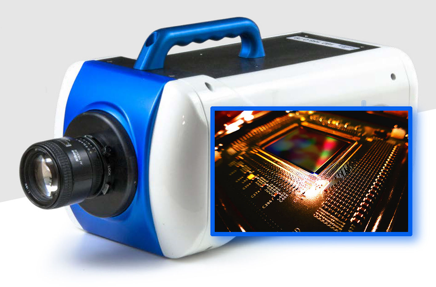 SI Sensors design novel CMOS image sensors for ultra-fast cameras that increase the velocity of the signal electrons allowing them to be swept out of the photodiode and be detected much faster. Developing ultra-high-speed CMOS image sensors is difficult because at very high frame rates, the integration time is extremely short and there is never enough light...
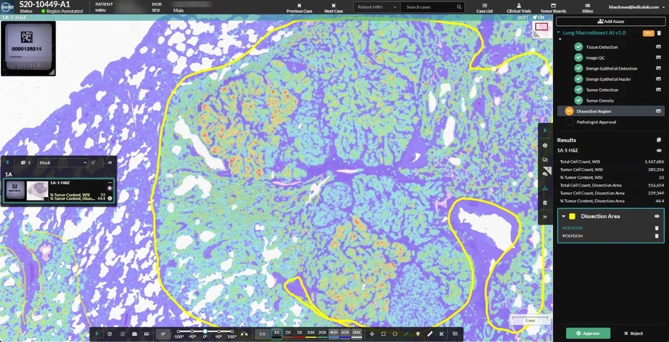 Indica Labs, the leading provider of AI-powered digital pathology solutions, proudly announces the launch of Lung Macrodissect AI, a groundbreaking tool that revolutionizes slide macrodissection and molecular pathology workflows. Lung Macrodissect AI represents a significant leap forward for the integration of artificial intelligence with digital pathology by offering pathologists a streamlined macrodissection workflow...
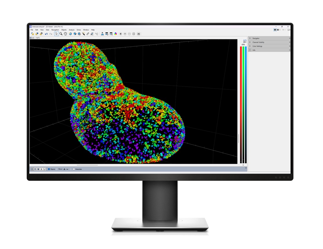 Flexibility in image analysis is key to addressing your diverse research questions. The ZEISS arivis software suite is designed to accelerate your research, offering advanced tools that ensure you reach reliable and reproducible results faster. No matter the size, format, or source of your image, ZEISS arivis can help you analyze it. And all without the need to code. Version 4.2 of arivis Pro is available now...
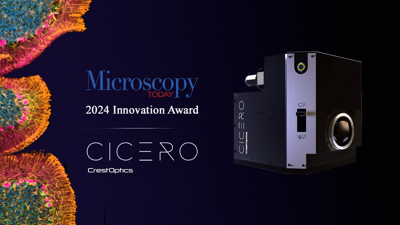 CrestOptics S.p.A., a manufacturer of high-end microscopy solutions and advanced systems for fluorescence microscopy and diagnostic applications, announce that its CICERO spinning disk confocal system has been named a ‘Top 10 Best Microscopy Innovation’ by Microscopy Today, an industry-leading microscopy publication from the Microscopy Society of America...
 Indica Labs announces the expansion of their reseller network in the Asia Pacific (APAC) region. New distributorship agreements have been inked with MRL Cybertec Corporation, Pathology Ware International, Minh Khang Technical Service & Trading Co. Ltd., and Histocenter, for distribution in the Philippines, Thailand, Vietnam, and Malaysia, respectively. The new partnerships build upon Indica’s previous commercial success in China and Japan where sales have grown substantially in recent years...
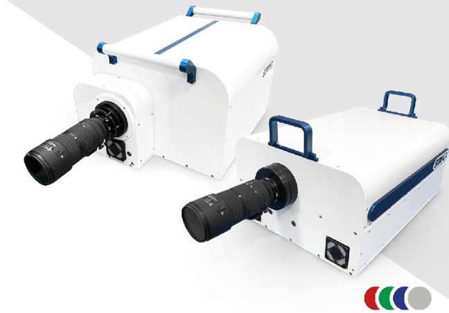 Specialised Imaging has introduced a new version of its market-leading SIM family of ultra high-speed framing camera that makes hyperspectral imaging simpler with the option of user interchangeable filters. Though hyperspectral imaging was possible on previous models, the new SIM design was driven by requests from the field of energetic materials research which required a camera with simple and fast exchange of filters in all its operating channels...
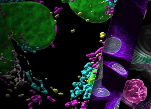 Oxford Instruments Andor has announced the release of Imaris 10.2, the latest version of its market-leading AI-powered image analysis software for microscopic images. In the last decade, Imaris has constantly evolved to improve the visualisation and analysis of large images. Past releases have introduced AI (machine learning) to improve object identification and classification, spatial measurements, correlative data visualisation, and other tools for more precise segmentation...
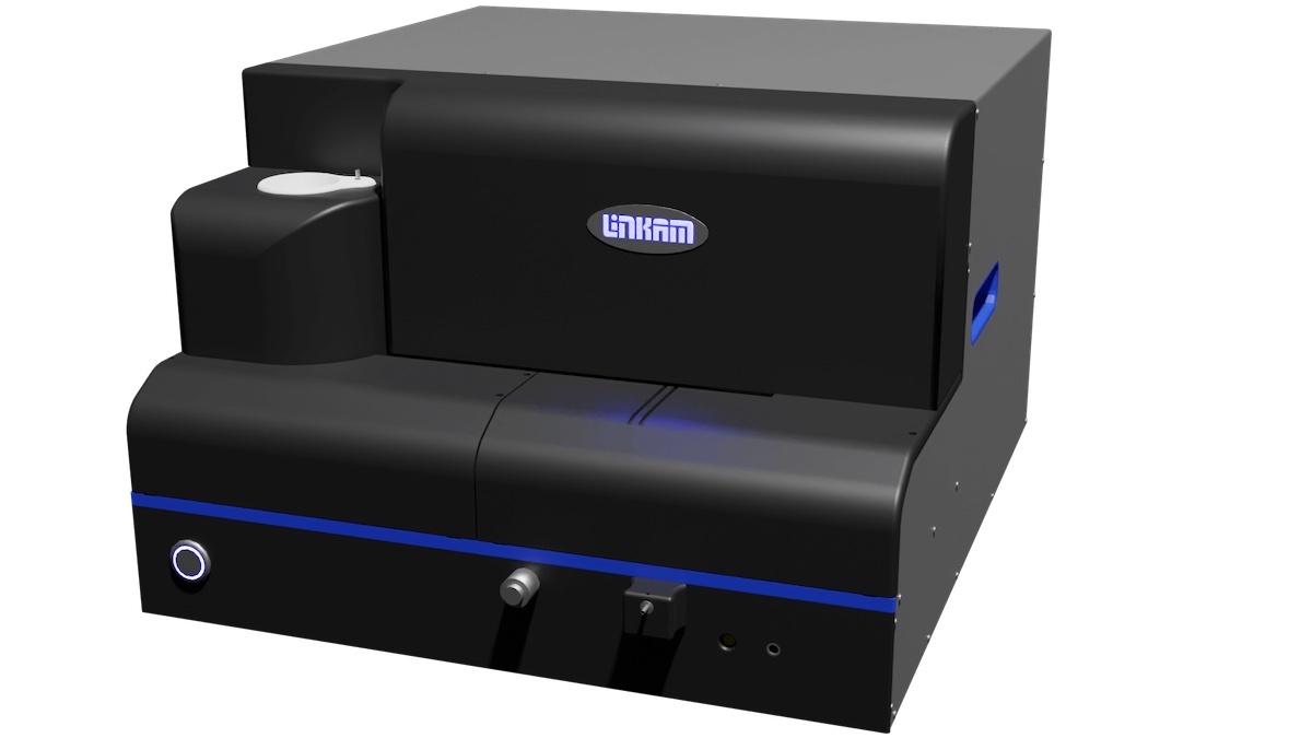 Linkam Scientific Instruments is showcasing its cryo-correlative portfolio at this year’s M&M conference, and will be hosting tutorials and workshops at the event. Linkam will also be presenting two posters during the conference. Linkam’s CryoGenium plunger system, one of the company’s latest developments, will be shown on the booth. The CryoGenium is a fully automated system which offers a range of features for users of cryo-EM, cryo-tomography, single particle tomography (SPT) and cryo-CLEM methods....
 TESCAN, a leading provider of scientific instrumentation, announces the launch of the TESCAN AMBER 2, the fourth generation of the TESCAN gallium FIB-SEM platform and the successor to the current AMBER generation, at the Microscopy & Microanalysis (M&M) Expo 2024. Alongside AMBER 2, TESCAN is also unveiling two new integrated innovative tools...
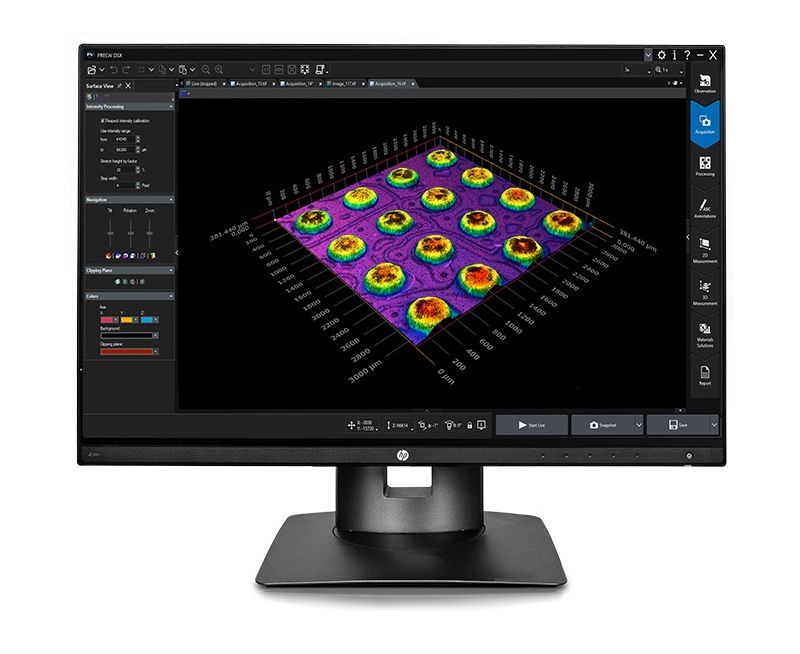 vident’s popular PRECiV™ image analysis software now powers our DSX1000 digital microscope, bringing new levels of ease of use and usability. With the launch of PRECiV v. 2.1.1, a single, unified software program can now be used to control all our manual, motorized, and digital industrial microscopes. PRECiV DSX makes using the DSX1000 digital microscope even simpler, providing seamless control over all motorized functions for greater speed and accuracy...
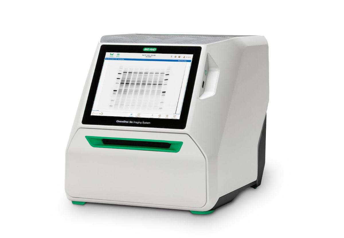 Bio-Rad Laboratories, Inc., a global leader in life science research and clinical diagnostics products, announce the launch of the ChemiDoc™ Go Imaging System, the latest addition to its portfolio of ChemiDoc Imaging Systems. The system offers rapid, reliable, and highly sensitive gel and western blot imaging on a benchtop scale. The ChemiDoc Go Imaging System uses complementary metal oxide semiconductor (CMOS) digital imaging to capture gel and western blot images with the same high sensitivity as larger instruments...
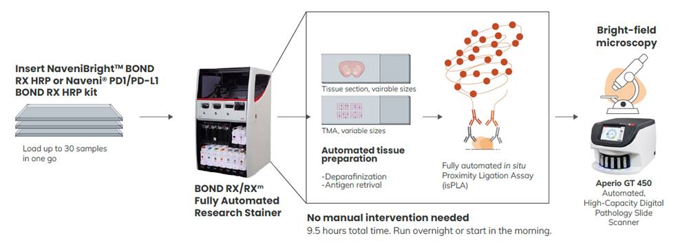 Leica Biosystems and Navinci Partner to Accelerate Cancer Research Through First-Ever Fully Automated In Situ Proximity Ligation Assays. Leica Biosystems, a global leader in anatomic pathology, and Navinci, a leader in developing innovative solutions for studying protein interactions, announce a strategic collaboration to drive innovation in cancer patient care. This partnership enables automated in situ proximity ligation assays on the BOND RX Fully Automated Research Stainer...
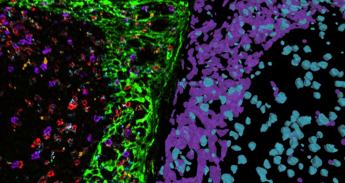 Leica Microsystems, a leading provider of microscopy and scientific instrumentation, has released version 14 of Aivia, its flagship image analysis solution. This update introduces a suite of new features and enhancements for accurate deep-learning based cell segmentation, automated phenotyping and spatial data analysis in 3D multiplexed images..
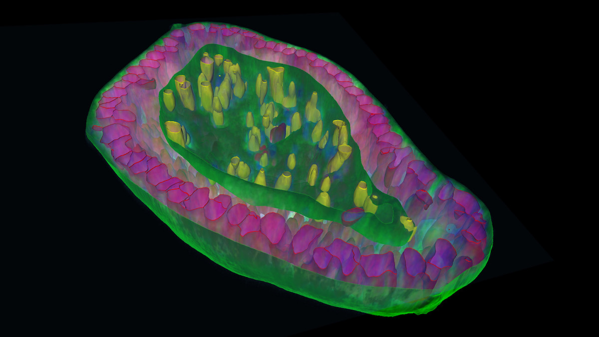 ZEISS has unveiled arivis Pro version 4.2, empowering researchers with unprecedented flexibility to tailor microscopy image analysis to their unique needs. This major release introduces advanced AI-powered segmentation tools, enhanced 3D analysis capabilities, and seamless handling of massive datasets – a universal solution optimized for any imaging workflow...
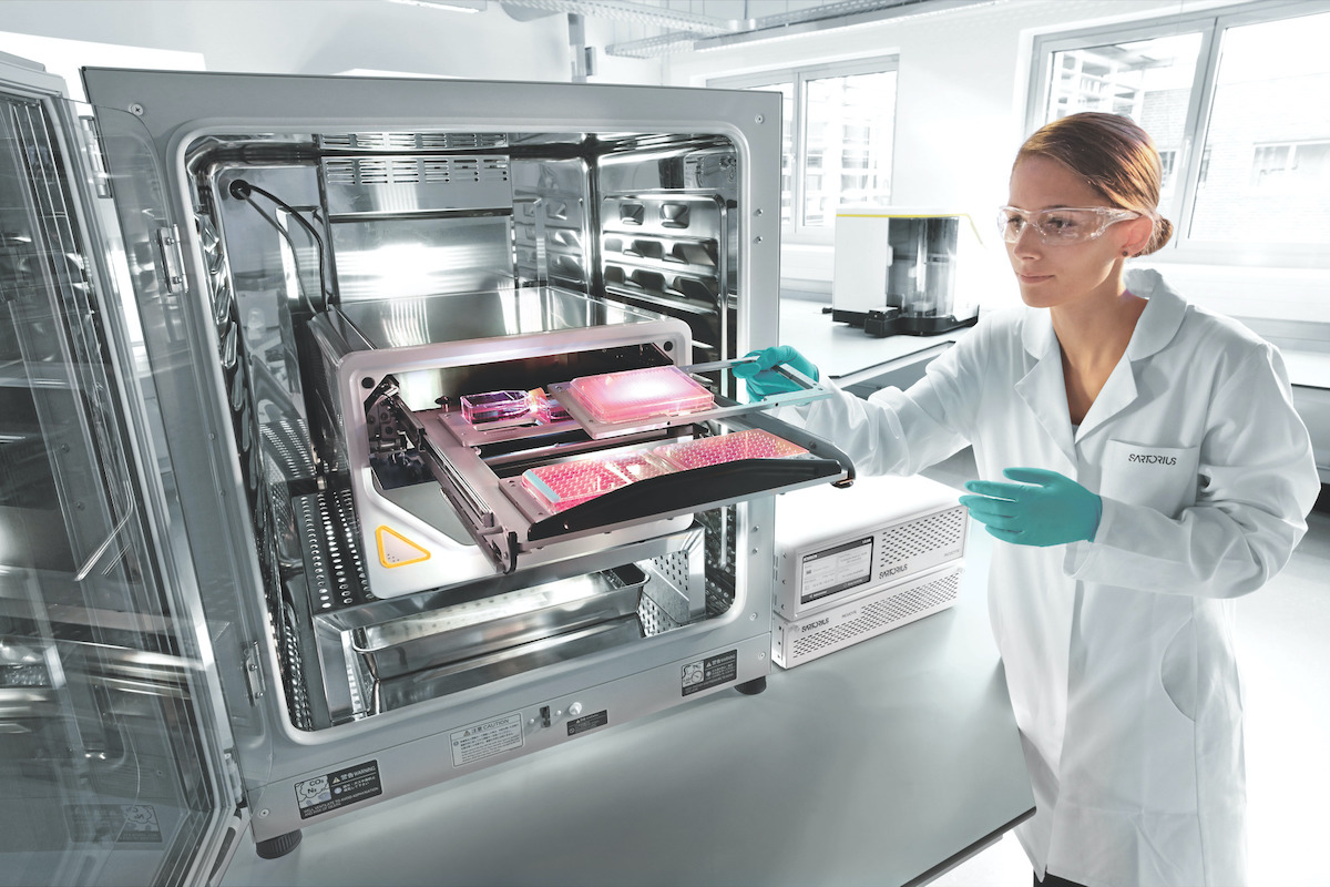 The life science group Sartorius is expanding its multidisciplinary collaboration with NVIDIA to help enable the development of new and better therapies, combining Sartorius’ in-depth knowledge of life sciences and bioprocessing with NVIDIA’s AI-powered computing platforms and software. "Biological interactions are exceptionally complex. Making better use of data by integrating life science expertise with AI solutions is a promising approach to simplify and accelerate biopharma drug discovery and manufacturing progress...
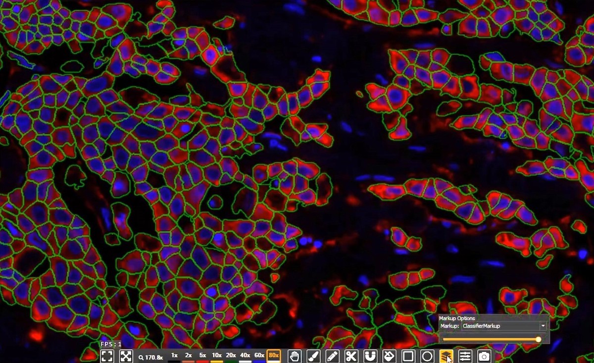 Indica Labs, the global leader in AI-powered digital pathology solutions, announce the release of the transformative new version of its industry leading life sciences software. The 4.0 versions of HALO®, HALO AI, and HALO Link offer researchers and pathologists unparalleled ease of use and performance, with even more opportunities to leverage advanced AI-powered image analysis in an open digital pathology ecosystem...
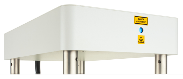 In recent years, Second-Harmonic Generation (SHG) microscopy has established itself as the technique of choice for the study of crystallized biomolecules such as starch, collagen and myosin. Unlike fluorescence-microscopy, SHG microscopy does not involve the creation of excited electronic states, so cell viability issues associated with heating and photo-bleaching are reduced. The report describes typical results from collagen in liver tissue samples generated by SHG microscopy...
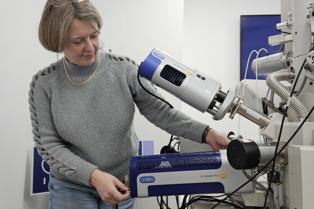 Oxford Instruments has been awarded the King’s Award for Enterprise for Innovation for the Symmetry detector. Symmetry enables a deeper understanding of a material’s structure down to the nanoscale level. Symmetry radically increased the speed with which such in-depth analyses can be performed, resulting in the technique being much more widely adopted. Symmetry uses a technique called electron backscatter diffraction (EBSD) to analyse surfaces for minuscule weaknesses or flaws in the crystalline structure...