
 Wai Chan, Managing Director of Specialised Imaging, commented “Over the last year we have achieved strong growth in our user base of Kirana high-speed digital video cameras. Delivering high resolution and high speed (up to 5 million frames / second) data acquisition in an affordable, yet no-compromise camera design the easy-to-use Kirana has appealed to many organizations investing in the benefits of ultra high-speed...
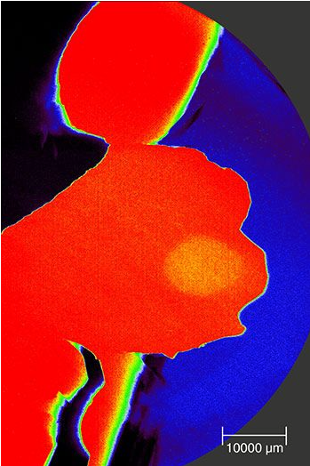 The inVia system enables users to study the widest range of samples with the broadest range of Raman imaging techniques. Renishaw's unique suite of complementary imaging options make it easy for users to get the chemical and structural information they need. On booth #2808, Renishaw's experts will explain the collective benefits of each technique and how the addition of transmission Raman provides maximum flexibility. Applications Scientist, Pippa Law, will give a...
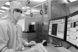 The application software analyses aerial images, taken by the photomask qualification tool ZEISS AIMS. It runs fully automated and provides immediate, standardized results. ZEISS has released the new application software ZEISS AutoAnalysis. The software solution provides fully automated analysis of ZEISS AIMS aerial images in parallel to the measurements as they are being captured. Manual interaction is no longer required, thus...
 JPK Instruments has moved to new offices in Berlin. Having outgrown their headquarters' premises of the last twelve years, JPK has moved across Berlin to a new suite of offices and laboratories together with larger manufacturing and service areas to meet the growing demands for their products worldwide. Having recently established offices in the USA, December 2014, JPK has invested in a new location in Berlin to provide increased levels...
 As camera manufacturers continue to increase sensor size, while simultaneously increasing pixel resolution, Resolve Optics custom large format lenses provide an optimised solution to enable these products to achieve their full performance potential opening the door to exciting new application solutions. As well as providing very high resolution over a large image format – Resolve Optics has designed diffraction-limited large format lenses with a large...
 The analysis of cultural heritage materials presents a number of challenges such as: limited and extremely small samples, complexity of sample structure, the importance of maintaining spatial integrity and, most notably, the rarity of the samples. These limitations present specific challenges for the identification of many traditional organic dyes, particularly in paintings that may have multiple original paint layers (depending on the artist’s...
 ZEISS is introducing a new compact confocal laser scanning microscope for high-end confocal imaging, ZEISS LSM 800. Tailored to the needs of a broad range of applications in individual research environments, the system complements the recently introduced ZEISS LSM 8 family. With highly sensitive GaAsP detector technology and fast linear scanning, ZEISS LSM 800 provides high image quality and offers enhanced productivity and throughput, as wel...
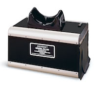 The CX-20 cabinet combines separate long-wave and short-wave 8-watt UV light sources with uniquely designed specular aluminum reflectors to assure maximum intensity and exceptional fluorescent contrast. An internal 25-watt white light bulb provides visible illumination. The unit has a removable bottom panel so it can be easily placed over 20 x 20 gels and TLC plates, or to provide greater illumination, over a Spectroline UV transilluminator...
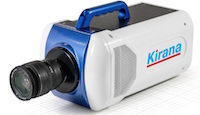 The Specialised Imaging Kirana camera provides REL, Inc with the ability to create high-resolution images of the ultra high-speed events that often occur with materials testing. Taking up to 5,000,000 frames per second, and shuttering every 100 nanoseconds, there is no compromise in capturing high quality data. The data is exported in 180 frames so each test can be easily saved and viewed. The initial applications undertaken...
 Uniquely combining high performance, leading edge features and affordability the omniDOC series provide an easy-to-use, yet powerful gel imaging system that satisfies the needs of most laboratories. The omniDOCi shares all the same features as the standard omniDOC but with the added benefit of wireless connectivity enabling the system to be run in a darkroom from a remote PC or tablet....
 Users of the SPARC technology at Chalmers University of Technology in Gothenburg, Sweden are applying cathodoluminescence measurements to study the properties of nanophotonic devices. Dr Ruggero Verre is a post-doctoral researcher in the Department of Applied Physics - Division of Bionanophotonics at the Chalmers University of Technology in Gothenburg in southern Sweden. His work in the group headed by...
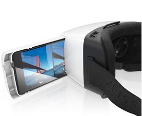 The attention of the young development team was sharply focused on user-friendliness, design and technology. To dive into virtual worlds, all the user needs to do is select an appropriate VR app and slide his or her smartphone into the headset using the tray provided. ZEISS VR ONE will initially be available worldwide with a tray for the Samsung Galaxy S5 smartphone and the iPhone 6. Other models are in the pipeline. The Computer Aided...
 Heading up this new organization is Dr Stefan Kaemmer who has been appointed General Manager of US Operations. Respected German technology leaders, JPK Instruments, are pleased to announce the opening of the first USA-based offices. With more than twelve years supplying nanoscale solutions worldwide for researchers in the bio, life and materials sciences, JPK is to open their offices in Southern California to support users across the...
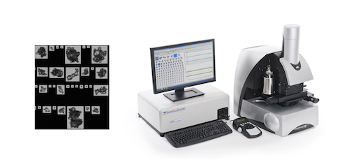 The ability of uncontrolled agglomeration to substantially impact the performance and value of powder products makes efficient agglomerate detection vital across a number of industries. ‘Identification of agglomerates using automated image analysis’ presents practical strategies for efficiently and robustly differentiating agglomerates from primary particles, to support product development, QC and process troubleshooting....
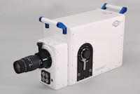 Incorporating a supplementary optical port, that uses a beamsplitter to deliver 50% of the primary image to a secondary image plane, allowing secondary instruments to share the same optical axis, thereby providing true undistorted datasets for simultaneous data measurement. Recording simultaneous ultra fast two-dimensional and time-resolved images is of significant interest to scientists in a growing number of fields of study including...
 The new kit uses imaging flow cytometry to obtain statistically significant quantitative assessment of NF?B translocation, as well as visual identification of the translocation at a single-cell level.
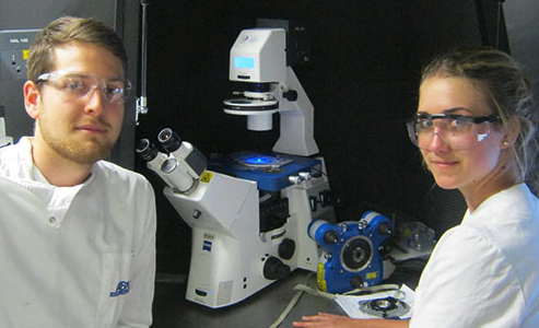 The Australian Research Council Centre of Excellence in Plant Cell Walls is a collaborative project involving the Universities of Adelaide, Melbourne and Queensland in partnership with South Australia State Government and seven international institutions. Their aim is to advance fundamental scientific understanding of plant cell wall biology. Cell wall composition determines the quality of most plant-based products used in modern human societies...
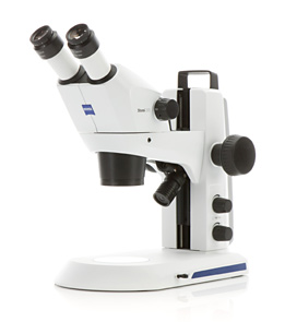 ZEISS is introducing two new compact Greenough stereo microscopes for education, lab routine and industrial inspection: ZEISS Stemi 305 and ZEISS Stemi 508. Users are able to observe their samples in true color, 3D, with high contrast and free of distortion or color fringes. ZEISS Stemi 305 offers a 5:1 zoom. The microscope is easy to use and everything is integrated: LED illumination, reflected and transmitted light illuminations and documentation. Users choose...
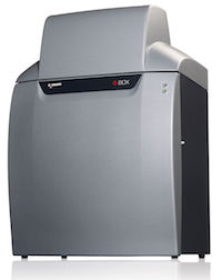 Researchers at the major university are using a G:BOX Chemi XRQ multi-task imager to precisely analyse agarose gels of mouse and rabbit DNA stained with the safe fluorescent dye SybrSafe®. They are also utilising the G:BOX Chemi XRQ to easily image both large and small SDS-PAGE gels and chemiluminescent Western blots of proteins. This is allowing scientists at the university to accurately detect proteins and is contributing to identifying potential therapeutic targets in...