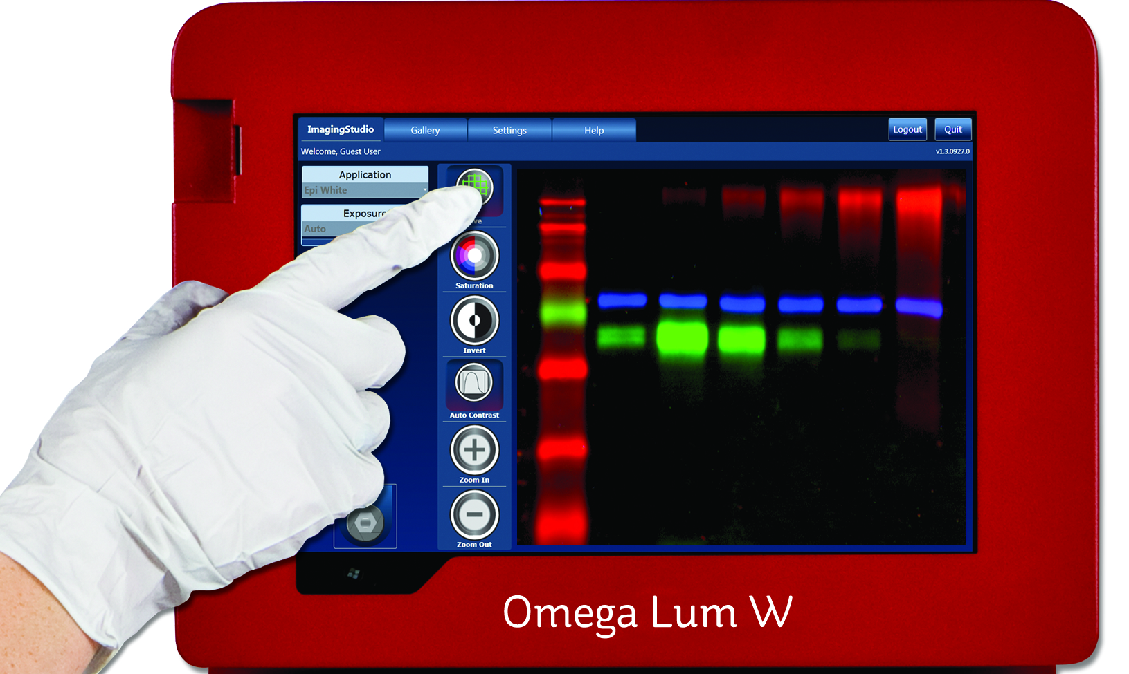
An exiciting new imaging system can be used for gel, multicolour R, G, B and chemiluminescence.
Eikonix, the Cambridge based gel documentation experts, are pleased to announce the introduction of the Omega Lum W analysis system. This exciting new product can be used for gel, multicolour R, G, B and chemiluminescent imaging and brings performance and features at a price which is now affordable to a much wider market. This small footprint system breaks the mould of the older traditional heavy and cumbersome gel documentation systems by utilising some innovative design work and incorporating an integral tablet for control....
MR Solutions and Summit Pharmaceuticals have reached an agreement where Summit Pharmaceuticals will be the sole supplier for MR Solutions’ preclinical, multi-modality cryogen free MRI imaging systems to the Japanese market.
Summit Pharmaceuticals, part of the Sumitomo Corporation, is a leading Japanese provider of drug discovery instruments and through this partnership, both companies will be able to offer a complete imaging portfolio of high field preclinical multi-modality MRI solutions including PET/MR and SPECT/MR, plus CT, optical systems, and bioresource materials. Based in Surrey, UK, MR Solutions pioneered the development of the world’s...
Gel Company celebrates 20 years of serving the life science market. Founded in 1995, Gel Company, based in San Francisco, develops manufactures, and supplies low cost products to the life science research community.
The company has set its goal to provide innovative, high quality products with an efficient, friendly service. The aim of Gel Company is to offer all its products at competitive prices. The focus is to develop, manufacture and supply innovative tools for proteomics, genomics, cell biology, liquid handling and microarray applications to scientists around the world.
Renishaw, a world leader in metrology and spectroscopy technologies, reports on the use of its inVia confocal Raman microscope in conjunction with a Bruker Dimension Icon atomic force microscope (AFM) to study electrochemical energy storage materials at the US Army Research Laboratory (ARL) in Maryland, USA.
Dr Collin Becker is a mechanical engineer at the US Army Research Laboratory (ARL) in Maryland, USA. His group studies materials for advanced lithium ion batteries and future-generation energy storage systems, such as sodium ion, magnesium ion and solid-state batteries. His research on lithium ion batteries focuses on developing high capacity anode materials to improve the overall energy density, rate of discharge and safety of...
JPK Instruments, a world-leading manufacturer of nanoanalytic instrumentation for research in life sciences and soft matter, reports on how their NanoWizard® AFM system is being combined with a toolbox of fluorescence microscopy techniques at the Weatherall Institute of Molecular Medicine located at Oxford University.
The Weatherall Institute of Molecular Medicine (WIMM) at Oxford University comprises various groups involved in molecular medicine research forming a “Research Hotel.” They all come from different medical departments and also different topics (immunology, haematology, oncology, neurobiology), but their joint efforts within one place and the possibility to use state-of-the-art facilities shall optimize research efforts and bolster...
EM Resolutions, manufacturers and suppliers of tools and accessories for users of electron microscopes, announce new standards for SEM calibration - the EM-Tec MCS Series.
EM Resolutions has extended its range of resolution and magnification standards for SEM with the introduction of the EM-Tec MCS and M series of magnification calibration standards. These fully featured practical calibration standards have been specially developed for magnification calibration or critical dimension measurements in table top SEM, standard SEM, FEGSEM, FIB, Auger, SIMS and reflected light microscope systems...
Syngene, a world-leading manufacturer of image analysis solutions, is pleased to announce its G:BOX Chemi XX6 multi-application-functional imager is being utilised by scientists at major human protein R&D company, Octapharma Biopharmaceuticals GmbH in Germany for analysing a range of large proteins.
This is allowing the researchers there to accurately characterise proteins which will be used as therapies to treat conditions such as Haemophilia A. Researchers in the Molecular Biochemistry Unit at Octapharma in Berlin are using a G:BOX Chemi XX6 system to analyse proteins of up to 20 million KDa on oversized agarose gels and blots of up to 22cm. These proteins have been isolated from the plasma of healthy individuals or are recombinant versions of these plasma proteins, and studying them is...
Olympus introduces its new Z-Drift Compensation IX3-ZDC2 system and updated cellSens software, for enhanced live cell and multi well plate imaging.
These new technologies deliver fast and accurate multi well time-lapse acquisition, stable focus during long time-lapse studies and an easier user interface. Taking well plate imaging to the next level, Olympus presents two innovative tools for the customisable IX3 inverted microscopes. The new IX3-ZDC2 module offers cutting-edge Olympus focal drift compensation technology, enabling accurate focus stability during long...
The OptiScan® III from Prior Scientific is a powerful, entry level system for controlling the OptiScan range of motorised microscope stages and focusing devices.
Compatible with a wide range of both upright and inverted microscopes, the compact OptiScan III uniquely can be used to drive a motorised stage and a focus motor while precision motor drive technology ensures quiet and smooth motion. A joystick provides users with precise and ergonomic control of their microscope accessories in the X, Y and Z axes. The controller is compatible with most image analysis software packages...
Next Generation Atom Probe Included Among 100 Most-Significant Developments in Research and Development
LEAP 5000 Atom Probe Offers Precise Atom-by-Atom, Sub-Nanometric Analysis of Metals, Semiconductors and Other Materials
CAMECA, a unit of AMETEK Materials Analysis, was recognized among the winners of the 54th Annual R&D 100 Awards by the editors of R&D magazine for its development of the LEAP 5000. Launched in August 2014, the LEAP 5000 is the latest generation of atom probe microscopes, which offer precise atom-by-atom identification, 3-D spatial positioning, and accurate atomic-scale reconstruction of a material’s microstructure...
Spectronics Corporation produces state-of-the-art products that technicians need to perform everyday essential laboratory applications.
One such product is Spectroline®CX-20 high-intensity UV viewing cabinet. Designed for peak efficiency, it guarantees maximum ultraviolet irradiance and fluorescent contrast. The CX-20 cabinet combines separate long-wave and short-wave 8-watt UV light sources with uniquely designed specular aluminum reflectors to assure maximum intensity and exceptional fluorescent contrast. An internal 25-watt white light bulb provides visible illumination...
EM Resolutions, manufacturers and suppliers of tools and accessories for users of electron microscopes, announce the availability of EM-Tec FIB grids which offer a secure way to attach TEM lamellas to the posts during lift-out procedures with FIB or SEM/FIB systems.
The new EM-Tec range of FIB lift-out grids from EM Resolutions provides a secure way to attach TEM lamella prepared with Focussed Ion Beam (FIB) instruments. Available in multiple post configurations, with a shape optimized for easy accessibility, EM-Tec FIB grids are compatible with all standard 3 mm TEM grid holders. EM-Tec FIB lift-out grids are available in three types: copper, molybdenum and unique, smooth-walled molybdenum...
Delivering new benchmarks in dynamic performance and accuracy, Queensgate’s NPC-D-5200 is a digital controller for use with the company’s highly advanced closed loop piezo actuators and stages.
Queensgate pioneered the use of capacitive sensors to provide precise positional feedback in closed loop; the NPC-D-5200 incorporates a precision capacitive measurement circuit and is updated with the stage position 120000 times per second, delivering high positional accuracy at speed. It has the capacity to address a broad spectrum of highly demanding alignment and metrology applications. By employing proprietary low noise...
The funds will be used to fuel international growth and to enter into the in-vitro diagnostics market.
Ovizio Imaging Systems, an innovative Belgian company developing cell counting solutions based on quantitative microscopy for life sciences applications, today announces it has secured a funding round of €8m ($9.1m) co-led by New Science Ventures, a US-based venture capital firm, and a private investor. This funding round...
LaVison BioTec, developers of advanced microscopy solutions for the life sciences, report on the work of Nicolas Renier, a Post-Doctoral Fellow in the laboratory of Marc Tessier-Lavigne at the Rockefeller University in New York where he applies light sheet microscopy to study axon rewiring in adult nervous systems.
Drs Nicolas Renier and Zhuhao Wu are post-doctoral fellows in the laboratory of Marc Tessier-Lavigne at the Rockefeller University in New York where they have co-developed methodologies to develop new imaging techniques applying light sheet microscopy to the study axon rewiring in adult nervous systems. It is hoped that this could lead to an understanding as to how experience or even pathologies such as Alzheimer's...
Complete workflow automation for long term cell assays
BioTek Instruments released the BioSpa™ 8 Automated Incubator, a unique platform that links microplate washers and dispensers with readers and imaging systems for unattended workflow automation. Real time temperature and CO2/O2 control and monitoring, plus humidity level monitoring and plate lid handling provide an ideal environment for cell-based and other assays, with minimal manual intervention...
Market leaders in temperature controlled microscopy, Linkam Scientific Instruments report on the use of their temperature controlled stages applied to CLEM and fluorescence microscopy to assist in endocytic sorting in the School of Biochemistry at the University of Bristol.
Dr Paul Verkade is a Reader in Cell Imaging in the School of Biochemistry at the University of Bristol where he also heads the Electron Microscopy unit of the Wolfson Bioimaging Facility. His current research focus is to develop techniques and tools for the use of Correlative Light Electron Microscopy (CLEM) studying endocytic sorting. Dr. Verkade is currently chair of the Electron Microscopy section of the Royal Microscopy Society and chair of...
Mazurek Optical Services are pleased to announce they have recently installed a suite of light microscopes in the School of Earth and Ocean Sciences at Cardiff University.
The microscopes have already been used in the undergraduate teaching laboratories by students returning for the new academic year. The School of Earth and Ocean Sciences at Cardiff University offers students a research-led experience across a wide raft of disciplines. They provide undergraduates with top class facilities that include dedicated IT labs, state-of-the-art analytical equipment and even their own research vessel. To coincide...
Deben, a leading provider of in-situ testing stages together with innovative accessories and components for electron microscopy, reports on the use of the Microtest 200 N tensile stage in the Department of Aeronautics at Imperial College.
London where it is used to measure load displacement curves on nanocellulose-reinforced polymer composites. Dr Koon-Yang Lee is a Lecturer in Composite Manufacturing in the Department of Aeronautics at Imperial College London. His young and dynamic research group focuses on the manufacturing of polymer (nano)composites, surface and interface engineering, particle stabilised emulsions and foams. His research is highly...
Prior Scientific has announced that the Core Imaging Facility at the UK's prestigious National Institute for Medical Research (NIMR) has chosen a Prior HLD117 linear motor stage to be the sample platform at the centre of a new system based around an Olympus IX83 microscope.
NIMR is one of the world’s leading medical research institutes. It is dedicated to studying important questions about biological processes that are relevant to all aspects of health. Research at NIMR covers a broad spectrum of basic biomedical science, including infectious diseases, immunology, cell and developmental biology, neuroscience and structural biology. The world-class facilities for research include biological imaging resources, the...
JPK Instruments, a world-leading manufacturer of nanoanalytic instrumentation for research in life sciences and soft matter, reports on the multiple applications where their NanoWizard® AFM system is being used at the Korean Institute of Chemical Technology to study soft materials such as biomolecules and polymers.
Established in 1976 for R&D of chemical technology in Korea, the Korean Research Institute of Chemical Technology (KRICT) has helped drive the growth of the country“s chemical industry. The focus is on the development of world-class key technologies. There are four key research fields: the development of eco-friendly chemical process technology; the development of high value-added green chemical materials; the discovery of...
Ovizio Imaging Systems, an innovative microscopy company specializing in automated cell culture monitoring systems, and Pall Life Sciences, a global leader in biopharmaceutical fluid management, today announce the signing of a long term global supply agreement for the iLine S microscope.
The financial terms of the agreement have not been disclosed. Ovizio has collaborated with Pall for several years to customize the iLine S microscope and will now further improve the technology. The microscope has been specifically designed for use with Pall’s technology and will be commercialized as part of Pall’s Xpansion® cell production platform; the first fully-closed bioreactor for large-scale production of adherent stem cells. The partnership...

 Summit Pharmaceuticals, part of the Sumitomo Corporation, is a leading Japanese provider of drug discovery instruments and through this partnership, both companies will be able to offer a complete imaging portfolio of high field preclinical multi-modality MRI solutions including PET/MR and SPECT/MR, plus CT, optical systems, and bioresource materials.
  The company has set its goal to provide innovative, high quality products with an efficient, friendly service. The aim of Gel Company is to offer all its products at competitive prices. The focus is to develop, manufacture and supply innovative tools for proteomics, genomics, cell biology, liquid handling and microarray applications to scientists around the world.
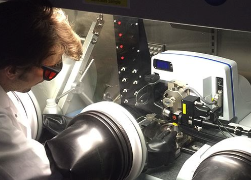 Dr Collin Becker is a mechanical engineer at the US Army Research Laboratory (ARL) in Maryland, USA. His group studies materials for advanced lithium ion batteries and future-generation energy storage systems, such as sodium ion, magnesium ion and solid-state batteries. His research on lithium ion batteries focuses on developing high capacity anode materials to improve the overall energy density, rate of discharge and safety of...
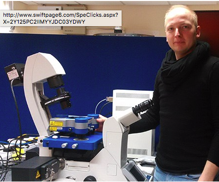 The Weatherall Institute of Molecular Medicine (WIMM) at Oxford University comprises various groups involved in molecular medicine research forming a “Research Hotel.” They all come from different medical departments and also different topics (immunology, haematology, oncology, neurobiology), but their joint efforts within one place and the possibility to use state-of-the-art facilities shall optimize research efforts and bolster...
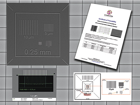 EM Resolutions has extended its range of resolution and magnification standards for SEM with the introduction of the EM-Tec MCS and M series of magnification calibration standards. These fully featured practical calibration standards have been specially developed for magnification calibration or critical dimension measurements in table top SEM, standard SEM, FEGSEM, FIB, Auger, SIMS and reflected light microscope systems...
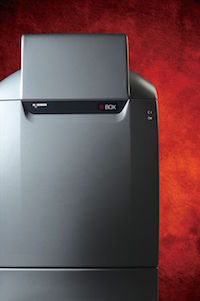 This is allowing the researchers there to accurately characterise proteins which will be used as therapies to treat conditions such as Haemophilia A.
 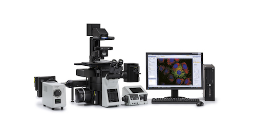 These new technologies deliver fast and accurate multi well time-lapse acquisition, stable focus during long time-lapse studies and an easier user interface. Taking well plate imaging to the next level, Olympus presents two innovative tools for the customisable IX3 inverted microscopes. The new IX3-ZDC2 module offers cutting-edge Olympus focal drift compensation technology, enabling accurate focus stability during long...
 Compatible with a wide range of both upright and inverted microscopes, the compact OptiScan III uniquely can be used to drive a motorised stage and a focus motor while precision motor drive technology ensures quiet and smooth motion. A joystick provides users with precise and ergonomic control of their microscope accessories in the X, Y and Z axes.
  CAMECA, a unit of AMETEK Materials Analysis, was recognized among the winners of the 54th Annual R&D 100 Awards by the editors of R&D magazine for its development of the LEAP 5000. Launched in August 2014, the LEAP 5000 is the latest generation of atom probe microscopes, which offer precise atom-by-atom identification, 3-D spatial positioning, and accurate atomic-scale reconstruction of a material’s microstructure...
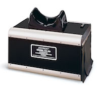 One such product is Spectroline
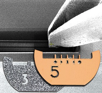 The new EM-Tec range of FIB lift-out grids from EM Resolutions provides a secure way to attach TEM lamella prepared with Focussed Ion Beam (FIB) instruments. Available in multiple post configurations, with a shape optimized for easy accessibility, EM-Tec FIB grids are compatible with all standard 3 mm TEM grid holders.
 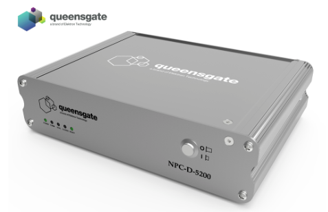 Queensgate pioneered the use of capacitive sensors to provide precise positional feedback in closed loop; the NPC-D-5200 incorporates a precision capacitive measurement circuit and is updated with the stage position 120000 times per second, delivering high positional accuracy at speed. It has the capacity to address a broad spectrum of highly demanding alignment and metrology applications. By employing proprietary low noise...
 Ovizio Imaging Systems, an innovative Belgian company developing cell counting solutions based on quantitative microscopy for life sciences applications, today announces it has secured a funding round of €8m ($9.1m) co-led by New Science Ventures, a US-based venture capital firm, and a private investor. This funding round...
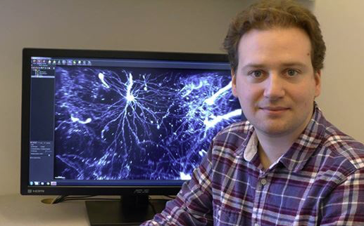
 Dr Paul Verkade is a Reader in Cell Imaging in the School of Biochemistry at the University of Bristol where he also heads the Electron Microscopy unit of the Wolfson Bioimaging Facility. His current research focus is to develop techniques and tools for the use of Correlative Light Electron Microscopy (CLEM) studying endocytic sorting. Dr. Verkade is currently chair of the Electron Microscopy section of the Royal Microscopy Society and chair of...
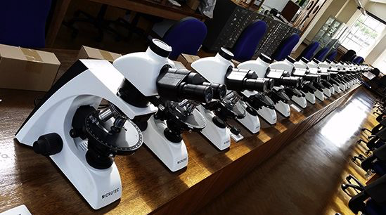 The microscopes have already been used in the undergraduate teaching laboratories by students returning for the new academic year. The School of Earth and Ocean Sciences at Cardiff University offers students a research-led experience across a wide raft of disciplines. They provide undergraduates with top class facilities that include dedicated IT labs, state-of-the-art analytical equipment and even their own research vessel. To coincide...
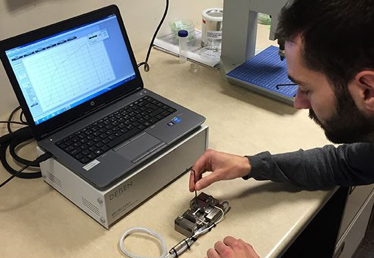 London where it is used to measure load displacement curves on nanocellulose-reinforced polymer composites. Dr Koon-Yang Lee is a Lecturer in Composite Manufacturing in the Department of Aeronautics at Imperial College London. His young and dynamic research group focuses on the manufacturing of polymer (nano)composites, surface and interface engineering, particle stabilised emulsions and foams. His research is highly...
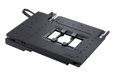 NIMR is one of the world’s leading medical research institutes. It is dedicated to studying important questions about biological processes that are relevant to all aspects of health. Research at NIMR covers a broad spectrum of basic biomedical science, including infectious diseases, immunology, cell and developmental biology, neuroscience and structural biology. The world-class facilities for research include biological imaging resources, the...
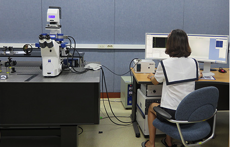 Established in 1976 for R&D of chemical technology in Korea, the Korean Research Institute of Chemical Technology (KRICT) has helped drive the growth of the country“s chemical industry. The focus is on the development of world-class key technologies. There are four key research fields: the development of eco-friendly chemical process technology; the development of high value-added green chemical materials; the discovery of...
 The financial terms of the agreement have not been disclosed. Ovizio has collaborated with Pall for several years to customize the iLine S microscope and will now further improve the technology. The microscope has been specifically designed for use with Pall’s technology and will be commercialized as part of Pall’s Xpansion® cell production platform; the first fully-closed bioreactor for large-scale production of adherent stem cells. The partnership...