Channels
Special Offers & Promotions
JPK Reports on the Use of AFM and Advanced Fluorescence Microscopy at the University of Freiburg
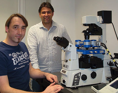
JPK Instruments, a world-leading manufacturer of nanoanalytic instrumentation for research in life sciences and soft matter, reports on how researchers at the University of Freiburg is combining the use of AFM and advanced fluorescence microscopy to characterize the local mechanical properties of cells.
 The research team of Junior Professor Dr Winfried Römer is located at the Centre for Biological Signalling Studies (BIOSS) at the University of Freiburg. Under the title of Synthetic Biology of Signalling Processes, the Römer team studies the interactions of human pathogens (bacteria, viruses) and pathogenic products (toxins) with various human cells by using a highly interdisciplinary research approach at the interface of biology, medicine, physics and chemistry. Within this work, the group is developing a unique platform for membrane research based on natural and synthetic membrane systems of different complexity in combination with state-of-the-art microscopy techniques.
In particular, the lab studies the cellular uptake of the human, opportunistic pathogen Pseudomonas aeruginosa in non-phagocytic cells. One major goal is to elucidate the early steps of the bacterial internalization process using a combination of two complementary microscopy techniques: AFM and fluorescence microscopy.
Postdoctoral worker Dr Josef Madl, who is implementing the combined measurements, says “For studying bacterial invasion we infect human lung cells with a GFP-tagged P. aeruginosa strain. The bacteria attach to the cell surface and, after some time, start to invade into the cells. We employ conventional AFM imaging to visualize bacteria on the cell surface with high 3D resolution. In addition, the Quantitative Imaging (QI™) mode can be utilized for obtaining both the topography and an elasticity map of the cell surface. Thereby we can determine if and how the bacterium affects the local mechanical properties of the plasma membrane during invasion. We can optimize our experiments further by using AFM in combination with fluorescence microscopy. This allows us to quickly identify regions-of-interest for AFM imaging by visualizing the bacteria on the cells via GFP fluorescence. The combined operation with fluorescence also enables us to highlight important structural components of the cells, for example the underlying cytoskeleton or the plasma membrane wrapping around an invading bacterium. Finally, we want to perform AFM in combination with the optical super-resolution method STORM. Thereby we can accurately measure the localization of various key players in the invasion process, such as actin or bacterial lectins, and correlate this information with the 3D topography and local mechanical properties even more precisely than with conventional diffraction-limited microscopy.”
Describing the benefits of JPK’s AFM compared to other SPM systems, Dr Madl found the NanoWizard® 3 very user-friendly. “The design of the instrument is practical. It has a high mechanical stability and is well-suited for life sciences research. A key feature for us is that it can be easily combined with an inverted optical microscope. For our purposes, the DirectOverlay™ algorithm and its integration in the software are very useful and important. Furthermore, JPK’s QI™ mode is very powerful. We are also impressed with the technical support we receive from JPK.”
more news from JPK instruments
Media Partners


